
60-year-old chronic smoker male came to the emergency with complaints of progressive hoarseness and difficulty in breathing for the last 2 months. What is the most probable diagnosis? How do you manage the stridor in this patient? What are the confirmatory ENT investigations to make the diagnosis?
"laryngeal neoplasms"[MeSH Terms] AND "airway obstruction"[MeSH Terms]
laryngoscopy glottic carcinoma vocal cord lesion
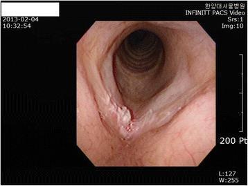
This clinical photograph captures a fiberoptic laryngoscopy view of the glottic larynx. The image demonstrates a well-defined pathological lesion situated on the anterior third of the right true vocal cord. The lesion exhibits an exophytic growth pattern with a markedly irregular, leukoplakic (whitish), and granular surface texture, which contrasts sharply with the smooth, pearly-pink appearance of the contralateral left vocal cord. There is visible involvement of the anterior commissure, and the surrounding mucosa shows signs of mild erythema, suggestive of localized inflammation. Anatomical landmarks including the vocal folds and the tracheal rings visible through the glottic opening are shown. This visual is characteristic of glottic squamous cell carcinoma or high-grade laryngeal dysplasia, illustrating common clinical findings in patients presenting with chronic hoarseness and a history of occupational carcinogen exposure.
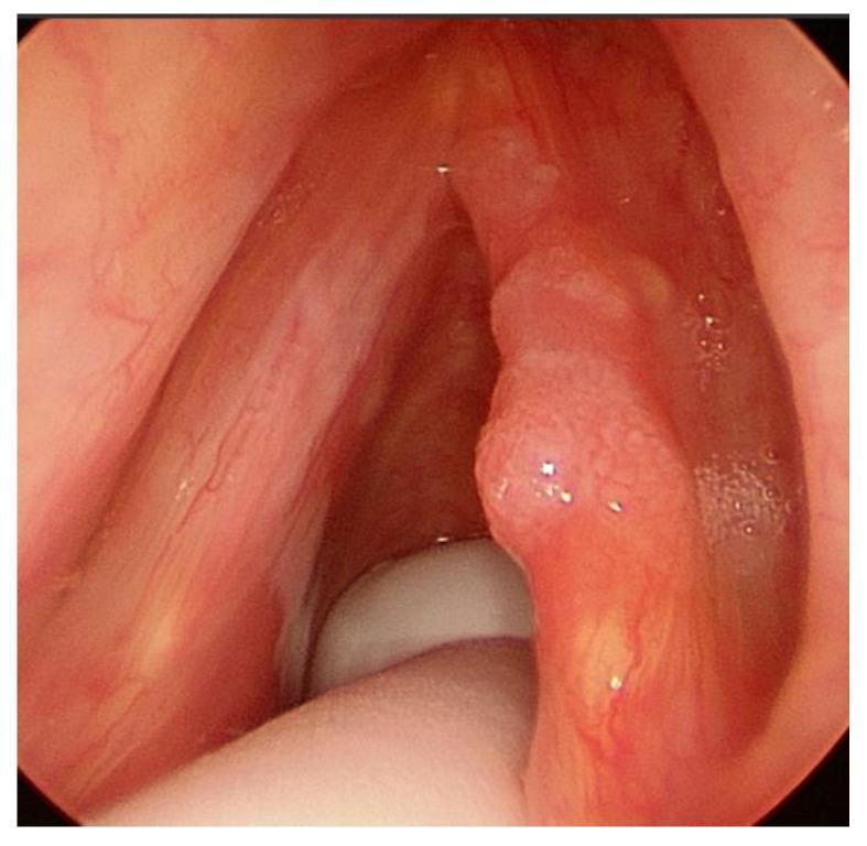
This clinical endoscopic image provides a laryngeal view of the vocal folds, demonstrating early-stage glottic malignancy. The right vocal cord features a prominent, exophytic mass with a nodular and irregular surface texture. The lesion appears flesh-colored to slightly erythematous and is localized to the mid-portion of the cord, consistent with a T1a glottic carcinoma. Notably, the anterior commissure remains uninvolved, with a clear boundary visible between the tumor and the anterior junction of the folds. In contrast, the left vocal cord appears healthy, exhibiting a smooth, pearly-white mucosal surface and normal vascular patterns. This comparison highlights the morphological characteristics of focal laryngeal neoplasia, such as surface irregularity and loss of the straight vocal fold edge, while illustrating the clinical staging criteria where tumor limitation to a single cord and lack of commissure involvement define the T1a classification. This visual is relevant for otolaryngology training regarding diagnostic laryngoscopy and head and neck oncology staging.
Most probable diagnosis
-
Age 60, male, and chronic smoking are the classic risk profile. Alcohol is a synergistic risk factor.
-
Progressive hoarseness for more than 2 weeks, especially with smoking, is laryngeal cancer until proven otherwise. The cord is involved early, so hoarseness is the presenting symptom.
-
Dyspnoea and stridor over 2 months suggest a bulky tumour, or a fixed cord with a narrowed glottic airway. This is at least T3 disease, or T4 if there is cartilage invasion. Stridor is a late sign.
-
Other causes of chronic hoarseness with stridor are:
- bilateral vocal cord palsy, for example from a thyroid, oesophageal or lung malignancy
- laryngeal tuberculosis
- a subglottic or tracheal tumour
- Reinke's oedema or papillomatosis (unlikely here)
These need to be excluded.
Management of stridor
- Sit him upright in the position of comfort. Give humidified oxygen and monitor SpO2.
- Keep him nil by mouth. Alert the ENT and anaesthesia teams early.
- Arrange a senior ENT surgeon, an anaesthetist and a difficult airway/tracheostomy set-up.
- Temporising medical measures buy time but do not treat a fixed tumour obstruction:
- nebulised adrenaline
- IV dexamethasone or hydrocortisone to reduce peritumoural oedema
- heliox, if available
- Avoid sedatives and avoid lying him flat.
- Stridor at rest, use of accessory muscles, suprasternal and intercostal retractions, cyanosis, exhaustion or altered sensorium mean he needs a definitive airway now.
- Mild stridor only on exertion may allow a brief, controlled work-up.
- Awake tracheostomy under local anaesthesia is the safest option in a tumour-obstructed airway. It is the standard for severe stridor.
- The tracheostomy is done through a low incision, below the tumour.
- Blind intubation can precipitate complete obstruction or bleeding from a friable tumour.
- Cummings lists tumour under "Airway obstruction" among the indications for tracheotomy (Box 7.1, Cummings Otolaryngology Head and Neck Surgery).
- Emergency cricothyroidotomy or needle cricothyroidotomy is the rescue option if he decompensates before a tracheostomy.
- Anaesthetic principles (Scott-Brown's Otorhinolaryngology):
- With glottic or periglottic obstruction, spontaneous respiration should be preserved.
- Inhalational induction with a non-irritant agent such as sevoflurane is favoured.
- Muscle relaxants should be avoided until the airway is secured.
- Awake fibreoptic intubation is of limited use here. The scope can block a narrow glottis ("corking the bottle") and cause bleeding or oedema.
- The anaesthetist should be ready for immediate front-of-neck access.
- Alternative in an operating theatre: Gentle direct laryngoscopy with a small endotracheal tube, or rigid bronchoscope ventilation, can be used. A tracheostomy is then done, with biopsy under the same anaesthetic if safe.
- Tracheostomy placement: Do it low and away from the likely laryngectomy field. Tracheostomy through a tumour or too high risks stomal recurrence. A tracheostomy done before definitive surgery usually does not worsen the prognosis. Definitive treatment, usually total laryngectomy or chemoradiation, is planned afterwards.
Confirmatory ENT investigations
- Indirect laryngoscopy (mirror) is the bedside screening test.
- Flexible fibreoptic nasopharyngolaryngoscopy is the first-line outpatient test, done carefully if stridor is present. It shows the lesion, its extent, cord mobility, subglottic and supraglottic spread, and the airway lumen.
- Videostroboscopy (when the airway is safe) checks mucosal wave and early infiltration. It is of little use in advanced disease.
- Rigid 70° or 90° telescopy gives a better view of the anterior commissure and subglottis.
- This allows biopsy of the lesion for histopathology (squamous cell carcinoma), assessment of the tumour extent, and palpation of the arytenoid, ventricle and subglottis.
- It is combined with tracheostomy if the airway is compromised.
- It is done together with pharyngo-oesophagoscopy and bronchoscopy to look for synchronous second primaries in smokers.
- Fine needle aspiration cytology of any palpable neck node.
- paraglottic and pre-epiglottic space invasion
- cartilage invasion (thyroid or cricoid)
- extralaryngeal spread
- nodal disease
- PET-CT is used in advanced disease for nodal and distant staging.
- Chest imaging (CXR or CT) looks for lung metastases or a synchronous primary.