I now have all the key information needed for a comprehensive exam-oriented answer. Let me compose the full response:
Preterm Labor - Comprehensive Exam Notes
(Source: Creasy & Resnik's Maternal-Fetal Medicine, Principles and Practice; Park's Textbook of Preventive and Social Medicine)
1. DEFINITION
- Preterm birth: Delivery between 20 0/7 and 36 6/7 weeks of gestation (< 259 days)
- Preterm labor: Regular uterine contractions with progressive cervical change before 37 completed weeks
- Subcategories by gestational age:
| Category | Gestational Age |
|---|
| Extremely preterm | < 28 weeks |
| Very preterm | 28-31+6 weeks |
| Moderate preterm | 32-33+6 weeks |
| Late preterm | 34-36+6 weeks |
- Subcategories by birth weight:
- LBW: < 2500 g
- VLBW: < 1500 g
- ELBW: < 1000 g
2. EPIDEMIOLOGY
- Preterm birth rate in the USA: ~10.23% (2019), rising since 2015
- Most common underlying cause of perinatal and infant morbidity and mortality in developed nations
- Consequences extend across the life course: neurodevelopmental, respiratory, gastrointestinal morbidities
- Spontaneous preterm labor accounts for ~50% of all preterm births; the rest are indicated (medically initiated) for maternal or fetal compromise
3. CLASSIFICATION
Two broad types:
-
Spontaneous preterm birth
- Spontaneous onset of labor (with intact membranes)
- Preterm prelabor rupture of membranes (pPROM)
- Preterm cervical insufficiency
- Uterine bleeding of uncertain origin (including abruption)
-
Provider-initiated (indicated) preterm birth
- Induction of labor or elective cesarean before 37 weeks for maternal/fetal indications
4. PATHOPHYSIOLOGY
Preterm parturition involves premature and often asynchronous activation of one or more steps in the normal parturition cascade:
- Cervical ripening - premature softening, effacement, dilation
- Membrane and decidual activation - cytokine release, prostaglandin production
- Coordinated uterine contractility - shift from quiescence to active contractions
Four key pathological pathways leading to preterm labor:
| Pathway | Mechanism |
|---|
| Infection/Inflammation | Ascending infection (BV, chorioamnionitis) → cytokines (IL-1β, TNF, IL-6) → prostaglandin synthesis → contractions |
| Uteroplacental ischemia/Hemorrhage | Abruption, placental dysfunction → decidual hemorrhage → thrombin activation → MMP release |
| Uterine overdistension | Multiple gestation, polyhydramnios → mechanical stretch → prostaglandins & oxytocin receptors upregulated |
| Stress/HPA activation | Psychosocial stress, CRH from placenta → early activation of fetal HPA axis |
- Proinflammatory agents (LPS, IL-1β) can stimulate preterm labor in animal models
- Infection/inflammation is identified in up to 25-40% of preterm births, especially at very early gestational ages
5. RISK FACTORS
A. Maternal Characteristics
| Risk Factor | Details |
|---|
| Prior preterm birth | Single strongest risk factor; ~15-50% recurrence |
| Multiple gestation | Preterm delivery in >50% of twins, >75% of triplets |
| Uterine anomalies | Bicornuate, septate uterus |
| Cervical insufficiency | Prior surgery (cone biopsy, LEEP, D&E), Ehlers-Danlos syndrome |
| Extremes of age | Adolescent pregnancy or advanced maternal age |
| Short inter-pregnancy interval | < 18 months |
| Race/Ethnicity | Black women 2-3x higher risk than white women |
B. Infections/Medical Conditions
- Urinary tract infection, malaria, HIV, syphilis, bacterial vaginosis
- Diabetes, hypertension, anaemia, asthma, thyroid disease
C. Lifestyle/Environmental
- Smoking, excess alcohol, recreational drugs
- Nutritional: undernutrition, obesity, micronutrient deficiencies
- Excess physical work/activity
D. Obstetric Factors
- Polyhydramnios, placenta previa, abruption
- Uterine fibroids, prior uterine surgery
- ART-conceived pregnancies (increased multiple gestation risk)
E. Genetic Factors
- Familial risk: if a woman's mother had a preterm delivery, her risk increases
- Polymorphisms in EBF1, EEFSEC, AGTR2 genes (uterine development, vascular control)
- Collagen disorders (Ehlers-Danlos) - associated with cervical insufficiency
6. DIAGNOSIS
Clinical Criteria
Traditional criteria (less sensitive in early labor):
-
Persistent uterine contractions (≥ 6/hour) + cervical dilation ≥ 3 cm or effacement ≥ 80%
-
Or: membrane rupture/bleeding
-
Problem: overdiagnosis in 40-70% of women diagnosed with preterm labor clinically
-
Contractions may be painful or painless depending on cervical resistance
Symptoms suggesting preterm labor:
- Pelvic pressure, increased vaginal discharge, backache, menstrual-like cramps (more by persistence than severity)
Diagnostic Tests
For women with contractions but CX dilation < 2 cm or effacement < 80%:
1. Fetal Fibronectin (fFN)
- Extracellular matrix protein normally found at choriodecidual junction
- Sampled from cervicovaginal fluid between 22-34 weeks
- Negative fFN (< 50 ng/mL): High negative predictive value (~99%) - delivery within 7 days very unlikely
- Positive fFN: lower predictive value but warrants further management
- Do NOT swab after digital exam, intercourse, or if membranes ruptured
2. Transvaginal Ultrasound (TVU) Cervical Length
| Cervical Length | Interpretation |
|---|
| ≥ 30 mm | Reassuring - low risk for imminent delivery |
| 15-30 mm | Intermediate - add fFN for further risk stratification |
| < 15 mm | High risk (sensitivity 75%, specificity 90%) |
| < 20 mm in twins | 10-fold positive LR for PTB < 32 weeks |
- Transabdominal measurement has poor reproducibility - always confirm with TVU
- Combining cervical length + fFN improves NPV to ~99%
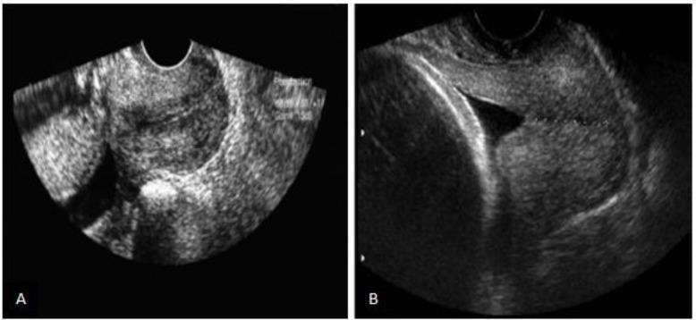
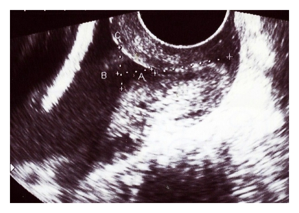
TVU showing cervical funneling (V/U-shaped), a sonographic marker of cervical incompetence and preterm birth risk
7. MANAGEMENT
Step 1 - Confirm Preterm Labor and Assess Gestational Age
- Detailed US for fetal presentation, weight, anomalies
- GBS culture, cervicovaginal fFN, TVU cervical length
- Rule out contraindications to tocolysis
Step 2 - Antenatal Corticosteroids (ACS)
THE SINGLE MOST IMPORTANT INTERVENTION
- Indicated: 24 0/7 to 33 6/7 weeks (also consider 34-36 6/7 weeks in select cases)
- Betamethasone 12 mg IM x2 doses 24 hours apart OR
- Dexamethasone 6 mg IM x4 doses 12 hours apart
- Benefits:
- Reduces neonatal mortality, RDS, IVH, NEC, patent ductus arteriosus
- Promotes surfactant synthesis, increases lung compliance, reduces vascular permeability
- Maturational effects on brain, kidneys, gut
Step 3 - Tocolysis
Goal: Delay delivery 48 hours to complete ACS and allow transfer to a tertiary center (Level III/IV). Tocolysis does NOT prevent preterm birth long-term.
Contraindications to Tocolysis
- Absolute: Severe preeclampsia/gestational hypertension, hemorrhage/abruption, intrauterine infection, maternal cardiac disease
- Fetal: Non-reassuring fetal status, lethal anomaly, IUGR with reverse diastolic flow
First-Line Tocolytic Agents
A. Calcium Channel Blockers (Nifedipine) - PREFERRED FIRST LINE
| Parameter | Detail |
|---|
| Mechanism | Inhibits voltage-dependent Ca²⁺ channels → ↓ intracellular Ca²⁺ → myometrial relaxation |
| Dose | 10-20 mg orally, repeat every 3-6 hours |
| Efficacy | Reduces delivery within 48 hours (RR = 0.30); ↑ latency vs. beta-agonists |
| Advantages | Fewer side effects than MgSO₄ or β-mimetics; better neonatal outcomes |
| Maternal SE | Hypotension, headache (20%), flushing (8%), dizziness, nausea |
| Caution | Do NOT combine with IV β-agonists (cardiovascular effects); do NOT combine with MgSO₄ (neuromuscular blockade) |
| Cochrane | Preferred over other agents - greater contraction suppression, fewer SE, improved neonatal morbidity (↓ NICU admission, NEC, RDS, IVH) |
B. NSAIDs - COX Inhibitors (Indomethacin)
| Parameter | Detail |
|---|
| Mechanism | Inhibits prostaglandin synthesis → ↓ uterine contractility |
| Dose | 50 mg oral loading dose, then 25-50 mg every 6 hours |
| Duration | Limit to 48 hours; use before 32 weeks only |
| Efficacy | Effective short-term tocolytic |
| Fetal SE | Premature ductal constriction, oligohydramnios, neonatal pulmonary hypertension |
| Ductal constriction | Occurs in up to 50% after ≥1 week; usually resolves within 24 hrs after stopping |
| Contraindications | Renal anomalies, oligohydramnios, ductal-dependent cardiac defects, TTTS, gestation ≥ 32 weeks |
| Monitoring | AFI + fetal renal anatomy before use; monitor AFI and ductal flow if >48 hr |
C. Beta-Agonists (Terbutaline, Ritodrine)
| Parameter | Detail |
|---|
| Mechanism | β₂ receptor agonism → ↑ cAMP → ↓ intracellular Ca²⁺ → myometrial relaxation |
| Dose | Terbutaline 0.25 mg SC every 20-30 min (up to 3 doses) |
| Efficacy | Effective in delaying delivery 48 hours and 7 days |
| Maternal SE | Tachycardia, palpitations, tremor, pulmonary edema, hypokalemia, hyperglycemia |
| Neonatal SE | Fetal tachycardia, neonatal hypoglycemia |
| Note | Greater side effects than nifedipine; less favored currently |
| Cochrane | Effective but greater side effects than other agents |
D. Magnesium Sulfate (MgSO₄)
| Parameter | Detail |
|---|
| Mechanism | Competes with Ca²⁺ at motor end plate and cell membrane → ↓ myometrial contractility when serum Mg 5-8 mg/dL |
| DTR lost | At 9-13 mg/dL |
| Respiratory depression | At 14 mg/dL |
| Excreted | Almost entirely by kidney; 75% during infusion, 90% within 24 hrs |
| Tocolytic efficacy | NOT supported - Cochrane (37 trials) found no benefit over placebo or other tocolytics |
| Neuroprotection | STRONGLY indicated at < 32 weeks - reduces cerebral palsy (RR 0.68), gross motor dysfunction |
| Neuroprotective dose | IV bolus 4-6 g over 30 min ± maintenance 1-2 g/hr for 12-24 hr |
| Maternal SE | Flushing, nausea, vomiting, headache, blurred vision, pulmonary edema |
| Contraindication | Myasthenia gravis |
Key exam point: MgSO₄ is NOT recommended as a tocolytic (ACOG) but IS recommended for fetal neuroprotection when delivery is anticipated < 32 weeks.
E. Oxytocin Receptor Antagonists (Atosiban)
- Blocks oxytocin receptors → decreased uterine contractions
- Not available in the USA; used in Europe
- Cochrane: inferior to calcium channel blockers and β-agonists for tocolysis
- Advantage: minimal maternal/fetal side effects
Summary Table: Tocolytics
| Drug | Mechanism | SE | Notes |
|---|
| Nifedipine | Ca²⁺ channel block | Hypotension, headache | First-line preferred |
| Indomethacin | COX inhibitor | Ductal constriction, oligohydramnios | Use < 32 wks, max 48 hr |
| Terbutaline | β₂ agonist | Pulmonary edema, tachycardia | More SE, less favored |
| MgSO₄ | Ca²⁺ antagonism | Flushing, respiratory depression | NOT a tocolytic; use for neuroprotection |
| Atosiban | Oxytocin antagonist | Minimal | Not available in USA |
Step 4 - Neuroprotection with MgSO₄
- All women at risk of delivery before 32 weeks
- Regimen: 4-6 g IV bolus over 30 min, then 1-2 g/hr for up to 24 hr
- Reduces moderate-severe cerebral palsy significantly
- Rescue dosing if >12 hours from last dose and delivery imminent
Step 5 - GBS Prophylaxis
- Antibiotics for GBS prophylaxis only (penicillin or ampicillin IV)
- Antibiotics for other specific pathogens (e.g., UTI)
- Routine broad-spectrum antibiotics for preterm labor with intact membranes have NOT been effective in prolonging pregnancy or reducing morbidity
Step 6 - Regionalized Care
| Level | Capabilities |
|---|
| Level I | Normal mothers and infants |
| Level II | Most maternal/infant complications; NICU for >1500 g |
| Level III | Sickest/smallest infants; complex maternal care |
| Level IV | ECMO, complex cardiothoracic surgery |
8. MAINTENANCE TOCOLYSIS
Does NOT reduce the rate of preterm birth - not recommended.
- Cochrane reviews of oral β-mimetics, terbutaline pump, oxytocin antagonists, and calcium channel blockers show no decline in preterm birth frequency with maintenance therapy
- Some evidence of nifedipine prolonging pregnancy (mean 5.35 days) but no reduction in preterm birth at various cutoffs
- Outpatient uterine contraction monitoring also NOT beneficial
9. PREVENTION
Primary Prevention
- Public education about avoidable risk factors (smoking, ART, short inter-pregnancy interval)
- Policies to reduce higher-order multiple gestations
- Nutritional support, treatment of infections (UTI, BV)
Secondary Prevention (Screening)
- Cervical length screening at 18-24 weeks in singleton pregnancies with prior PTB
- CL < 25 mm: offer vaginal progesterone (17-hydroxyprogesterone caproate or micronized progesterone)
- Reduces PTB risk by ~30-40%
- Fetal fibronectin screening in high-risk women
- Cerclage: Indicated for cervical insufficiency with prior PTB and short cervix; NOT beneficial for multiple gestations
Tertiary Prevention (Management of Active Preterm Labor)
- Tocolysis + ACS + neuroprotection + regionalized care (as above)
10. COMPLICATIONS / NEONATAL OUTCOMES
Directly Related to Prematurity
| System | Complication |
|---|
| Respiratory | Respiratory Distress Syndrome (RDS), Bronchopulmonary Dysplasia (BPD) |
| CNS | Intraventricular Hemorrhage (IVH), Periventricular Leukomalacia (PVL), Cerebral Palsy |
| GI | Necrotizing Enterocolitis (NEC) |
| Cardiovascular | Patent Ductus Arteriosus (PDA) |
| Metabolic | Hypoglycemia, hypothermia, hyperbilirubinemia |
| Infection | Sepsis (especially GBS, gram-negative) |
| Eyes | Retinopathy of Prematurity (ROP) |
- Consequences extend across life course: NCDs (hypertension, diabetes) in later life
- Morbidity and mortality inversely proportional to gestational age
11. QUICK EXAM MNEMONICS
Risk Factors - "PUMPED MOM":
- Prior PTB
- Uterine anomalies
- Multiple gestation
- Placental problems (previa, abruption)
- Extremes of age
- Distension (polyhydramnios)
- Medical conditions (DM, HTN)
- Obesity/nutrition
- Microbial infections (BV, UTI)
Key Cut-offs to Remember:
- Preterm = < 37 weeks (20-36+6)
- Fetal fibronectin: positive if > 50 ng/mL
- Cervical length reassuring: ≥ 30 mm; high risk: < 15 mm
- Indomethacin: use ONLY < 32 weeks, max 48 hours
- MgSO₄ neuroprotection: < 32 weeks
- ACS: 24-33+6 weeks (consider 34-36+6)
- Respiratory depression from MgSO₄: at 14 mg/dL
- DTR loss from MgSO₄: at 9-13 mg/dL
12. HIGH-YIELD EXAM POINTS
- Preterm birth is the #1 cause of perinatal and infant morbidity/mortality in developed nations
- Antenatal corticosteroids are the most important single intervention; reduce RDS, IVH, NEC, mortality
- Nifedipine is the preferred first-line tocolytic (Cochrane evidence)
- MgSO₄ is NOT a tocolytic but is the agent for neuroprotection < 32 weeks
- Indomethacin: limit to < 32 weeks, max 48 hours (ductal constriction risk)
- Maintenance tocolysis is ineffective - do not use
- fFN negative = very high NPV (~99%) for PTB within 7 days; useful for ruling OUT true preterm labor
- Antibiotics: only for GBS prophylaxis in preterm labor with intact membranes; NOT for prolonging pregnancy
- Cerclage: beneficial for cervical insufficiency in singletons; NOT beneficial in multiple gestations
- Atosiban not available in USA but widely used in Europe
- Overdiagnosis of preterm labor occurs in 40-70% with clinical criteria alone
- Prior PTB is the single strongest clinical risk factor for recurrence
Sources: Creasy & Resnik's Maternal-Fetal Medicine: Principles and Practice, 8th edition; Park's Textbook of Preventive and Social Medicine; MgSO₄ neuroprotection: Cochrane 2024 update PMID 38830233