
Type 1 hypersensitivity reaction
I'll start by reading the medical library skill, then search for comprehensive information on Type 1 hypersensitivity.
Reading File
Now let me search the library in parallel for comprehensive coverage of Type 1 hypersensitivity:
Finding Sources
Finding Sources
Finding Sources
Reading File
Good. Now let me pull the deeper immunology and clinical content from multiple books in parallel:
Reading File
Reading File
Finding Sources
Excellent - rich sources found. Now let me read the key detailed sections from Robbins, Cellular & Molecular Immunology, and Jawetz simultaneously:
Reading File
Reading File
Reading File
Now let me get the mast cell mediators and clinical details from Robbins and Cellular & Molecular Immunology, plus fetch the key diagram:
Reading File
Reading File
Loading Image
Excellent content. Let me get the late-phase reaction details and clinical manifestations/treatment from Robbins, plus the mast cell mediators diagram:
Reading File
Loading Image
I now have comprehensive material from multiple authoritative textbooks plus two excellent diagrams. Let me compile the full response.
Type I Hypersensitivity Reaction (Immediate Hypersensitivity)
Type I hypersensitivity is an IgE-mediated immune reaction that occurs within seconds to minutes of re-exposure to an antigen (allergen) in a previously sensitized individual. It is the most prevalent type of hypersensitivity and encompasses conditions like anaphylaxis, allergic asthma, allergic rhinitis (hay fever), urticaria, and atopic dermatitis.
Overview: Sequence of Events
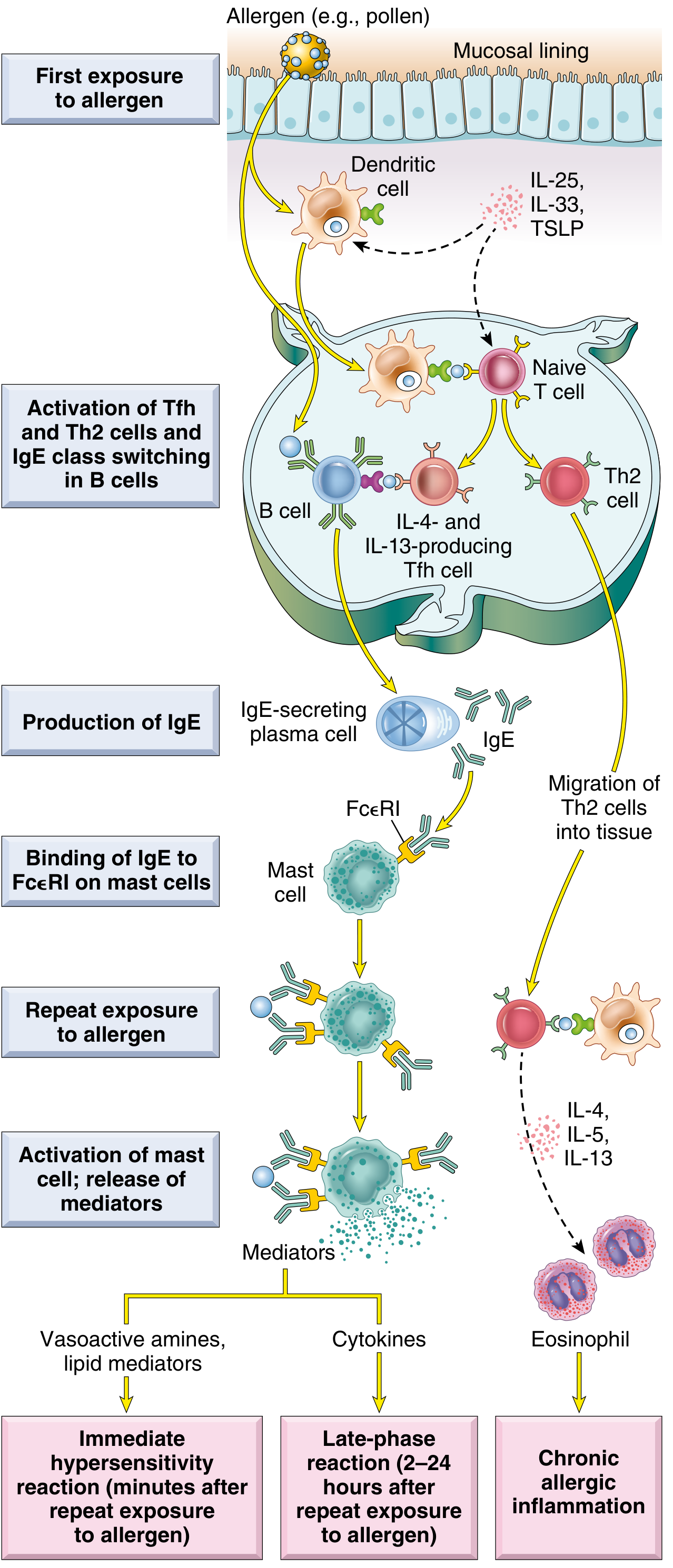
Fig. 6.14 - Robbins, Cotran & Kumar Pathologic Basis of Disease
Step 1: Sensitization Phase (First Exposure)
When an allergen (e.g., pollen, house dust mite proteins, bee venom, peanut proteins) enters the body for the first time:
- Antigen presentation: Dendritic cells process and present allergen peptides to naive CD4+ T cells. Epithelial cytokines - IL-25, IL-33, and TSLP - promote a type 2 immune environment.
- Th2 differentiation: Naive T cells differentiate into Th2 cells and T follicular helper (Tfh) cells, which produce IL-4 and IL-13.
- IgE class switching: IL-4 and IL-13 drive B cells to undergo class-switch recombination, producing allergen-specific IgE from plasma cells.
- Mast cell sensitization: IgE binds via its Fc portion to high-affinity FcεRI receptors on mast cells and basophils. These IgE-coated cells are now "sensitized" and ready to react.
- Cellular and Molecular Immunology, p. 1303-1307
Key point: No clinical symptoms occur at this stage. The individual has no adverse reaction on first exposure (with rare exceptions).
Step 2: Elicitation Phase (Re-exposure - "Challenge")
On re-exposure to the same allergen:
- The allergen (being multivalent) cross-links adjacent IgE antibodies on the mast cell surface.
- This brings the underlying FcεRI receptors together, triggering intracellular signal transduction cascades.
- The result: mast cell activation and degranulation - release of preformed and newly synthesized mediators.
"IgE-coated mast cells are said to be sensitized because they react to subsequent encounters with antigen. If the antigen is multivalent, it binds to and cross-links adjacent IgE antibodies, bringing the underlying Fce receptors together." - Robbins, Cotran & Kumar Pathologic Basis of Disease, p. 200
Mast Cell Mediators
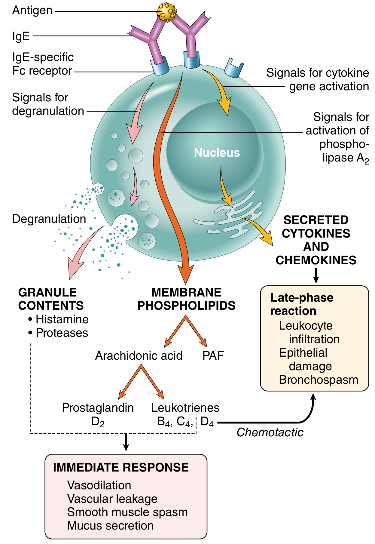
Fig. 6.15 - Robbins, Cotran & Kumar Pathologic Basis of Disease
A. Preformed Granule Mediators (Released within seconds)
| Mediator | Action |
|---|---|
| Histamine | Vasodilation, increased vascular permeability, smooth muscle contraction, mucus secretion |
| Tryptase / Chymase (neutral proteases) | Tissue damage; generate kinins and C3a from precursors |
| Heparin / Chondroitin sulfate (proteoglycans) | Package and store amines in granules; anticoagulant |
| Serotonin | Vasoactive effects |
Plasma histamine peaks ~10 minutes after anaphylaxis onset; returns to normal within 1 hour. Serum tryptase is elevated in anaphylaxis (and chronically elevated in systemic mastocytosis). Urinary histamine may remain elevated for 24 hours. - Quick Compendium of Clinical Pathology
B. Newly Synthesized Lipid Mediators (Produced within minutes)
Mast cell activation triggers phospholipase A2, converting membrane phospholipids to arachidonic acid, which feeds two pathways:
-
Cyclooxygenase pathway → Prostaglandin D2 (PGD2): Most abundant mast cell cyclooxygenase product; causes intense bronchospasm and increases mucus secretion.
-
5-Lipoxygenase pathway → Leukotrienes:
- LTC4 and LTD4: Most potent vasoactive and spasmogenic agents known; thousands of times more active than histamine on a molar basis in causing bronchospasm and increasing vascular permeability.
- LTB4: Highly chemotactic for neutrophils, eosinophils, and monocytes.
-
Platelet-activating factor (PAF): Derived directly from membrane phospholipids; contributes to vascular and bronchospastic effects.
-
Robbins, Cotran & Kumar Pathologic Basis of Disease, p. 200-201
C. Cytokines (Produced over hours)
| Cytokine | Role |
|---|---|
| TNF, IL-1, chemokines | Promote leukocyte recruitment (late-phase reaction) |
| IL-4 | Amplifies Th2 response |
| IL-5 | Most potent eosinophil-activating cytokine |
Phase Breakdown: Immediate vs. Late-Phase Reaction
Immediate Reaction (seconds to ~1 hour)
- Driven by histamine, LTC4, LTD4, PGD2
- Manifestations: vasodilation, vascular leakage, smooth muscle spasm, mucus secretion
- Clinically: bronchospasm, urticaria, rhinorrhea, anaphylaxis
Late-Phase Reaction (2-24 hours after allergen exposure)
-
Leukocytes (especially eosinophils) are recruited by eotaxin and mast cell-derived chemokines.
-
IL-5 from Th2 cells activates eosinophils, which release:
- Proteolytic enzymes
- Major basic protein (MBP) and eosinophil cationic protein - cause tissue damage
- Galectin-10 - forms Charcot-Leyden crystals (seen in sputum of asthmatics)
-
The late-phase reaction is a major driver of symptoms in asthma - this is why antihistamines alone are insufficient for asthma; corticosteroids are required.
-
Robbins, Cotran & Kumar Pathologic Basis of Disease, p. 201
Atopy and Genetic Predisposition
Atopy = hereditary predisposition to mount IgE responses against common environmental antigens. Features:
- Elevated serum IgE levels
- More IL-4-producing Th2 cells than general population
- Positive family history in ~50% of atopic individuals
- Genetic linkage to polymorphisms in IL-3, IL-4, IL-5, IL-9, IL-13, GM-CSF genes, and HLA loci on chromosome 6
- The atopic triad (atopic march): atopic dermatitis (infancy) → allergic rhinitis → asthma (childhood)
"A positive family history of allergy is found in almost 50% of atopic individuals." - Robbins, Cotran & Kumar Pathologic Basis of Disease
Nature of Allergens
Allergens are typically proteins or chemicals bound to proteins (e.g., pollen, house dust mite proteins, animal dander, shellfish, peanuts, penicillin as a hapten). Characteristics:
-
Molecular weight: 5-70 kD
-
Stable, glycosylated, soluble in body fluids
-
Many are enzymes (e.g., cysteine protease of house dust mite, PLA2 in bee venom)
-
T cell-independent antigens (polysaccharides) cannot elicit type I reactions unless protein-conjugated
-
Cellular and Molecular Immunology, p. 1306
Clinical Manifestations
| Form | Manifestation |
|---|---|
| Systemic anaphylaxis | Bronchospasm, cardiovascular collapse, shock - potentially fatal |
| Atopic asthma | Bronchospasm, mucus hypersecretion, airway inflammation |
| Allergic rhinitis (hay fever) | Sneezing, rhinorrhea, nasal congestion |
| Urticaria / Angioedema | Whealing, swelling |
| Atopic dermatitis (eczema) | Skin inflammation |
| Food allergy | GI symptoms (abdominal pain, diarrhea), systemic anaphylaxis |
"In the most extreme systemic form, called anaphylaxis, mast cell-derived mediators can restrict airways to the point of asphyxiation and produce cardiovascular collapse leading to shock, which together may result in death." - Cellular and Molecular Immunology, p. 1303
Non-IgE-Mediated (Nonatopic) Triggers
About 20-30% of immediate hypersensitivity reactions are not IgE-mediated and do not involve Th2 cells. Triggers include:
- Temperature extremes (heat, cold)
- Exercise
- Drugs (codeine, morphine, contrast media)
- Complement anaphylatoxins (C3a, C5a)
- Bee venom components (melittin)
These are termed anaphylactoid reactions and are clinically indistinguishable from true anaphylaxis.
Note: Anaphylactoid reactions and hereditary angioedema are NOT classified as Type I hypersensitivity reactions. - Quick Compendium of Clinical Pathology
Diagnosis
| Test | Details |
|---|---|
| Skin prick test | Measures specific IgE attached to skin mast cells; most convenient and specific screening method |
| Serum specific IgE (sIgE) | Safer alternative (no risk of anaphylaxis); valid from 6 weeks of age; preferred in cardiovascular disease or severe sensitivity |
| Serum tryptase | Elevated in acute anaphylaxis; chronically elevated in mastocytosis |
| Plasma histamine | Peaks ~10 min; returns to normal within 1 hour |
| Urinary histamine | Elevated for up to 24 hours post-reaction |
- Tietz Textbook of Laboratory Medicine, p. 3972
Treatment
| Approach | Rationale |
|---|---|
| Epinephrine (adrenaline) | First-line for anaphylaxis; reverses bronchospasm and cardiovascular collapse |
| Antihistamines (H1 blockers) | Effective for rhinitis and urticaria (immediate reaction only); insufficient for asthma |
| Corticosteroids | Target late-phase reaction; required for asthma management |
| Leukotriene receptor antagonists | Block LTC4/LTD4 effects (e.g., montelukast for asthma) |
| Allergen avoidance | Best long-term prevention |
| Allergen immunotherapy (desensitization) | Repeated low-dose allergen exposure to induce tolerance; shifts IgG/IgA response, reduces IgE |
| Anti-IgE (Omalizumab) | Biologic that binds free IgE, reducing mast cell sensitization |
"The best prevention relies on the identification of the antigen (detected by skin test or IgE antibody serology) and subsequent avoidance." - Jawetz, Melnick & Adelberg's Medical Microbiology
Th2 Cytokines and Their Role
| Cytokine | Source | Role in Type I Hypersensitivity |
|---|---|---|
| IL-4 | Th2, Tfh cells | IgE class switching in B cells; Th2 amplification |
| IL-13 | Th2 cells | IgE class switching; mucus production |
| IL-5 | Th2 cells | Eosinophil activation and recruitment |
| IL-10 | Regulatory | Anti-inflammatory; modulates allergic response |
Th2-type responses are associated with promotion of IgE and eosinophilic responses in atopy - Tietz Textbook of Laboratory Medicine
Summary
Type I hypersensitivity is a two-phase process:
- Sensitization: First allergen exposure → Th2/IgE production → mast cell sensitization (no symptoms)
- Elicitation: Re-exposure → IgE cross-linking → mast cell degranulation → immediate reaction (histamine, leukotrienes) + late-phase reaction (eosinophils, cytokines)
The central players are IgE, mast cells, and Th2 cytokines (especially IL-4, IL-5, IL-13). The reaction can range from trivial (mild rhinitis) to fatal (anaphylactic shock).
Sources: Robbins, Cotran & Kumar Pathologic Basis of Disease | Cellular and Molecular Immunology (Abbas et al.) | Jawetz, Melnick & Adelberg's Medical Microbiology | Tietz Textbook of Laboratory Medicine | Quick Compendium of Clinical Pathology
This is a shared conversation. Sign in to Orris to start your own chat.