
Cataract in k khurana book
Reading File
Now let me search the library for the Khurana book and cataract content simultaneously.
Finding Sources
Finding Sources
The library doesn't have a book specifically titled "Khurana" (K.K. Khurana's Ophthalmology), but it does have Kanski's Clinical Ophthalmology which is the standard ophthalmology reference with extensive cataract content. Let me search more broadly and also check all ophthalmology books available.
Finding Sources
Finding Sources
The library has Kanski's Clinical Ophthalmology (not K.K. Khurana). These are both major ophthalmology textbooks - K.K. Khurana's "Comprehensive Ophthalmology" is not in this library. Let me read the comprehensive cataract content from Kanski (which covers the same material) and inform the user.
Reading File
Reading File
Reading File
Reading File
Reading File
Reading File
I now have comprehensive cataract content. Let me also fetch a couple of key clinical images to enrich the answer.
Loading Image
Loading Image
I have all the information needed. Here is the complete, detailed answer on Cataract from the ophthalmology textbook:
Note: The library does not contain K.K. Khurana's Comprehensive Ophthalmology directly. The answer below is drawn from Kanski's Clinical Ophthalmology: A Systematic Approach (10th ed.), which covers the same cataract content at an equivalent or greater depth. Topics align closely with the Khurana syllabus.
CATARACT
Source: Kanski's Clinical Ophthalmology, 10th Edition
Introduction & Definition
Cataract is any opacity of the crystalline lens. It is the commonest cause of reversible visual disability in the world. There is no medical treatment to prevent development or progression of cataract. Phacoemulsification with intraocular lens (IOL) implantation is the treatment of choice and is the commonest surgical procedure performed worldwide.
Anatomy of the Lens
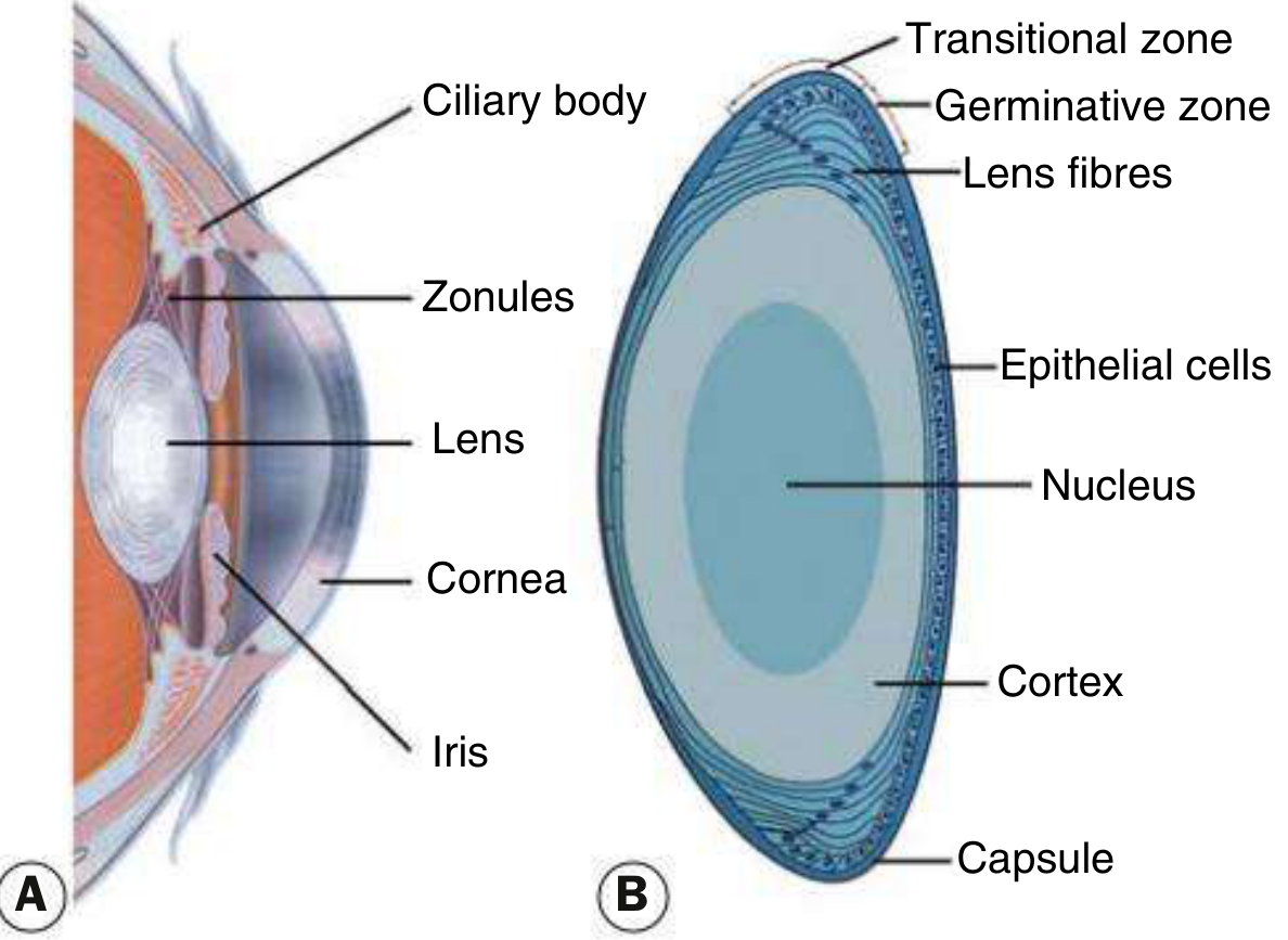
- Zonules hold the lens in position
- Capsule - an acellular membrane enclosing the lens
- Epithelial cells - cuboid cells beneath the anterior capsule extending to the equator; cells in the germinative zone divide continuously and differentiate into lens fibers
- Lens substance - central nucleus surrounded by cortical material
- Primary function - to focus light on the retina
Effect on Vision
Cataracts often develop slowly; in early stages the vision alters subtly. The effect depends on:
- (a) The type of cataract
- (b) Its location (central opacities affect vision more)
- (c) Whether it is unilateral or bilateral
ACQUIRED CATARACT
Age-Related Cataract
1. Subcapsular Cataract
- Anterior subcapsular cataract: lies directly under the lens capsule; associated with fibrous metaplasia of the lens epithelium
- Posterior subcapsular opacity (PSC): lies just in front of the posterior capsule; granular or plaque-like on oblique slit-lamp; appears black and vacuolated on retroillumination. Vacuoles are swollen migratory lens epithelial cells (Wedl/bladder cells). Located at the nodal point - has a particularly profound effect on vision. Patients suffer from glare (e.g., from oncoming car headlights); symptoms increase with miosis (near work, bright light)
2. Nuclear Sclerotic Cataract
- An exaggeration of normal ageing change
- Associated with myopia (increased refractive index of nucleus) - some elderly patients can read without glasses again = "second sight of the aged"
- Characterized by a yellowish hue from deposition of urochrome pigment
- Best assessed with oblique slit-lamp beam
- When advanced, nucleus appears brown (brunescent); in rare cases, black
3. Cortical Cataract
- May involve anterior, posterior, or equatorial cortex
- Opacities start as clefts and vacuoles between lens fibers due to cortical hydration
- Progress to cuneiform (wedge-shaped) or radial spoke-like opacities, often starting in the inferonasal quadrant
- Glare is a common symptom
4. Christmas Tree Cataract
- Uncommon; characterized by polychromatic needle-like formations in the deep cortex and nucleus
Cataract Maturity
| Type | Feature |
|---|---|
| Immature | Lens partially opaque |
| Mature | Lens completely opaque |
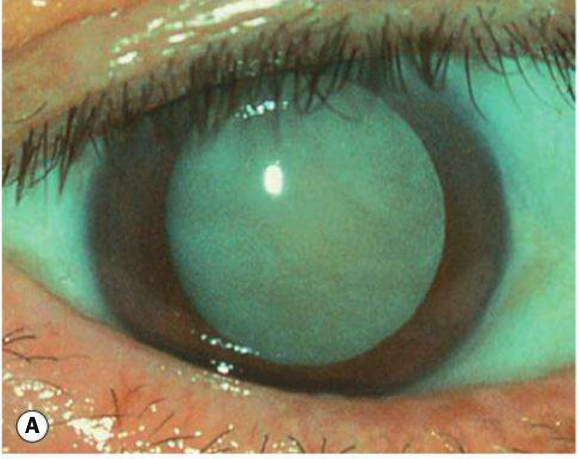
| Hypermature | Shrunken, wrinkled anterior capsule due to leakage of water out |
| Morgagnian | Hypermature; liquefied cortex with nucleus sinking inferiorly |
Cataract in Systemic Disease
Diabetes Mellitus
- Hyperglycaemia → high glucose in aqueous → diffuses into lens → metabolized to sorbitol (by aldose reductase) → accumulates → secondary osmotic overhydration
- Mild: fluctuation in refractive index (hyperglycaemia = myopia; hypoglycaemia = hypermetropia)
- Classic diabetic cataract (young patients): snowflake cortical opacities - rare, may mature within days or resolve spontaneously
- Age-related cataract occurs sooner in diabetics
- Nuclear sclerosis is common and progresses rapidly
- ~1 in 5 cataract surgery patients have diabetes
Myotonic Dystrophy
- ~90% develop fine iridescent cortical opacities in the third decade (sometimes like Christmas tree cataract)
- Evolve into wedge-shaped cortical and subcapsular opacities, often star-like, by the fifth decade
Neurofibromatosis Type 2 (NF2)
- Early cataract in >60% of patients
- Opacities: posterior subcapsular, capsular, cortical or mixed; tend to develop in early adulthood
Secondary (Complicated) Cataract
Develops as a result of other primary ocular disease:
- Chronic anterior uveitis - most common cause; also related to steroid use. Earliest finding: polychromatic lustre at the posterior lens pole. Progresses to posterior and anterior opacities
- Acute congestive angle closure - forms glaukomflecken (small anterior grey-white subcapsular opacities); represent focal lens epithelial infarcts; pathognomonic of previous acute angle closure
- High myopia - posterior subcapsular opacity and early-onset nuclear sclerosis
Traumatic Cataract
Causes include:
- Penetrating trauma - direct damage to capsule and lens fibers
- Blunt trauma - classic flower-shaped (rosette) opacity
- Intralenticular metallic foreign body
- Electric shock and lightning strike
- Infrared radiation - "glassblower's cataract" (anterior capsular opacity)
- Ionizing radiation
CONGENITAL CATARACT
Incidence: ~3 in 10,000 live births
- 1/3 associated with systemic disease
- 1/3 inherited
- 1/3 of unknown cause
- 2/3 bilateral; cause identifiable in ~half
- Autosomal dominant (AD) inheritance is the most common aetiological factor
- Unilateral cataracts are usually sporadic
Associated Metabolic Disorders
| Disorder | Key Feature |
|---|---|
| Galactosaemia | AR; absent GALT enzyme; 'Oil droplet' opacity; early death if galactose not withheld; early exclusion may reverse changes |
| Lowe syndrome (oculo-cerebro-renal) | X-linked recessive; OCRL1 gene; cataract universal; congenital glaucoma in ~50%; female carriers have insignificant cortical opacities |
| Mannosidosis | AR; α-mannosidase deficiency; spoke-like opacities in posterior lens cortex |
| Hypo/pseudohypoparathyroidism | Cataract among potential features |
| Hypo/hyperglycaemia | Cataract among potential features |
Associated Intrauterine Infections
| Infection | Cataract feature |
|---|---|
| Rubella | ~15% bilateral/unilateral pearly nuclear or diffuse cataract |
| Toxoplasmosis | Cataract with chorioretinitis, microphthalmos, optic atrophy |
| Cytomegalovirus (CMV) | Part of TORCH syndrome ophthalmic spectrum |
MANAGEMENT OF ACQUIRED CATARACT
Indications for Surgery
- Visual improvement - most common; when opacity causes difficulty in daily activities
- Medical indications - cataract adversely affecting eye health (e.g., phacolytic or phacomorphic glaucoma; to visualize fundus for monitoring/treatment)
- Clear lens exchange - for refractive error correction (replacing a healthy lens)
Preoperative Considerations
- Systemic alpha-blockers (e.g., tamsulosin) - associated with intraoperative floppy iris syndrome (IFIS); stop or manage accordingly
- Anticoagulants/antiplatelets - follow local protocol
- Routine blood tests and ECG not usually required for local anaesthesia
- Diabetics: monitor retina before and after surgery (risk of worsening macular oedema)
Surgical Technique - Phacoemulsification
The modern standard is phacoemulsification with IOL implantation - uses ultrasound energy to emulsify and aspirate lens material through a small incision, followed by insertion of a foldable intraocular lens.
- Kanski's Clinical Ophthalmology: A Systematic Approach, 10th Edition, Ch. 10 (Lens)
This is a shared conversation. Sign in to Orris to start your own chat.