
What is liver cerhosis
liver cirrhosis histology gross pathology
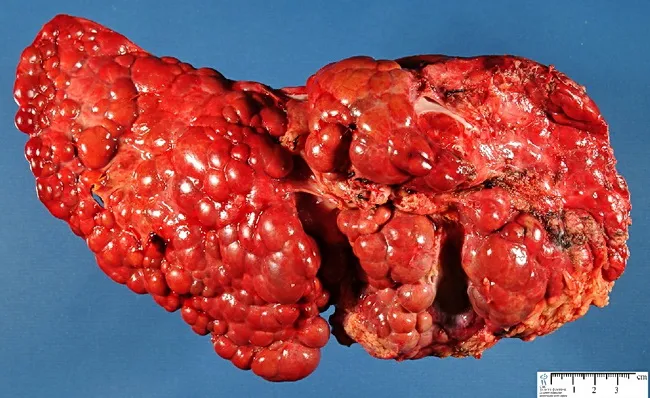
Gross pathology photograph of the liver showing advanced cirrhosis with coarse, nodular surface. The specimen demonstrates macronodular cirrhosis, in which regenerative nodules exceed 3 mm in diameter, a histologic and macroscopic hallmark of fibrosis and architectural disarray associated with chronic tyrosinemia type I. The context is hereditary infantile tyrosinemia due to fumarylacetoacetate hydrolase deficiency, a metabolic liver disease that progresses to cirrhotic remodeling and risk of hepatocellular carcinoma if untreated. The hepatic nodularity and firm consistency reflect extensive parenchymal scarring with bridging fibrous septa linking nodules. Color is a deep reddish-brown, with variable areas of congested or mottled parenchyma; surface nodularity may obscure normal lobular anatomy. This image provides a macroscopic correlate to the cellular and architectural changes described in histology, including regenerative nodules embedded in dense fibrous tissue. Clinically, such findings support severe liver disease in tyrosinemia and justify monitoring for portal hypertension and hepatocellular carcinoma risk. In educational and research contexts, this photograph is valuable for teaching gross hepatic morphology, cirrhotic evolution in metabolic disorders, and the distinction between macronodular vs micronodular cirrhosis. Potential uses include medical education, pathology slide correlation, and diagnostic radiology-pathology correlation studies. These features reinforce differential considerations for cirrhosis etiologies in pediatrics worldwide today.
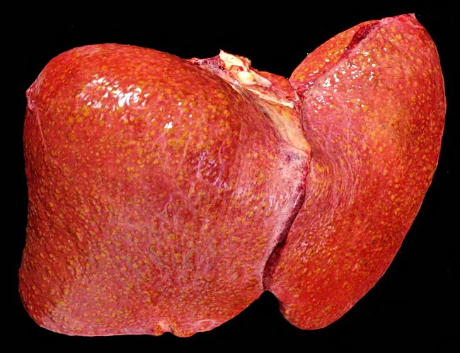
Gross pathology photograph of a human liver showing cirrhotic nodularity with coarse surface, reflecting end-stage hepatic disease. The image depicts hepatocellular nodules separated by fibrous septa, typical of chronic liver injury. In Wilson disease, impaired copper transport due to ATP7B mutation leads to copper deposition in hepatocytes, contributing to inflammation, steatosis in early disease, and progression to chronic hepatitis and cirrhosis. The macroscopic features include enlarged, nodular liver with a firm texture. While histology is not shown, cirrhosis features such as bridging fibrosis and regenerative nodules would be expected. This image serves as an educational reference for macroscopic evaluation of cirrhosis within copper storage disorders and complements histopathology with architectural assessment. Clinically significant for teaching end-stage liver disease, differential diagnosis with viral hepatitis and alcohol-related liver disease, and correlation with systemic copper accumulation; useful in pathology, hepatology, medical education, and radiology training for recognizing advanced liver damage in Wilson disease. This description highlights the relationship between macroscopic appearance and underlying pathophysiology: copper-induced hepatocellular injury triggers stellate cell activation, extracellular matrix deposition, nodularity, and eventual loss of normal lobular architecture. In practice, gross pathology complements biochemical copper quantification, hepatic copper staining (rhodanine) and ATP7B genotyping in diagnosis. The image can also aid educational case libraries and digital pathology databases for differential diagnosis training.
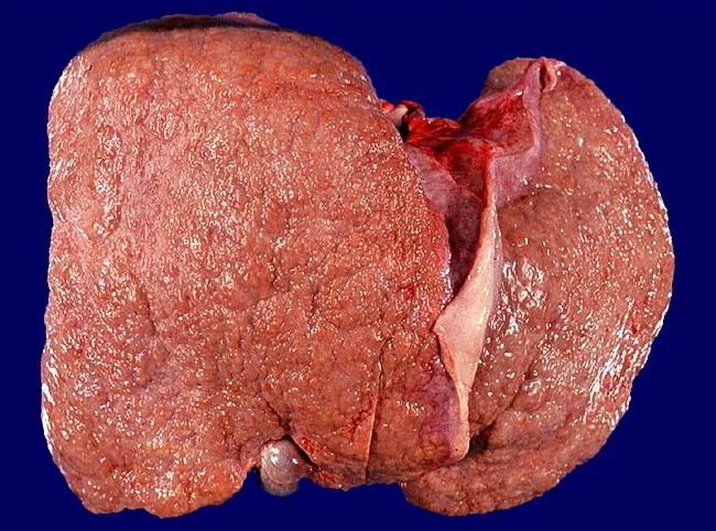
This is a gross pathology photograph of a human liver showing diffuse micronodular cirrhosis with nodules smaller than 3 mm on the surface, consistent with alcoholic liver disease. The organ appears enlarged and firm, with a coarse, irregular exterior and a nodular regenerative pattern that distorts normal lobular architecture. Coloration is mottled brownish-red, reflecting parenchymal degeneration and fibrotic remodeling; the capsule surface is intact but irregular. The microscopic correlate would reveal widespread hepatic fibrosis with fibrous septa and numerous tiny regenerative nodules; histology would show steatosis, ballooning degeneration, inflammation, and eventual cirrhotic architecture. The described micronodularity (<3 mm) indicates advanced fibrosis rather than macro-nodular cirrhosis, and underscores the progression from steatosis to steatohepatitis to cirrhosis in long-term alcohol exposure. Clinically, this morphology signals portal hypertension risk, reduced synthetic function, and potential progression to hepatic failure; timely management includes alcohol cessation, nutritional support, surveillance for varices, and evaluation for liver transplantation in end-stage disease. This image is informative for education on liver gross anatomy, correlation with histopathology, and teaching about alcohol-related liver disease staging. It is appropriate for pathology, hepatology, and radiology-pathology correlation. Note the dominant nodularity and preserved capsule, features commonly seen in alcoholic micronodular cirrhosis for teaching and research applications.
Liver Cirrhosis
Definition
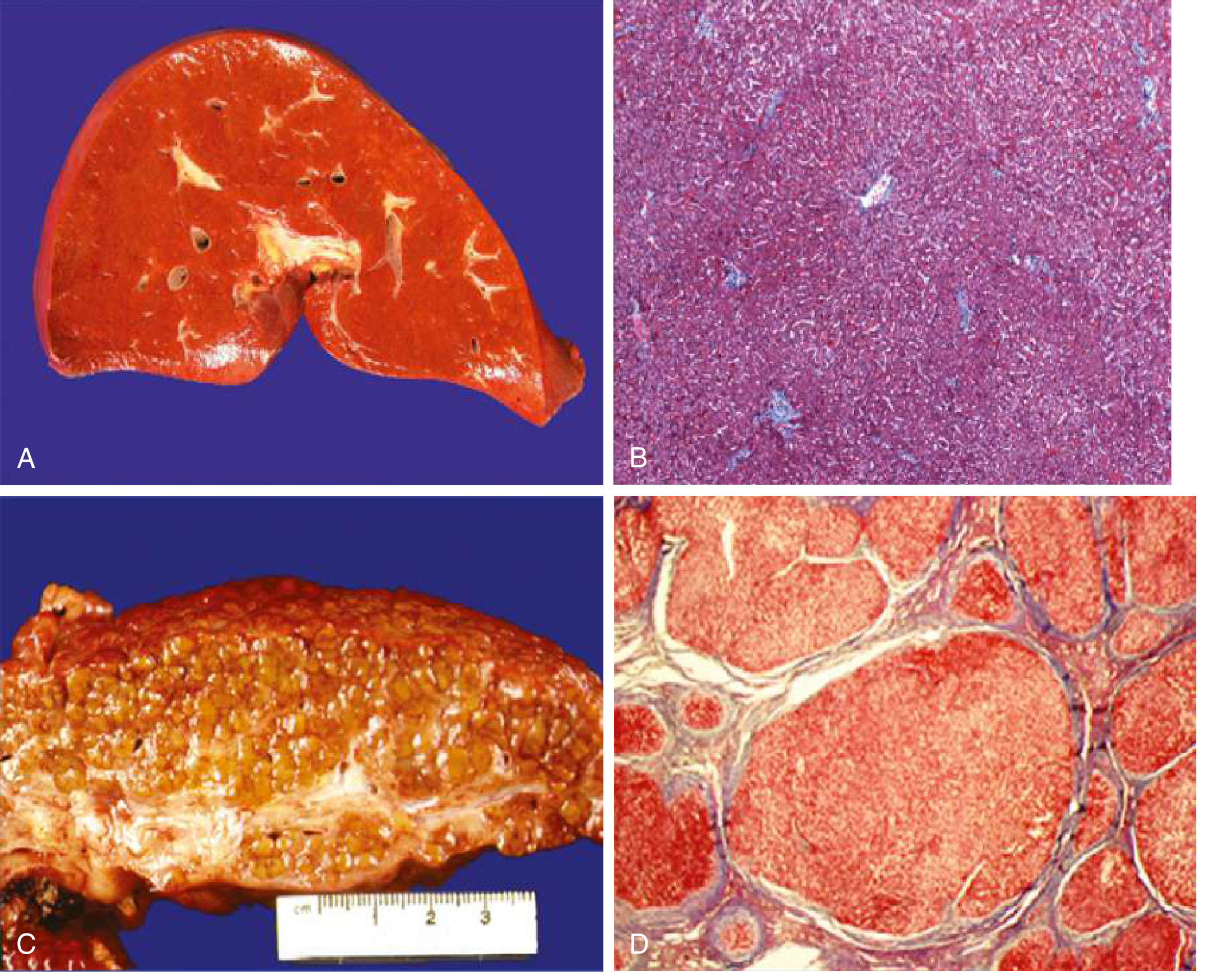
Gross and Microscopic Appearance
Pathogenesis - How Does Cirrhosis Develop?
- Normally, stellate cells are quiescent and reside in the space of Disse (between hepatocytes and sinusoidal endothelial cells), storing vitamin A.
- On liver injury, they become activated - they lose vitamin A, proliferate, and transform into contractile myofibroblasts.
- They secrete large amounts of extracellular matrix - collagen types I and III, sulfated proteoglycans, and glycoproteins.
- This leads to collagen deposition in the space of Disse, causing "capillarization" of the sinusoids - the sinusoidal endothelial cells lose their fenestrae, reducing exchange between plasma and hepatocytes.
- Goldman-Cecil Medicine, p. 1614
Causes
| Cause | Notes |
|---|---|
| Alcohol-related liver disease | Leads to micronodular cirrhosis initially |
| Non-alcoholic fatty liver disease (NAFLD) | Increasingly common with obesity epidemic |
| Chronic Hepatitis B | Major cause in Asia and Africa |
| Chronic Hepatitis C | Major cause in Western countries |
-
Cholestatic diseases: Primary biliary cirrhosis, Primary sclerosing cholangitis
-
Biliary obstruction: Biliary atresia, Cystic fibrosis
-
Metabolic disorders: Hemochromatosis, Wilson disease, Alpha-1-antitrypsin deficiency, Glycogen storage diseases
-
Vascular: Budd-Chiari syndrome, Right-sided heart failure
-
Drugs/toxins
-
Cryptogenic cirrhosis (no clear cause identified)
-
Goldman-Cecil Medicine, Table 139-1, p. 1615
Stages: Compensated vs. Decompensated
| Stage | Features |
|---|---|
| Compensated | Patient may be asymptomatic; liver still functioning adequately |
| Decompensated | Development of ascites, variceal hemorrhage, encephalopathy, or jaundice |
Complications
1. Portal Hypertension
- Results from increased resistance to portal flow (fibrosis + regenerative nodule compression = fixed component; vasoconstriction due to intrahepatic NO deficiency = functional component) AND increased portal venous inflow from splanchnic vasodilation.
- Leads to: Esophageal/gastric varices, splenomegaly with hypersplenism, portosystemic collaterals
2. Ascites
- Results from reduced plasma proteins (lower oncotic pressure) + raised portal capillary pressure - fluid and protein leak into peritoneal cavity.
- The kidneys respond by retaining salt and water (similar to reduced plasma volume states), which worsens fluid accumulation.
- Can be complicated by spontaneous bacterial peritonitis (SBP) and hepatorenal syndrome.
- Guyton & Hall Textbook of Medical Physiology, p. 552
3. Hepatic Encephalopathy
- Results from both portal hypertension (portosystemic shunting of nitrogenous compounds) and liver insufficiency.
4. Jaundice
- Due to the liver's inability to excrete bilirubin (liver insufficiency).
5. Pruritus
- Severe chronic jaundice triggers itching that can be debilitating.
6. Coagulopathy
- Loss of liver synthetic function reduces clotting factor production.
7. Hepatocellular Carcinoma (HCC)
- Cirrhosis is a major risk factor for HCC development, especially in viral hepatitis-related disease.
Key Points to Remember
- Cirrhosis is not a specific diagnosis - it is a morphologic endpoint with variable causes and prognoses.
- Not all chronic liver disease ends in cirrhosis (e.g., biliary tract diseases may not).
- Not all cirrhosis leads to end-stage liver failure (e.g., treated autoimmune hepatitis or cured Hep C may retain adequate function despite cirrhosis).
- Regression is possible: With disease remission (e.g., antiviral treatment), fibrosis can regress - scars thin, fragment, and nodules coalesce into larger islands of regenerating parenchyma. - Robbins & Kumar, p. 598
- When the cause is unknown, the term cryptogenic cirrhosis is used.