
Frequent motion without passing flatus
intestinal obstruction abdominal X-ray dilated bowel loops
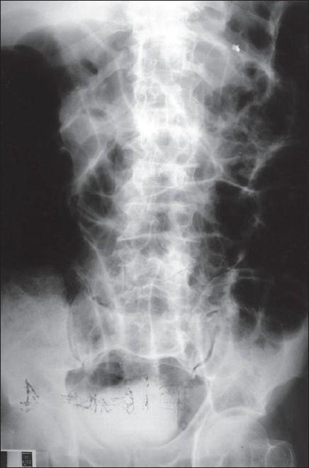
A plain abdominal X-ray (radiography) in an anteroposterior projection demonstrating signs of intestinal obstruction. The image reveals multiple centrally located, dilated loops of bowel characterized by radiolucent (dark) air-filled segments. These distended loops are arranged in a 'ladder-like' pattern with visible valvulae conniventes extending across the full width of the lumen, which is characteristic of a small bowel obstruction. The periphery of the abdomen shows generalized gas distension, though some regions on the left are obscured by radiopaque densities. In the lower pelvic region, linear and fragmented radiopaque foreign materials are visible, possibly surgical clips or markers. The lumbar spine and pelvic bones provide anatomical context, while punctate radiopaque densities scattered in the abdominal field may suggest dystrophic calcifications. This diagnostic image is intended for medical education on the radiological features of mechanical bowel obstruction and the use of abdominal flat plates in acute clinical settings.
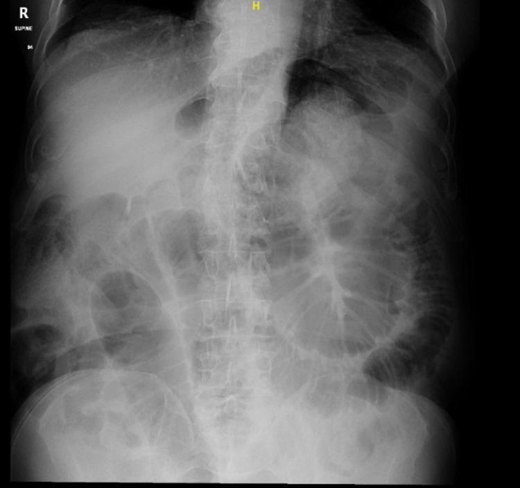
A supine plain film abdominal X-ray (diagnostic image) demonstrating multiple dilated loops of small bowel, concentrated primarily in the central and lower abdominal regions. The intestinal loops exhibit characteristic valvulae conniventes (plicae circulares) that span the entire diameter of the lumen, distinguishing them as small bowel. The distribution is disorganized and spans from the mid-abdomen toward the pelvic cavity. Notable features include the absence of visible pneumoperitoneum (no free air under the diaphragm) and a lack of significant gas in the distal colon or rectum, findings highly suggestive of a mechanical small bowel obstruction (SBO). Anatomical landmarks such as the lumbar vertebrae and pelvic bones are visible, providing orientation. This imaging is clinically significant for evaluating acute abdominal pain and suspected bowel obstruction, providing early diagnostic evidence for further cross-sectional imaging or surgical intervention.
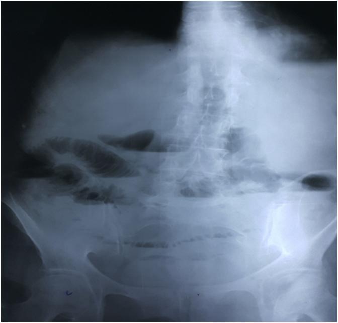
This diagnostic image is a plain erect abdominal X-ray (radiography) demonstrating classic radiographic features of an intestinal obstruction. The most prominent finding is the presence of multiple dilated loops of small bowel containing horizontal air-fluid levels, concentrated in the central and upper abdominal regions. The dilated loops exhibit visible valvulae conniventes (plicae circulares), which are thin mucosal folds that span the entire diameter of the bowel lumen, identifying these as small intestinal loops. A significant clinical sign is the complete absence of gas within the large bowel and rectum, indicating a high-grade or complete mechanical obstruction proximal to the colon. The lumbar spine and pelvic bones are visible, providing anatomical context for the bowel loop distribution. This image is a primary educational resource for recognizing the 'step-ladder' pattern of small bowel obstruction and differentiating small bowel from large bowel dilation in an acute clinical setting.
Frequent Motions Without Passing Flatus
Why This Happens - The Physiology
- Gas cannot pass beyond the obstructing point because peristalsis cannot overcome the mechanical block
- Flatus is therefore absent - gas accumulates proximal to the block, causing distension
- However, liquid stool and contents distal to the obstruction can still be evacuated by residual muscular activity
- This explains why patients may pass stool (or even have "diarrhea-like" frequent motions) initially while being completely unable to pass any flatus
Classification of Constipation in Obstruction
| Type | Definition | Implies |
|---|---|---|
| Absolute constipation | Neither faeces nor flatus passed | Complete intestinal obstruction - cardinal feature |
| Relative constipation | Only flatus passed (no stool) | Partial obstruction |
| Frequent motions, no flatus | Stools passed but no gas | Distal contents evacuating past point of obstruction |
- Bailey and Love's Short Practice of Surgery 28th Ed., p. (Constipation section)
"This may be classified as absolute (i.e. neither faeces nor flatus is passed) or relative (where only flatus is passed). Absolute constipation is a cardinal feature of complete intestinal obstruction. Some patients may pass flatus or faeces after the onset of obstruction as a result of the evacuation of the distal bowel contents." - Bailey & Love's Short Practice of Surgery 28th Ed.
Clinical Context
- Pain - colicky, crampy, periumbilical (small bowel) or hypogastric (large bowel); progresses to constant pain with strangulation
- Vomiting - early and bilious in proximal obstruction; delayed and feculent in distal/colonic obstruction
- Abdominal distension - more prominent in distal obstructions
- High-pitched "tinkling" or "rushing" bowel sounds - pathognomonic of mechanical obstruction (contrast with absent sounds in ileus)
- Constant (non-colicky) severe pain not relieved by opiates
- Fever, tachycardia, leukocytosis >20,000/mm³
- Peritonism (guarding, rebound tenderness)
- Metabolic acidosis, elevated lactate
Common Causes of Mechanical Obstruction
| Site | Common Causes |
|---|---|
| Small bowel | Adhesions (post-op, #1 cause), hernias, Crohn's disease, intussusception, volvulus |
| Large bowel | Carcinoma of colon (#1 cause), diverticular disease, volvulus (sigmoid, caecal), faecal impaction |
Investigation
-
Plain AXR (erect + supine) - first-line; look for:
- Step-ladder pattern of dilated small bowel loops (>2.5 cm) with air-fluid levels
- Absence of gas in colon/rectum (supports complete obstruction)
- "Coffee-bean" sign in sigmoid volvulus
Here is the classic radiographic appearance of mechanical small bowel obstruction - the step-ladder pattern of dilated loops with no gas distally: -
CT abdomen with contrast - gold standard; identifies site, cause, and complications (ischemia, perforation)
-
Labs - CBC, electrolytes, creatinine, lactate (elevated lactate/acidosis = ischemia)
Management Principles
- IV fluids - correct electrolyte disturbance (hypokalemia, metabolic alkalosis common from vomiting)
- NGT decompression - relieves proximal distension
- NBM (nil by mouth)
- Urinary catheter - monitor output
- Avoid enemas - they evacuate distal contents and confuse the clinical picture without relieving the obstruction
- Surgical intervention - mandatory for complete obstruction, strangulation, or failure of conservative management
Relevant medicine in partial obstruction
Medical Management of Partial Bowel Obstruction
1. General Supportive Measures (Non-Drug)
| Measure | Purpose |
|---|---|
| Nil by mouth (NBM) | Bowel rest, reduces luminal secretion |
| IV fluid resuscitation (isotonic, e.g. 0.9% NaCl or Hartmann's) | Corrects dehydration from vomiting and third-space losses |
| Electrolyte replacement | Correct hypokalemia, hypochloremia (from vomiting) |
| Nasogastric tube (NGT) decompression | Decompresses stomach, reduces nausea, prevents aspiration |
| Urinary catheter | Monitor urine output and fluid balance |
- Schwartz's Principles of Surgery 11th Ed.
"Fluid resuscitation is integral to treatment. Isotonic fluid should be given intravenously... The stomach should be continuously evacuated of air and fluid using a nasogastric (NG) tube. Effective gastric decompression decreases nausea, distention, and the risk of vomiting and aspiration." - Schwartz's Principles of Surgery 11th Ed.
2. Gastrografin (Water-Soluble Contrast) - Dual Diagnostic AND Therapeutic Role
- Being hyperosmolar, it draws fluid into the bowel lumen
- This increases intraluminal fluid, reduces bowel wall edema, and promotes peristalsis
- The net effect is relief of partial adhesive obstruction in many cases
- 60-100 mL via NGT
- Serial X-rays taken at intervals (typically 4, 8, 24 hours)
- If contrast reaches the colon within 24 hours → likely to resolve non-operatively
- If contrast does not pass beyond obstruction, or patient does not pass flatus/feces within 12-24 hours → surgery should be considered
- Assalia et al (1994): Gastrografin reduced time to first bowel movement (6.2 vs 23.3 hours), operative rate (10% vs 21%), and hospital stay (2.2 vs 4.4 days)
- Choi et al (2002): Gastrografin appeared to reduce need for surgery by 74%
- Meta-analysis (Abbas et al): Reduced length of stay by ~2 days, time to resolution by ~28 hours, odds ratio for surgery 0.44
"Use of water-soluble contrast not only predicts likelihood of success of nonoperative management but also reduces the need for surgery (odds ratio 0.44), length of stay by about 2 days, and time to resolution by about 28 hours, without an increase in morbidity or mortality." - Schwartz's Principles of Surgery 11th Ed.
3. Disease-Specific Medical Therapy
Crohn's Disease - Partial/Complete SBO
- Corticosteroids (IV) - reduce transmural inflammation causing the partial obstruction
- IV fluids + NGT decompression as above
- Bowel rest ± TPN - reduces luminal antigen load and disease activity
"Almost all cases of complete or high-grade partial small bowel obstruction from Crohn's disease respond to conservative management... treated with nasogastric decompression, intravenous hydration, and steroid therapy. This program allows for resolution of the acute episode of obstruction in a vast majority of cases." - Maingot's Abdominal Operations
Post-Operative Partial SBO
- >90% resolve spontaneously with bowel rest + IV fluids
- No specific drug needed; time and supportive care are sufficient - Sabiston Textbook of Surgery
Radiation Enteropathy-associated pSBO
- Managed conservatively with bowel rest, nutritional support (TPN if needed)
- Elemental/low-residue enteral feeds when tolerated
- Medical management is often difficult and frustrating in this group - Maingot's
4. Analgesia and Antiemetics (Symptomatic)
| Drug Class | Examples | Role |
|---|---|---|
| Antiemetics | Metoclopramide, ondansetron, cyclizine | Relieve nausea/vomiting |
| Analgesics | IV/IM opioids (morphine, pethidine) | Pain relief; do NOT withhold - does not mask peritonism signs in experienced hands |
| Anticholinergics | Hyoscine butylbromide (Buscopan) | Reduces colicky spasm |
5. Antibiotics
- Not routinely indicated in uncomplicated partial SBO
- Given only if there is concern for bowel ischemia, bacterial translocation, fever, or leukocytosis suggesting impending strangulation
- Broad-spectrum cover (e.g. co-amoxiclav, or piperacillin-tazobactam + metronidazole) if surgery is planned or ischemia suspected - Schwartz's Principles of Surgery 11th Ed.
6. When to Escalate to Surgery
- No improvement in symptoms after 48 hours of NGT + IV fluids → surgery
- Contrast fails to reach colon within 24 hours on Gastrografin challenge → surgery
- Any signs of strangulation (fever, tachycardia, peritonism, rising lactate/WCC) → urgent surgery
"Most patients with partial small obstruction whose symptoms do not improve within 48 hours after initiation of nonoperative therapy should be considered for surgery." - Schwartz's Principles of Surgery 11th Ed.
Summary
| Intervention | Drug/Agent | Role |
|---|---|---|
| Fluid resuscitation | Normal saline / Hartmann's | Correct dehydration, third-space losses |
| NGT decompression | - | Decompress, prevent aspiration |
| Gastrografin | Diatrizoate meglumine | Diagnostic + therapeutic (osmotic) - key intervention |
| Corticosteroids | IV hydrocortisone/prednisolone | Crohn's-related obstruction only |
| Antiemetics | Metoclopramide, ondansetron | Symptomatic relief |
| Analgesia | Opioids (cautious), hyoscine | Pain/colic relief |
| Antibiotics | Broad-spectrum | Only if ischemia/surgery anticipated |
| Avoid | Enemas, prokinetics, barium | Can worsen obstruction or cause peritonitis |
Loose stool with flatus how to approach
diarrhea approach algorithm flowchart diagnosis
Summary : This flowchart presents an algorithm for the diagnosis and treatment of deep vein thrombosis (DVT) during pregnancy, as recommended by the European Society of Cardiology (ESC). It outlines the stepwise approach from initial suspicion to diagnosis and management, including the use of low-molecular-weight heparin (LMWH), ultrasound imaging, and specialist consultation. flowchart: # Nodes : • Start (rounded rectangle): "Suspicion of pregnancy-related Deep vein thrombosis" • Decision/Action (rounded rectangle, yellow): "Administration of first therapeutic dose of LMWH if anticipated diagnostic delay (Class IIa)" • Process (rounded rectangle): "Venous ultrasound compression ultrasound of infrainguinal veins and ultrasound of iliac veins" • Decision (split into three branches): "Positive", "Inconclusive", "Negative" • Process (rounded rectangle): "Serial venous ultrasound scans (2 exams within 7 days) or MR-venography" (for inconclusive results) • Decision (split from serial scans): "Positive", "Negative" • Terminal (rectangle): "Diagnosis of DVT" • Terminal (rectangle): "No DVT" • Management (rectangle): "Therapeutic dose LMWH (Class I)" • Management (rectangle): "Consultation with Pregnancy Heart Team including a vascular specialist and a haematologist (Class I)" # Connectors : • Downward arrows connect each step in sequence. • From "Venous ultrasound", three branches: – Positive → "Diagnosis of DVT" – Inconclusive → "Serial venous ultrasound scans or MR-venography" – Negative → "No DVT" • From "Serial venous ultrasound scans or MR-venography": – Positive → "Diagnosis of DVT" – Negative → "No DVT" • From "Diagnosis of DVT", two parallel arrows to: – "Therapeutic dose LMWH (Class I)" – "Consultation with Pregnancy Heart Team including a vascular specialist and a haematologist (Class I)" • Both management steps are joined by "AND". # Layout : • Top-down, single entry point. • Branching at ultrasound result (positive/inconclusive/negative). • Re-merging after serial scans. • Parallel management steps after DVT diagnosis. • Color coding: yellow for urgent action, purple for diagnostic steps, green for management. # Analysis : • The algorithm prioritizes early treatment with LMWH if diagnostic delay is expected. • Ultrasound is the primary diagnostic tool, with serial imaging or MR-venography for inconclusive cases. • Negative imaging at any stage rules out DVT. • Confirmed DVT leads to both therapeutic anticoagulation and multidisciplinary specialist consultation. • The flowchart emphasizes safety, rapid diagnosis, and coordinated care for pregnant patients with suspected DVT.
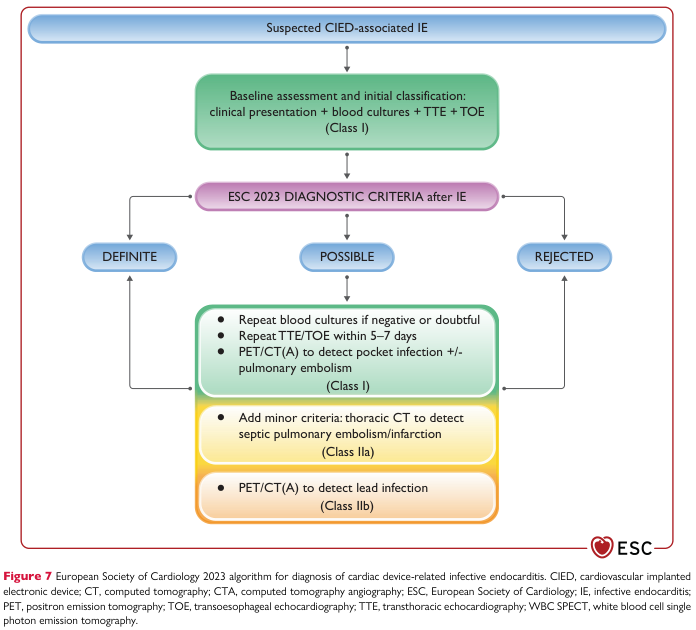
Summary : This flowchart presents the European Society of Cardiology (ESC) 2023 algorithm for diagnosing cardiac device-related infective endocarditis (CIED-associated IE), outlining the stepwise approach from initial suspicion to definitive, possible, or rejected diagnosis, and subsequent recommended investigations. flowchart: # Nodes : • Suspected CIED-associated IE (rounded rectangle, top) • Baseline assessment and initial classification: clinical presentation + blood cultures + TTE + TOE (rounded rectangle, green) • ESC 2023 DIAGNOSTIC CRITERIA after IE (rounded rectangle, purple) • Three outcome nodes: DEFINITE (rounded rectangle, blue), POSSIBLE (rounded rectangle, blue), REJECTED (rounded rectangle, blue) • For POSSIBLE branch, three investigation nodes: – Repeat blood cultures if negative or doubtful; Repeat TTE/TOE within 5–7 days; PET/CT(A) to detect pocket infection +/- pulmonary embolism (rounded rectangle, green, Class I) – Add minor criteria: thoracic CT to detect septic pulmonary embolism/infarction (rounded rectangle, yellow, Class IIa) – PET/CT(A) to detect lead infection (rounded rectangle, orange, Class IIb) # Connectors : • Downward arrow from Suspected CIED-associated IE to Baseline assessment and initial classification. • Downward arrow from Baseline assessment to ESC 2023 DIAGNOSTIC CRITERIA after IE. • Three arrows from ESC 2023 DIAGNOSTIC CRITERIA after IE to DEFINITE, POSSIBLE, and REJECTED. • From POSSIBLE, downward arrows to each of the three investigation nodes. • Arrow from investigation nodes (Class I, IIa, IIb) back to DEFINITE node, indicating possible reclassification after further testing. # Layout : • Vertical flow from top (suspicion) to bottom (diagnosis and further testing). • Three branches from diagnostic criteria: left (DEFINITE), center (POSSIBLE with further investigations), right (REJECTED). • Investigation steps for POSSIBLE diagnosis are stacked vertically and color-coded by recommendation class (Class I: green, Class IIa: yellow, Class IIb: orange). • Feedback loop from further investigations to DEFINITE diagnosis. # Analysis : • The algorithm emphasizes a structured, stepwise approach: initial assessment with clinical, microbiological, and echocardiographic data, followed by application of ESC diagnostic criteria. • If diagnosis is POSSIBLE, additional investigations (repeat cultures, imaging) are recommended, with the possibility of upgrading to DEFINITE diagnosis. • Color-coding highlights the strength of recommendations for each investigation (Class I strongest, Class IIb weakest). • The flowchart visually reinforces the importance of repeated and multimodal testing in ambiguous cases, and provides clear decision pathways for clinicians.
Approach to Loose Stool WITH Flatus
Step 0 - What the Symptom Tells You Immediately
Step 1 - Define the Diarrhea
| Duration | Category | Most Likely Cause |
|---|---|---|
| ≤14 days | Acute | Infection (virus, bacteria, protozoa) in ~80% of cases |
| 14-30 days | Persistent | Giardia, C. difficile, post-infectious, early IBD |
| >30 days | Chronic | Non-infectious (IBD, malabsorption, functional, neoplasm) |
- Yamada's Textbook of Gastroenterology 7th Ed.
Step 2 - History (Most Productive Part of the Evaluation)
A. Characterize the Stool
| Feature | Suggests |
|---|---|
| Watery, large volume, no blood | Secretory (cholera, VIPoma) or osmotic cause |
| Greasy, foul-smelling, floats | Steatorrhoea - malabsorption (celiac, pancreatic insufficiency) |
| Bloody/mucoid ("dysentery") | Inflammatory/invasive - Shigella, E. histolytica, Campylobacter, IBD |
| Small-volume, urgent, with tenesmus | Left colon/rectal pathology |
| Nocturnal diarrhea | Organic disease (IBD, VIPoma) - never functional |
| Persists with fasting | Secretory cause (VIPoma, bile acid diarrhea) |
| Resolves with fasting | Osmotic cause (lactase deficiency, FODMAP) |
B. Epidemiological Clues (Yamada's - Table 35.1 pattern)
| Clue | Think |
|---|---|
| Recent travel | Traveller's diarrhea - ETEC, Giardia, E. histolytica |
| Eating at restaurant / catered event | Food poisoning - S. aureus, Bacillus cereus (within hours), Salmonella (6-48h) |
| Outbreak in group setting (cruise, school) | Norovirus |
| Recent antibiotic use | C. difficile |
| Immunocompromised | Cryptosporidium, CMV, MAC, Isospora |
| Day-care exposure | Shigella, Giardia, Cryptosporidium, rotavirus |
| Raw/undercooked meat | E. coli O157:H7, Salmonella, Campylobacter |
| Shellfish | Vibrio species, hepatitis A |
| Duration >2 weeks without improvement | Giardia most likely; also E. histolytica, Cryptosporidium |
C. Drug History
- Antibiotics (direct mucosal injury or C. difficile)
- Magnesium-containing antacids
- Metformin, colchicine, SSRIs
- Stimulant laxatives (senna, bisacodyl - check for laxative abuse)
- NSAIDs (colitis, enteropathy)
- Chemotherapy agents
D. Red Flags ("Must Not Miss")
- Bloody stools
- Fever ≥38.5°C
- Nocturnal diarrhea waking from sleep
- Significant weight loss (>5 kg)
- Age >50 with new-onset altered bowel habit
- Immunocompromised host
- Elderly patient (≥70 years)
Step 3 - Physical Examination
| Finding | Significance |
|---|---|
| Fever, tachycardia | Infectious/inflammatory; assess for sepsis |
| Dehydration (dry mucosa, skin turgor, sunken eyes, oliguria) | Severity assessment - guide fluid therapy |
| Abdominal tenderness | Localise - RIF (Crohn's, Yersinia, appendicitis), LIF (UC, ischemic colitis) |
| Perianal examination | Fissures, fistulae, skin tags → Crohn's disease |
| Extraintestinal signs | Arthritis, uveitis, erythema nodosum, aphthous ulcers → IBD |
| Lymphadenopathy, wasting | Malignancy, HIV-related |
| Rectal exam | Exclude fecal impaction (which paradoxically causes overflow "diarrhea") |
Step 4 - Investigations
First-Line (All Patients with Non-Self-Limiting Diarrhea)
| Test | Purpose |
|---|---|
| Stool microscopy | Ova, cysts, parasites (Giardia, E. histolytica, Cryptosporidium) |
| Stool culture / PCR multiplex panel | Bacteria (Salmonella, Shigella, Campylobacter, E. coli O157, Yersinia, Vibrio); PCR > culture - faster, more sensitive |
| C. difficile toxin (EIA/PCR) | If recent antibiotics, hospitalization, or elderly |
| Fecal leukocytes / Fecal calprotectin | Raised = inflammatory diarrhea; guides need for further workup |
| Stool for occult blood | Inflammatory/invasive pathogens, malignancy |
| FBC, U&E, CRP | Severity, dehydration, inflammatory state |
| LFTs, TFTs | Thyrotoxicosis (causes motility diarrhea) |
Indications for Stool Microbiology (Harrison's, 22nd Ed.)
- Profuse diarrhea with dehydration
- Overtly bloody stools
- Fever ≥38.5°C
- Duration >48 h without improvement
- Recent antibiotic use
- Community outbreak
- Severe abdominal pain in patients >50 years
- Elderly (≥70) or immunocompromised
For Persistent/Chronic Diarrhea - Additional Tests
| Test | Purpose |
|---|---|
| Stool osmolality + electrolytes → Osmotic gap | Gap = 290 - 2(Na+K); >125 mOsm/kg = osmotic; <50 = secretory |
| 72-hour fecal fat | >7 g/day = steatorrhoea/malabsorption |
| Fecal elastase | Pancreatic exocrine insufficiency |
| Celiac antibodies (tTG-IgA, anti-endomysial) | Celiac disease |
| Colonoscopy + biopsies | IBD, microscopic colitis, malignancy; if duration >4 weeks |
| Upper GI endoscopy + duodenal biopsy | Celiac, Whipple's, Giardia on histology |
| Serum VIP, gastrin, calcitonin, 5-HIAA (urinary) | Neuroendocrine tumours (VIPoma, gastrinoma, carcinoid) |
| CT abdomen/pelvis | Mass lesion, lymphadenopathy, bowel wall thickening, fistula |
Step 5 - Mechanistic Classification of Chronic Diarrhea
| Mechanism | Key Feature | Common Causes |
|---|---|---|
| Secretory | Persists with fasting; large volume; no osmotic gap | VIPoma, carcinoid, bile acid diarrhea, laxative abuse, Addison's |
| Osmotic | Stops with fasting; osmotic gap >125; bloating + flatulence | Lactase deficiency, FODMAP, lactulose, Mg antacids |
| Steatorrheal (malabsorptive) | Greasy, bulky, foul stool; positive Sudan stain; fat >7 g/day | Celiac, chronic pancreatitis, SIBO, Crohn's |
| Inflammatory/Exudative | Blood/mucus in stool; raised CRP, calprotectin, WBC | IBD (Crohn's, UC), microscopic colitis, radiation colitis |
| Dysmotility | IBS pattern; urgency; alternating; no nocturnal symptoms | IBS, post-vagotomy, post-cholecystectomy, hyperthyroidism |
Step 6 - Management Algorithm
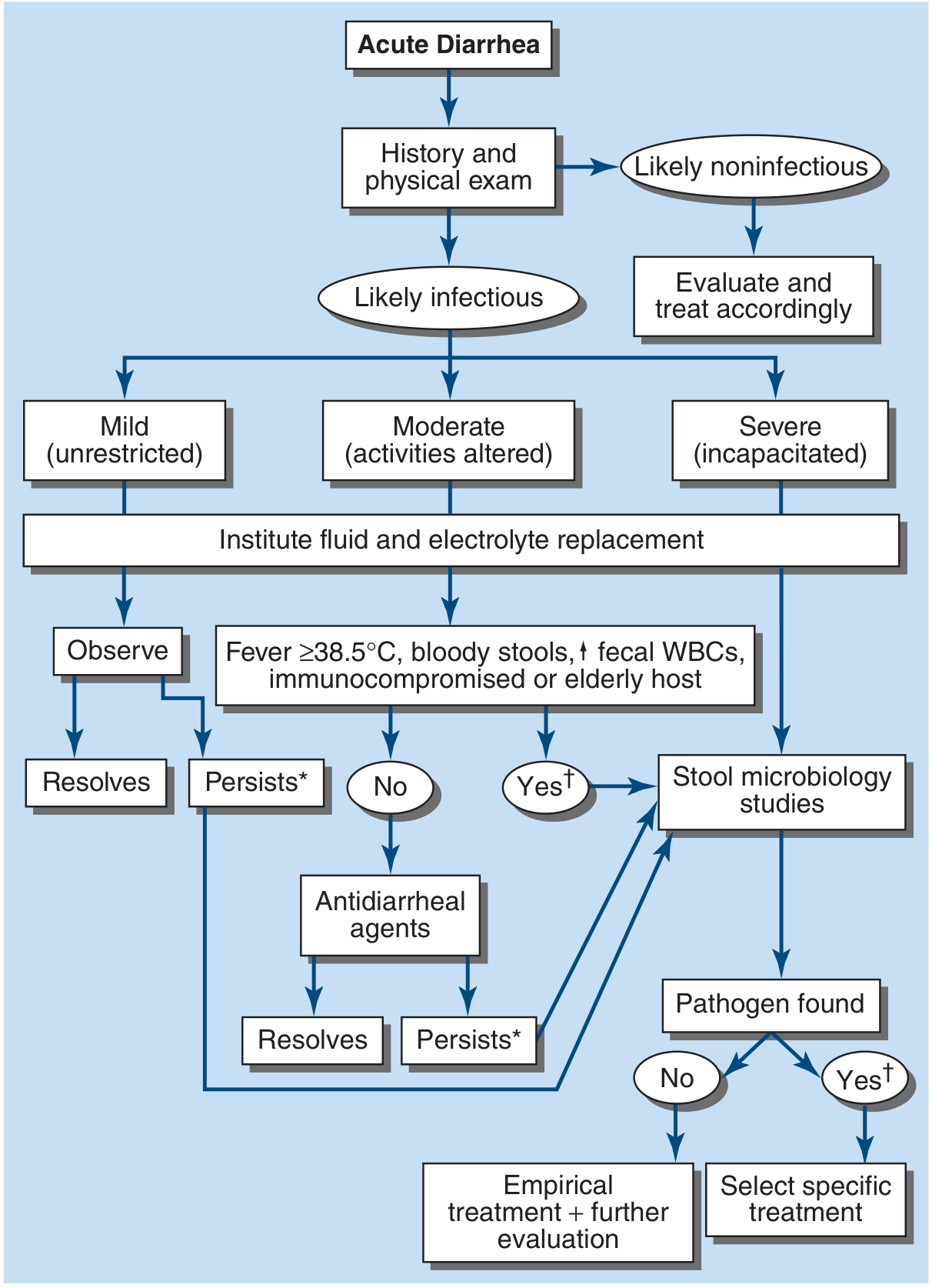
- All patients - oral fluid and electrolyte replacement (ORS); IV if severely dehydrated
- Mild, non-febrile, non-bloody - observe; antidiarrheal agents (loperamide) if no fever/dysentery
- Fever ≥38.5°C, bloody stool, fecal WBCs, immunocompromised, elderly - send stool microbiology studies
- Empirical antibiotics when moderate-severe febrile diarrhea:
- Azithromycin 500 mg single dose (first-line, esp. Campylobacter-endemic areas)
- Ciprofloxacin 500 mg BD for 3-5 days (quinolone-sensitive areas)
- Metronidazole 250-400 mg QID for 7 days (suspected Giardia)
- Pathogen found - select specific treatment based on organism
- No pathogen found + persists - empirical treatment + further evaluation (endoscopy, imaging)
Summary - One-Line Approach
Loose stool + flatus = patent bowel = diarrhea. Classify by duration (acute vs. chronic), characterize the stool (watery vs. fatty vs. bloody), take epidemiological history, examine for red flags, send targeted investigations (stool culture/PCR ± fecal calprotectin for acute; osmotic gap/colonoscopy/malabsorption tests for chronic), and treat - fluids always, antimotility only if non-inflammatory, antibiotics selectively.
Infective diarrhea treatment
Treatment of Infective Diarrhea
The Fundamental Principle
"Even for patients who contact a physician, the majority of acute bacterial diarrheal infections will resolve spontaneously without specific antibiotics." - Goldman-Cecil Medicine
1. Rehydration - Universal for All Patients
Oral Rehydration Therapy (ORT)
- Sodium chloride 3.5 g
- Trisodium citrate 2.9 g (or sodium bicarbonate 2.5 g)
- Potassium chloride 1.5 g
- Glucose 20 g
- (Reduced-osmolality ORS is now standard - 245 mOsm/L)
- ½ teaspoon salt + 6 level teaspoons sugar in 1 litre of clean water
- <6 months: 10 mg/day for 14 days
-
6 months: 20 mg/day for 14 days
- Reduces severity, duration, and recurrence of diarrhea in children in low-income settings
IV Rehydration
- Severe dehydration (cannot tolerate oral intake)
- Profuse losses (e.g. cholera - "rice-water stools")
- Vomiting preventing ORS
- Altered consciousness
2. Symptomatic (Antidiarrheal) Therapy
Loperamide (Antimotility)
- Dose: 4 mg initially, then 2 mg after each unformed stool; max 16 mg/day
- Indication: Mild-moderate watery, non-febrile, non-bloody diarrhea
- Reduces stool frequency and cramps; useful in traveller's diarrhea
- CONTRAINDICATED in:
- Febrile dysentery (bloody/mucoid stools)
- Suspected Shiga toxin-producing E. coli (STEC/O157:H7) - increases risk of haemolytic uraemic syndrome (HUS)
- Children with invasive diarrhea
- Suspected C. difficile colitis
Bismuth Subsalicylate
- Dose: 524 mg every 30-60 min as needed, max 8 doses/day (or 4200 mg/day)
- Reduces vomiting and diarrhea in mild cases; useful in viral gastroenteritis and traveller's diarrhea
- Avoid in: Immunocompromised, renal impairment (risk of bismuth encephalopathy), aspirin allergy, children (Reye's syndrome risk)
3. Antibiotic Therapy
Indications for Empirical Antibiotics
| Indication | Threshold |
|---|---|
| Fever | ≥38.5°C with moderate-severe diarrhea |
| Bloody/mucoid stools (dysentery) | Any severity with fever |
| Stool WBCs / raised fecal calprotectin | Suggests invasive organism |
| ≥6 unformed stools in 24 hours | Severe disease |
| Dehydration | Moderate-severe |
| Immunocompromised host | Any severity |
| Elderly patient (≥70 years) | Any severity |
| Mechanical heart valves / vascular grafts | Any severity |
| Traveller's diarrhea | Moderate-severe |
| Duration >48 hours without improvement |
Empirical Antibiotic Choices (Goldman-Cecil Medicine / Harrison's 22nd Ed.)
| Setting | Drug | Dose/Duration |
|---|---|---|
| Empirical severe/febrile acute diarrhea | Azithromycin | 500 mg once daily × 3 days OR 1 g single dose |
| Empirical (alternative) | Ciprofloxacin | 500 mg BD × 3-5 days |
| Levofloxacin | 500 mg OD × 3 days | |
| Traveller's diarrhea (non-invasive) | Rifaximin | 200 mg TDS × 3 days (non-absorbed; for non-invasive disease only) |
| Traveller's diarrhea (invasive/febrile) | Azithromycin | 1000 mg single dose or 500 mg OD × 3 days |
| Suspected Giardia (empirical) | Metronidazole | 250 mg QDS × 7 days |
Note: Quinolone (fluoroquinolone) resistance is widespread in South and Southeast Asia (Campylobacter) - prefer azithromycin in travellers returning from these regions.
4. Pathogen-Specific Treatment
Viral Gastroenteritis
| Virus | Treatment |
|---|---|
| Norovirus | Supportive care only |
| Rotavirus | Supportive care; ORS; prevention by vaccine |
| Adenovirus, Astrovirus, Sapovirus | Supportive care only |
Bacterial Infections
| Pathogen | Antibiotic | Dose/Duration | Notes |
|---|---|---|---|
| Campylobacter | Azithromycin (first-line) | 500 mg OD × 3 days | Fluoroquinolone resistance high; use azithromycin |
| Ciprofloxacin | 500 mg BD × 3-5 days | Only if sensitivity confirmed | |
| Shigella (Dysentery) | Azithromycin | 500 mg/day × 3-5 days | Treat ALL Shigella - reduces symptoms + eradicates from stool |
| Ciprofloxacin | 500 mg BD × 3-5 days | If susceptible | |
| Ceftriaxone (IV) | 50 mg/kg/day (paeds) | For severe/parenteral route | |
| Salmonella (non-typhi) | Usually no antibiotics | - | Prolongs faecal carriage, doesn't shorten illness |
| Ciprofloxacin / Ceftriaxone | If high-risk | High-risk = immunocompromised, extremes of age, haemoglobinopathy, bacteremia | |
| Typhoid (Salmonella typhi) | Azithromycin | 1 g OD × 5-7 days (uncomplicated) | Treat ALL cases |
| Ceftriaxone (IV) | 2 g OD × 10-14 days | Severe/complicated; multidrug-resistant strains | |
| Ciprofloxacin | 500 mg BD × 10-14 days | Only if fully susceptible (resistance common) | |
| Vibrio cholerae | Doxycycline | 300 mg single dose | Single dose reduces duration + shedding |
| Azithromycin | 1 g single dose | Preferred in children, pregnancy | |
| Ciprofloxacin | 500 mg BD × 3 days | Alternative | |
| TMP-SMX / Tetracycline / Erythromycin | Alternative regimens | Per local sensitivity | |
| ETEC (Traveller's diarrhea) | Rifaximin | 200 mg TDS × 3 days | Non-invasive only |
| Azithromycin | 1 g single dose | If invasive features | |
| STEC / E. coli O157:H7 | Avoid antibiotics | - | Risk of precipitating HUS; also avoid loperamide |
| Yersinia | Usually self-limited | - | Treat if severe: ciprofloxacin or TMP-SMX |
| C. difficile | See below |
Clostridioides difficile (CDI)
| Severity | Treatment | Duration |
|---|---|---|
| Non-severe (first episode) | Oral vancomycin 125 mg QDS OR Fidaxomicin 200 mg BD | 10 days |
| If unavailable: Metronidazole 500 mg TDS (non-severe only) | 10 days | |
| Fulminant (hypotension, shock, ileus, megacolon) | Vancomycin 500 mg QDS PO/NGT + IV Metronidazole 500 mg TDS | ≥10 days; individualise |
| If ileus: add vancomycin rectal enemas | ||
| Recurrent | Fidaxomicin preferred over vancomycin (lower recurrence rate) | |
| Consider faecal microbiota transplant (FMT) for multiply recurrent CDI |
- Sabiston Textbook of Surgery / Harrison's Principles 22nd Ed.
Parasitic Infections
| Parasite | Drug of Choice | Dose |
|---|---|---|
| Giardia lamblia | Tinidazole (first-line) | 2 g single dose PO |
| Metronidazole | 250-500 mg TDS × 7-10 days | |
| Nitazoxanide | 500 mg BD × 3 days | |
| Albendazole (alternative) | 400 mg OD × 5 days | |
| Entamoeba histolytica | Metronidazole (tissue) | 750 mg TDS × 5-10 days |
| Then: Diloxanide furoate (luminal) | 500 mg TDS × 10 days | |
| OR Paromomycin (luminal) | 500 mg TDS × 7 days | |
| OR Iodoquinol | 650 mg TDS × 20 days | |
| Cryptosporidium | Nitazoxanide | 500 mg BD × 3 days |
| In HIV: HAART + nitazoxanide + azithromycin | Until immune reconstitution | |
| Cyclospora | TMP-SMX DS (160/800 mg) | BD × 7-10 days |
| Ciprofloxacin (alternative) | 500 mg BD × 7-10 days |
- Tintinalli's Emergency Medicine / Rosen's Emergency Medicine
5. Special Considerations
Probiotics
- Not useful as treatment for established acute infectious diarrhea. - Goldman-Cecil Medicine
- Some evidence for reducing duration of viral diarrhea in children (modest benefit)
Zinc (Children)
- WHO recommends zinc supplementation for all children with diarrhea in resource-limited settings - reduces duration and recurrence
Immunocompromised Patients
- Antibiotic coverage indicated regardless of severity or causative organism
- Think of unusual pathogens: Cryptosporidium, Isospora, CMV colitis, MAC, Microsporidium
- Low threshold for colonoscopy/biopsy
Pregnancy
- Avoid quinolones and tetracyclines
- Azithromycin is the preferred antibiotic
- Paromomycin preferred over metronidazole for Giardia/Entamoeba in first trimester
Summary Table
| Component | Drug | When to Use |
|---|---|---|
| Rehydration | ORS / IV 0.9% NaCl or Ringer's | ALL patients |
| Zinc | 10-20 mg/day × 14 days | Children in developing world |
| Antidiarrhoeal | Loperamide 4 mg then 2 mg | Non-febrile, non-bloody only |
| Bismuth subsalicylate | 524 mg q30-60 min | Mild/viral; traveller's diarrhea |
| Empirical antibiotic | Azithromycin 500 mg OD × 3d | Severe/febrile/bloody diarrhea |
| C. difficile | Oral vancomycin 125 mg QDS × 10d | Post-antibiotic diarrhea |
| Giardia | Tinidazole 2 g single dose | Confirmed/suspected Giardia |
| Amoeba | Metronidazole + luminal agent | Confirmed E. histolytica |
| Cholera | ORS + doxycycline 300 mg stat | Severe watery diarrhea + epidemiology |
| STEC O157 | Avoid antibiotics + loperamide | Risk of HUS |