
bartholin vs skene cyst
Reading File
Bartholin cysts arise from obstruction of the Bartholin (greater vestibular) gland duct, located at the 4-5 and 7-8 o'clock positions of the vaginal introitus. Skene cysts arise from the Skene (paraurethral) glands near the urethral meatus. Both are benign vulvar cysts but differ in location, presentation, and management.
Searching sources...
Finding Sources
Finding Sources
Finding Sources
Reading File
Reading File
Finding Sources
Reading File
Reading File
Reading File
Loading Image
Reading File
Here is a detailed comparison of Bartholin vs Skene cysts:
Bartholin Cyst vs Skene (Paraurethral) Cyst
Gland Origin & Anatomy
| Feature | Bartholin Cyst | Skene Cyst |
|---|---|---|
| Gland | Bartholin (greater vestibular) gland | Skene (paraurethral / lesser vestibular) gland |
| Male homologue | Bulbourethral (Cowper's) gland | Prostate gland |
| Location | Posterior vestibule, posterolateral to the vaginal introitus | Adjacent to / just lateral to the urethral meatus |
| Clock position | 4-5 o'clock and 7-8 o'clock | Flanking the urethral meatus (anterior vulva) |
| Normal function | Secretes mucus for vaginal lubrication (starts at puberty, involutes with age) | Secretes fluid for vaginal lubrication |
Pathogenesis
Bartholin cyst: Obstruction of the Bartholin duct by an inflammatory process leads to accumulation of mucus - cysts are lined by transitional or squamous epithelium. Infection of the gland can produce acute adenitis and progress to a Bartholin abscess. - Robbins & Cotran Pathologic Basis of Disease
Skene cyst: Cystic dilation of the Skene gland duct, again typically from obstruction. Lined with transitional or stratified squamous epithelium. Not uncommonly noted in neonates and young to middle-aged women. Unlike urethral diverticulum (an important differential), Skene cysts do not communicate with the urethral lumen. - Campbell-Walsh-Wein Urology
Clinical Features
| Feature | Bartholin Cyst | Skene Cyst |
|---|---|---|
| Age group | Reproductive age (any age) | Neonates, young to middle-aged women |
| Size | Can reach 3-5 cm | Usually small, but can enlarge |
| Symptoms (cyst) | Often asymptomatic; pain and local discomfort when large | Often asymptomatic |
| If infected (abscess) | Rapidly enlarging, extremely painful, fluctuant mass at 4 or 8 o'clock position | Tender, inflamed; purulent fluid may be expressible from the ductular orifice |
| Urinary symptoms | Rare | Dysuria, urinary obstruction (if large enough to compress urethra) |
| Dyspareunia | Can occur | Can occur |
| Systemic symptoms | Fever/chills are uncommon | Rare |
Here is what a Skene gland cyst looks like clinically - note the translucent, rounded cystic structure near the urethra:
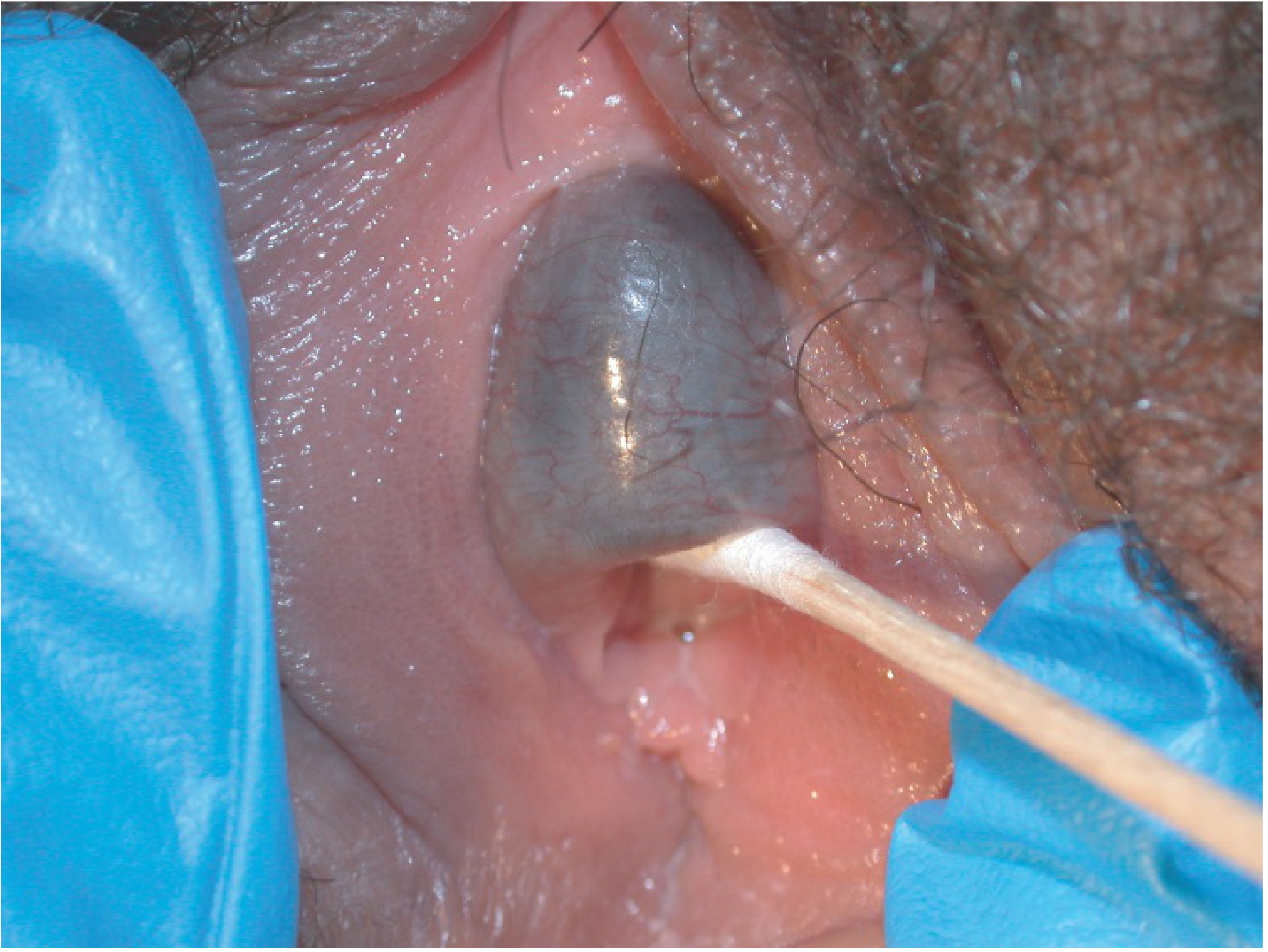
Skene gland cyst (Berek & Novak's Gynecology)
Microbiology (for abscesses)
- Bartholin abscess: E. coli is the most common isolate; many are polymicrobial. N. gonorrhoeae and C. trachomatis are less common but possible. - Tintinalli's Emergency Medicine
- Skene abscess: May involve similar organisms; N. gonorrhoeae can infect Skene glands in gonorrhea (alongside Bartholin gland involvement).
Key Differential Diagnoses
- Bartholin cyst must be distinguished from: labial abscess (requires gynecologic consult), Bartholin gland carcinoma (especially in women >40 - examine cyst wall for neoplastic epithelium)
- Skene cyst must be distinguished from: urethral diverticulum (UD communicates with the urethral lumen, is located mid-to-proximal urethra, does NOT distort the urethral meatus - Skene cysts are distal and distort the meatus), vaginal wall cysts, urethral prolapse
Management
| Treatment | Bartholin Cyst/Abscess | Skene Cyst/Abscess |
|---|---|---|
| Asymptomatic cyst | Observation | Observation |
| Abscess - first-line | Word catheter (stab incision, balloon inflated with 2-3 mL saline, left in place 4-6 weeks for epithelialization) | Incision and drainage, aspiration |
| Recurrent/large cyst | Marsupialization (incision on vaginal sidewall, suture cyst wall to vaginal mucosa with 4-0 Vicryl) | Marsupialization or surgical excision |
| Definitive | Excision (if marsupialization fails; send for histology if >40 yr) | Simple excision (curative) |
| Antibiotics | Broad-spectrum if abscess not yet walled-off | As needed for superinfection |
Quick Memory Hook
- Bartholin = Backside (posterior introitus), Big (up to 5 cm), Bulbourethral homologue - treat with Word catheter
- Skene = Surrounds urethra (anterior), Small but can obstruct, Simple excision is curative; homologue of the prostate
Sources: Robbins & Cotran Pathologic Basis of Disease; Tintinalli's Emergency Medicine; Berek & Novak's Gynecology; Campbell-Walsh-Wein Urology; Sabiston Textbook of Surgery; Pfenninger & Fowler's Procedures for Primary Care
bronchitis
Reading File
Bronchitis is inflammation of the bronchial mucosa, existing in two distinct forms: acute (self-limited, usually viral) and chronic (defined clinically as productive cough for at least 3 months per year for 2 consecutive years, a key component of COPD). Let me pull comprehensive textbook content on both.
Searching sources...
Finding Sources
Finding Sources
Reading File
Reading File
Reading File
Reading File
Reading File
Reading File
Reading File
Loading Image
Finding Sources
Here is a complete overview of bronchitis, covering both acute and chronic forms:
Bronchitis
Overview
Bronchitis = inflammation of the bronchial mucosa. Comes in two distinct clinical entities that differ in etiology, definition, pathology, and management.
| Acute Bronchitis | Chronic Bronchitis | |
|---|---|---|
| Definition | Self-limited inflammation of large airways without pneumonia | Productive cough ≥3 months/year for ≥2 consecutive years (no other identifiable cause) |
| Primary cause | Viral (up to 90%) | Cigarette smoke (90% of patients are smokers) |
| Definition type | Clinical/temporal | Clinical (not anatomic) |
ACUTE BRONCHITIS
Epidemiology
- ~5% of adults develop it annually
- ~100 million ambulatory care visits per year in the US
- Ninth most common ED diagnosis in the US
- Incidence peaks in fall and winter (correlates with viral season)
Etiology / Pathogens
Viral (up to 90% of cases):
- Influenza A and B
- Rhinovirus
- Coronavirus
- Parainfluenza virus
- RSV (respiratory syncytial virus)
- Human metapneumovirus
- Adenovirus
Bacterial (6-15% of cases):
- Bordetella pertussis, Mycoplasma pneumoniae, Chlamydia pneumoniae - in immunocompetent adults
- Haemophilus influenzae, Streptococcus pneumoniae, Moraxella catarrhalis - more common in COPD patients and smokers
Pathophysiology
Epithelial infection of the bronchi causes inflammation and thickening of the bronchial/tracheal mucosa, leading to:
- Airflow obstruction
- Bronchial hyperresponsiveness
- Decreased FEV1
- Clinical: cough, wheezing, dyspnea
- Sputum discoloration (yellow/green) = cellular debris, NOT reliably bacterial - Tintinalli's Emergency Medicine
Clinical Features
- Cough (hallmark) - with or without sputum, lasts 10-20 days on average, can persist >1 month
- Mild fever, dyspnea, headache, rhinorrhea, mild constitutional symptoms
- Symptoms begin like an upper respiratory infection but cough persists beyond 5 days
- Wheezing may be present due to bronchial hyperresponsiveness
- Purulent sputum is a poor predictor of bacterial infection
Diagnosis
- Clinical diagnosis - no routine lab workup needed in healthy patients
- Diagnose when: acute cough >5 days + no evidence of pneumonia, asthma exacerbation, COPD exacerbation, or CHF
- Chest X-ray only if pneumonia is suspected (fever, tachycardia, tachypnea, hypoxia, focal auscultatory findings suggest pneumonia)
- Rule out pneumonia: normal vitals + normal lung exam = pneumonia unlikely
- Cough persisting >3 weeks warrants investigation for other causes
- Routine sputum cultures are NOT necessary
Treatment
| Intervention | Recommendation |
|---|---|
| Antibiotics | NOT routinely recommended (IDSA "Choosing Wisely" campaign); only if strong suspicion of B. pertussis or atypical organisms; minimal benefit (~½ day shorter cough duration); significant adverse effects outweigh benefit |
| β2-agonists | NOT routinely; ONLY if airflow obstruction (wheezing) is present - associated with lower symptom scores and faster cough resolution |
| Oral corticosteroids | NOT recommended in patients without COPD or asthma |
| Antitussives (benzonatate) | May provide modest cough relief |
| Expectorants (guaifenesin) | May provide modest relief |
| Antihistamines / mucolytics | Limited evidence |
| Supportive care | Air humidifiers, antipyretics, rest |
Despite guidelines recommending against antibiotics, 71% of acute bronchitis patients received them in one US study (1996-2010) - Fishman's Pulmonary Diseases and Disorders
"Red flag" symptoms requiring urgent re-evaluation: hemoptysis, worsening dyspnea, weight loss, dysphagia, persistent fever.
CHRONIC BRONCHITIS
Definition
Productive cough with sputum for at least 3 months in at least 2 consecutive years, in the absence of any other identifiable cause. Definition is clinical (vs emphysema, which is defined anatomically). - Robbins & Cotran Pathologic Basis of Disease
Etiology / Risk Factors
- Cigarette smoking - primary cause (90% are smokers); damages airway epithelial cells AND impairs ciliary action (prevents mucus clearance, increases infection risk)
- Air pollutants: sulfur dioxide, nitrogen dioxide
- Occupational dusts: grain, cotton, silica
- Infection: does NOT initiate chronic bronchitis, but maintains it and causes acute exacerbations
Pathogenesis
- Mucus hypersecretion - earliest feature; involves inflammatory mediators (histamine, IL-13); hypertrophy of submucosal mucous glands in trachea/bronchi; goblet cell hyperplasia in small bronchi/bronchioles
- Acquired CFTR dysfunction - smoking causes CFTR dysfunction → abnormally dehydrated, viscous mucus → worsens severity
- Inflammation - neutrophils, lymphocytes, macrophages (notably NO eosinophils, unlike asthma)
- Small airway disease (chronic bronchiolitis) - peribronchial fibrosis, smooth muscle hypertrophy, mucus plugging → airflow obstruction
- Infection - secondary role; maintains and exacerbates inflammation
Morphology / Histopathology
- Grossly: hyperemic, edematous mucosa; mucinous or mucopurulent secretions filling airways
- Reid Index (key pathological marker):
- = Thickness of submucosal mucous gland layer / Thickness of bronchial wall (epithelium to cartilage)
- Normal: 0.4
- Increased in chronic bronchitis (proportional to severity and duration)
- Goblet cell metaplasia in small airways
- Inflammatory infiltrate (lymphocytes, macrophages, ± neutrophils)
- Smooth muscle hypertrophy, extracellular matrix deposition
- Severe cases: bronchiolitis obliterans (complete fibrotic obliteration of bronchiolar lumen)
- Emphysematous changes often coexist
Clinical Spectrum
- Early stages: mucoid sputum, no airflow obstruction
- Asthmatic bronchitis: intermittent bronchospasm and wheezing (hyperresponsive airways)
- Advanced: chronic outflow obstruction (COPD), usually with emphysema
- End-stage: hypoxemia → pulmonary hypertension → cor pulmonale
Complications of Long-standing Chronic Bronchitis
- Chronic obstructive pulmonary disease (COPD)
- Pulmonary hypertension
- Cor pulmonale (right heart failure from chronic hypoxia)
- Recurrent pulmonary infections / acute exacerbations (AECB)
Key Distinctions: Acute vs Chronic
| Acute | Chronic | |
|---|---|---|
| Duration | Days to weeks (self-limited) | Months to years (persistent) |
| Cause | Virus (90%) | Smoking (90%) |
| Cough | Lasts 10-20 days | Defined by ≥3 months/year |
| Sputum | May or may not be present | Persistent productive cough |
| Histology | Transient mucosal inflammation | Reid index >0.4, goblet cell hyperplasia, fibrosis |
| Antibiotics | Not indicated (unless specific pathogen) | Not for stable disease; yes for AECB with purulent sputum |
| Reversibility | Complete | Progressive if smoking continues |
| COPD link | No | Yes - component of COPD |
Sources: Robbins & Cotran Pathologic Basis of Disease; Robbins & Kumar Basic Pathology; Tintinalli's Emergency Medicine; Fishman's Pulmonary Diseases and Disorders; Textbook of Family Medicine 9e
external and internal hemorrhoids
Reading File
Hemorrhoids are vascular cushions of the anal canal that become pathological when they engorge, prolapse, or bleed. The key dividing line is the dentate (pectinate) line - internal hemorrhoids arise above it (no somatic pain fibers), external hemorrhoids arise below it (somatic innervation = painful). Let me pull the full textbook content.
Searching sources...
Finding Sources
Finding Sources
Reading File
Reading File
Reading File
Reading File
Reading File
Reading File
Finding Sources
Reading File
Loading Image
Here is a comprehensive breakdown of internal vs external hemorrhoids:
Hemorrhoids: Internal vs External
What Are Hemorrhoids?
Hemorrhoids are submucosal vascular beds (dilated vascular channels / collateral vessels) located in the anal and rectal canal. They are a normal part of anatomy - they assist with defecation and the sensation of anorectal fullness. Pathology arises when they engorge, prolapse, thrombose, or bleed.
Prevalence: ~4.4% of the US population (~10 million people) complain of hemorrhoidal disease; up to 25% of the adult population are affected. ~1.9 to 3.5 million physician visits annually in the US.
Typical locations: Right anterior, right posterior, and left lateral positions. Hemorrhoids at other locations should raise concern for carcinoma, lymphoma, or condyloma.
The Key Anatomical Dividing Line: The Dentate (Pectinate) Line
| Internal Hemorrhoids | External Hemorrhoids | |
|---|---|---|
| Location relative to dentate line | Above the dentate line | Below the dentate line |
| Venous plexus | Superior hemorrhoidal plexus (within distal rectum) | Inferior hemorrhoidal plexus (perianal) |
| Epithelium | Columnar or transitional mucosa | Squamous epithelium |
| Innervation | Autonomic only - NOT painful | Somatic innervation - painful |
| Portal vs caval | Portal system | Caval (systemic) system |
INTERNAL HEMORRHOIDS
Pathophysiology
Internal hemorrhoid symptoms occur due to loss of connective tissue support and resulting protrusion/prolapse of vascular tissue, rendering it susceptible to trauma from straining or hard stool passage. Associated with elevated circulating matrix metalloproteinases. More likely in patients with constipation, loose stools, or prolonged toilet sitting.
Risk Factors
- Constipation and straining (elevated intraabdominal/venous pressure)
- Low-fiber diet
- Prolonged sitting on toilet
- Pregnancy (mechanical compression of superior rectal vein + increased progesterone)
- Portal hypertension
- Obesity, aging, genetic factors
Symptoms
- Painless, bright-red rectal bleeding - hallmark; blood coats outside of stool, drips into toilet, or is seen on toilet paper; can be dark blood or clots if blood pools in rectum
- Prolapse - tissue protruding from anus
- Mucus or blood on underwear (from prolapse)
- Perianal itching (from moisture caused by prolapse)
- Pain is NOT typical - pain only occurs with thrombosis or strangulation
Grading (Classification)
| Grade | Description | Typical Treatment |
|---|---|---|
| Grade I | Bleed; may be enlarged; do not prolapse | Medical (fiber, fluids) |
| Grade II | Prolapse with defecation; reduce spontaneously | Medical ± rubber band ligation |
| Grade III | Prolapse and require manual reduction | Rubber band ligation or surgery |
| Grade IV | Remain prolapsed (irreducible) | Surgical excision |
Diagnosis
- Clinical history + physical examination
- Anoscopy (beveled or slotted anoscope) - gold standard for visualization and grading
- Flexible sigmoidoscopy/colonoscopy if bleeding source unclear, symptoms persist, or patient >40 years (to rule out colorectal cancer)
Treatment
Conservative / Medical (Grade I-II first line):
- Dietary fiber 20-30 g/day + 6-8 glasses of fluid daily (fiber supplements reduce symptoms by 53%)
- Stool softeners (docusate sodium), avoid straining
- Polyethylene glycol 3350 for constipation
- Topical agents: phenylephrine/mineral oil/petrolatum or glucocorticoid creams (short-term; caution - risk of candidiasis)
- Phlebotonics (flavonoids, plant extracts e.g., diosmin) - improve venous tone, reduce capillary permeability, superior to placebo for acute symptoms; excellent safety profile
Office Procedures (Grade I-III, failed medical therapy):
- Rubber band ligation (RBL) - most common office procedure; bands placed just proximal to dentate line (distal placement on squamous mucosa = pain, band must be removed immediately); tissue becomes necrotic and sloughs; contraindicated in anticoagulated patients
- Infrared photocoagulation (especially Grade I with persistent bleeding)
- Injection sclerotherapy
- Cryotherapy
- Doppler-guided hemorrhoidal artery ligation
Surgery (Grade IV, or failed office procedures):
- Formal hemorrhoidectomy
- Stapled hemorrhoidopexy (procedure for prolapsing hemorrhoids, PPH)
EXTERNAL HEMORRHOIDS
Pathophysiology
External hemorrhoids = dilated vessels of the inferior hemorrhoidal plexus below the dentate line, covered by squamous epithelium with somatic innervation. What is visible at the anal verge as "external hemorrhoids" is often redundant skin (anal tags) from previous episodes of external hemorrhoid inflammation, edema, and thrombosis.
Symptoms
- Usually asymptomatic when not thrombosed
- Skin tags: no bleeding, difficult hygiene, itching and irritation
- Thrombosed external hemorrhoid = the painful acute presentation
Thrombosed External Hemorrhoid - Special Entity
- Acute onset of severe perianal pain
- Visible purplish/bluish nodule at the anal verge with taut overlying skin
- Bluish discoloration from underlying clot
- Overlying skin may ulcerate and bleed (usually 1-2 days, may coincide with pain relief)
Clinical photo of thrombosed external hemorrhoid and excision:
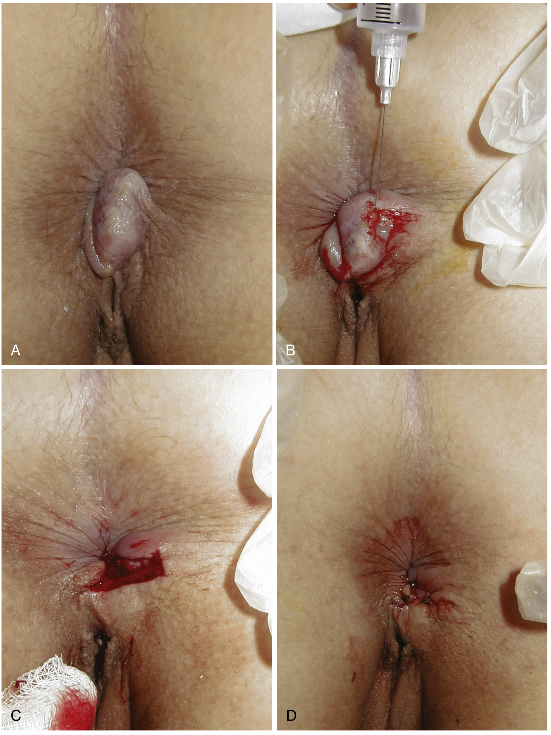
Office excision of thrombosed external hemorrhoid - Sleisenger & Fordtran's GI and Liver Disease
Treatment of thrombosed external hemorrhoid:
- If painless - reassurance; swelling subsides over several weeks
- If painful AND symptoms <72 hours - surgical excision under local anesthesia (1% lidocaine with epinephrine) is beneficial; enucleation preferred over simple incision (due to high recurrence with simple incision)
- If symptoms >72 hours - pain is already subsiding; observation preferred (pain resolves in 4-7 days regardless; outcome at 2 months is the same whether or not excision is performed)
- Conservative: warm sitz baths, stool softeners, topical analgesics (benzocaine, dibucaine, pramoxine)
Side-by-Side Comparison
| Feature | Internal | External |
|---|---|---|
| Location | Above dentate line | Below dentate line |
| Plexus | Superior hemorrhoidal | Inferior hemorrhoidal |
| Covering | Columnar/transitional mucosa | Squamous epithelium |
| Pain | Painless (unless thrombosed/strangulated) | Painful (somatic innervation) |
| Bleeding | Painless bright-red rectal bleeding | Minimal or none (unless ulcerated) |
| Key presentation | Prolapse, bleeding | Thrombosis (acute pain + blue nodule) |
| Grading system | Grade I-IV (by degree of prolapse) | Not formally graded |
| First-line treatment | Fiber + fluids + RBL | Observation or excision (if thrombosed <72 h) |
| Portal HTN link | Yes (portal venous drainage) | No (systemic venous drainage) |
Important Notes
- Patients often attribute unrelated anal symptoms (fissures, pruritus ani, warts) to hemorrhoids - careful exam is essential
- Hemorrhoids coexist with other anal pathology - do not assume hemorrhoids are the sole cause
- When hemorrhoids develop in the setting of portal hypertension, the implications are more serious
- In patients >40 years with hemorrhoidal bleeding, colonoscopy should be considered to exclude colorectal malignancy
Sources: Sleisenger & Fordtran's Gastrointestinal and Liver Disease; Textbook of Family Medicine 9e; Robbins & Kumar Basic Pathology; Clinical Gastrointestinal Endoscopy 3e; Yamada's Textbook of Gastroenterology; Creasy & Resnik's Maternal-Fetal Medicine
This is a shared conversation. Sign in to Orris to start your own chat.