

1000090923.jpg
Explain all
Reading File
This is a medical pathology exam list (Paper I). Let me search the medical library for all these topics simultaneously.
Searching sources...
Finding Sources
Finding Sources
Finding Sources
Finding Sources
Reading File
Reading File
Finding Sources
Finding Sources
Reading File
Finding Sources
Finding Sources
Finding Sources
Reading File
Reading File
I now have strong textbook content to answer all topics. Let me compile the full explanation now.
Paper I - Complete Explanation of All Topics
(Source: Robbins & Kumar Basic Pathology; Robbins, Cotran & Kumar Pathologic Basis of Disease)
1. Define Shock
Shock is a state of systemic tissue hypoperfusion due to reduced cardiac output and/or reduced effective circulating blood volume, leading to cellular hypoxia. At first the injury is reversible; prolonged shock causes irreversible tissue injury and is often fatal.
Three major types:
| Type | Example | Mechanism |
|---|---|---|
| Cardiogenic | Myocardial infarction, arrhythmia, cardiac tamponade | Failure of myocardial pump |
| Hypovolemic | Hemorrhage, severe burns | Inadequate blood/plasma volume |
| Septic | Gram-positive/gram-negative/fungal infections | Vasodilation, endothelial injury, DIC, cytokine cascade |
Other types include neurogenic shock (loss of vascular tone from spinal cord injury or anesthesia) and anaphylactic shock (IgE-mediated, systemic vasodilation + vascular permeability).
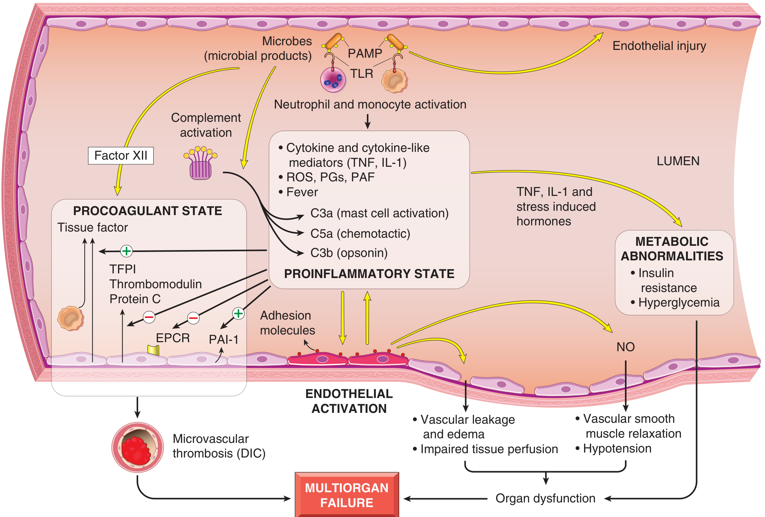
2. Septic Shock - Pathophysiology
Septic shock is the most common and complex type. It accounts for ~2% of all hospital admissions; mortality is 20-30%.
Trigger: Gram-positive bacteria (most common) > gram-negative bacteria > fungi.
Key pathogenic steps:
- Microbial PAMPs (pathogen-associated molecular patterns) activate Toll-like receptors (TLRs) on macrophages, neutrophils, dendritic cells, and endothelial cells.
- This triggers release of TNF, IL-1, IL-6, IL-12, IL-18, reactive oxygen species (ROS), prostaglandins.
- Endothelial activation and injury - upregulation of adhesion molecules, vascular leakage, systemic vasodilation (NO-mediated).
- Complement activation - C3a, C5a (anaphylatoxins) worsen inflammation.
- Coagulation activation - widespread thrombin activation leads to DIC (disseminated intravascular coagulation).
- Metabolic derangements - hyperglycemia initially, then hypoglycemia; lactic acidosis.
- If unchecked → multiorgan failure (lungs, kidneys, liver, brain).
A counterinflammatory immunosuppressive phase follows the initial hyperinflammatory state, leaving patients susceptible to secondary infections.
3. Warburg Effect
The Warburg Effect (aerobic glycolysis) is the phenomenon by which cancer cells preferentially use glycolysis for energy production even in the presence of oxygen, rather than the more efficient oxidative phosphorylation.
- Normal cells: use glycolysis only in low oxygen; switch to OXPHOS in oxygen.
- Cancer cells: use glycolysis + convert pyruvate to lactate even when O₂ is available.
- Why? Rapidly proliferating tumor cells need biosynthetic precursors (nucleotides, amino acids, lipids) more than ATP - glycolysis intermediates feed these biosynthetic pathways.
- Clinical use: This is the basis of PET scanning - tumors are detected by their high uptake of ¹⁸F-FDG (fluorodeoxyglucose).
4. Dystrophic Calcification
Dystrophic calcification is the deposition of calcium salts in dead or dying tissue, with normal serum calcium levels.
Sites:
- Areas of necrosis: coagulative, caseous, or liquefactive
- Atheromatous plaques (advanced atherosclerosis)
- Aging or damaged heart valves
- Tuberculous lymph nodes (can convert to "stone")
Mechanism: Cellular injury → membrane damage → intracellular calcium overload → phosphate release → calcium phosphate precipitation.
Morphology (H&E stain): Basophilic, amorphous granular deposits, intracellular or extracellular. Lamellated concentric structures called psammoma bodies may form (seen in papillary thyroid carcinoma, meningioma, serous ovarian tumors).
Key point: Serum calcium is NORMAL (differentiates from metastatic calcification, which occurs in normal tissues due to hypercalcemia).
5. Anaphylactic Shock
Anaphylactic shock is a severe, life-threatening systemic hypersensitivity reaction mediated by IgE antibodies.
Mechanism (Type I hypersensitivity):
- First exposure (sensitization): antigen stimulates IgE production → IgE binds to mast cells and basophils.
- Re-exposure: antigen cross-links IgE on mast cells → degranulation → release of histamine, leukotrienes, prostaglandins.
- These cause:
- Systemic vasodilation (drop in BP)
- Increased vascular permeability (urticaria, angioedema)
- Bronchospasm (wheezing, respiratory distress)
- GI symptoms
Common triggers: Bee stings, nuts, shellfish, penicillin, latex.
Treatment: Epinephrine (1st line), IV fluids, antihistamines, corticosteroids.
6. Distress Syndrome (ARDS - Acute Respiratory Distress Syndrome)
ARDS is characterized by rapid-onset diffuse alveolar damage causing severe hypoxia, not explained by cardiac failure.
Triggers: Sepsis (most common), pneumonia, trauma, aspiration, massive transfusion, pancreatitis.
Pathophysiology:
- Injury to alveolar epithelium (type I & II pneumocytes) and endothelium
- Massive neutrophil infiltration
- Release of proteases, ROS → damage alveolar-capillary membrane
- Protein-rich edema floods alveoli
- Hyaline membrane formation (hallmark on histology) - eosinophilic fibrin deposits lining alveoli
- Impaired surfactant production (type II cell injury) → atelectasis
Berlin Definition criteria: Acute onset, bilateral infiltrates on CXR, PaO₂/FiO₂ ratio <300 mmHg, not fully explained by cardiac failure.
Stages: Exudative (days 1-7) → Proliferative (1-3 weeks) → Fibrotic (beyond 3 weeks).
7. Decompression Sickness (Caisson Disease)
Decompression sickness occurs when a person exposed to high atmospheric pressure (divers, tunnel workers) ascends too rapidly.
Mechanism:
- At high pressure, nitrogen dissolves in blood and tissues (Henry's Law: gas solubility ∝ pressure).
- Rapid ascent → pressure drops → nitrogen comes out of solution as gas bubbles in tissues and vessels.
- Bubbles cause: obstruction of small vessels, tissue ischemia, focal necrosis.
Manifestations:
- "The Bends" - joint/muscle pain (nitrogen bubbles in joints)
- Pulmonary symptoms: "the chokes" (dyspnea, chest pain)
- Neurological: stroke-like symptoms
- Chronic: Aseptic (avascular) necrosis of bone - classically femoral head
Treatment: Hyperbaric oxygen (recompression) to re-dissolve bubbles.
8. Embolism
An embolus is an intravascular mass (solid, liquid, or gas) carried in blood from one location to obstruct another.
Types:
| Type | Description |
|---|---|
| Thromboembolism | Most common (95%+). DVT → pulmonary embolism |
| Fat embolism | After long bone fractures or liposuction; fat globules occlude pulmonary/cerebral vessels |
| Air/Gas embolism | IV line errors, surgery; >100 mL air can be fatal |
| Amniotic fluid embolism | Obstetric emergency; DIC + respiratory failure |
| Tumor embolism | Hematogenous spread of cancer |
| Septic embolism | Infected thrombi from infective endocarditis |
Pulmonary embolism (PE):
- Source: DVT of leg veins (90%)
- Effects depend on size: saddle embolus → sudden death; medium → hemorrhagic infarction; small → often silent
- Classic triad: dyspnea, chest pain, hemoptysis (rare to have all three)
9. Amyloidosis
Amyloidosis is a disease caused by extracellular deposition of abnormal insoluble fibrillar proteins (amyloid) that disrupt tissue architecture and function.
Structure: All amyloid proteins share a beta-pleated sheet configuration - this is why they stain with Congo red and show apple-green birefringence under polarized light.
Types:
| Type | Protein | Associated Condition |
|---|---|---|
| AL (Primary) | Immunoglobulin light chains | Multiple myeloma, plasma cell dyscrasias |
| AA (Secondary) | Serum amyloid A (SAA) | Chronic inflammatory diseases (RA, TB, osteomyelitis) |
| Aβ amyloid | Amyloid precursor protein | Alzheimer disease |
| Transthyretin (ATTR) | Transthyretin | Familial amyloidosis, senile cardiac amyloidosis |
| Aβ₂M | β₂-microglobulin | Long-term hemodialysis |
Organs affected: Kidney (proteinuria → nephrotic syndrome), heart (restrictive cardiomyopathy), liver (hepatomegaly), spleen ("sago spleen" - periarteriolar deposits), tongue (macroglossia in AL type), peripheral nerves.
10. Hodgkin Lymphoma (HL) vs Non-Hodgkin Lymphoma (NHL)
Hodgkin Lymphoma (HL)
- Hallmark cell: Reed-Sternberg (RS) cell - large binucleate cell with "owl-eye" nucleoli, surrounded by reactive lymphocytes, plasma cells, eosinophils.
- Origin: Germinal center B cells (usually EBV-driven)
- Bimodal age distribution: Young adults (15-35) and elderly (>55)
- Spread: Contiguous lymph node spread (predictable)
- Types (WHO): Classic HL (nodular sclerosis, mixed cellularity, lymphocyte-rich, lymphocyte-depleted) and Nodular lymphocyte predominant HL
- Most common type: Nodular sclerosis HL
- Treatment: Generally very good prognosis; ABVD chemotherapy ± radiation
Non-Hodgkin Lymphoma (NHL)
- No RS cells
- Heterogeneous group of B-cell, T-cell, and NK-cell lymphomas
- Spread: Non-contiguous; more likely to be widespread at diagnosis
- Common types:
- Diffuse Large B-cell Lymphoma (DLBCL) - most common NHL; aggressive
- Follicular Lymphoma - indolent; t(14;18) translocation → BCL-2 overexpression → blocked apoptosis
- Burkitt Lymphoma - c-MYC translocation t(8;14); "starry sky" pattern; linked to EBV
- Mantle Cell Lymphoma - t(11;14); cyclin D1 overexpression
- Marginal Zone Lymphoma (MALT) - H. pylori-associated gastric lymphoma
11. Multiple Myeloma
Multiple myeloma is a malignant proliferation of plasma cells (terminally differentiated B cells) in the bone marrow, producing a monoclonal immunoglobulin (M-protein/paraprotein).
Features (CRAB criteria):
- C - hyperCalcemia (from bone destruction; causes nausea, confusion, polyuria)
- R - Renal failure (Bence-Jones protein = light chains damage tubules; also hypercalcemia)
- A - Anemia (marrow infiltration by plasma cells)
- B - Bone lesions ("punched out" lytic lesions on X-ray; pathological fractures)
Additional features:
- Bence-Jones proteins (free light chains) in urine - pathognomonic
- Recurrent infections (suppressed normal immunoglobulins)
- Rouleaux formation of RBCs on blood smear
- Hyperviscosity syndrome
Diagnosis: Bone marrow biopsy (>10% plasma cells), serum protein electrophoresis (M-spike), serum free light chain assay.
12. SLE (Systemic Lupus Erythematosus)
SLE is a multisystem autoimmune disease characterized by production of autoantibodies against nuclear antigens, forming immune complexes that deposit in multiple organs.
Key autoantibodies:
- Anti-dsDNA and Anti-Sm - specific for SLE (diagnostic)
- Antinuclear antibody (ANA) - sensitive but not specific
- Anti-phospholipid antibodies - cause thrombosis, miscarriage (antiphospholipid syndrome)
Clinical features (SOAP BRAIN MD): Serositis, Oral ulcers, Arthritis, Photosensitivity, Blood disorders (anemia, thrombocytopenia, leukopenia), Renal (lupus nephritis), ANA positive, Immunological (anti-dsDNA), Neurological (seizures, psychosis), Malar rash, Discoid rash.
Pathogenesis:
- Defective clearance of apoptotic cells → nuclear antigens exposed
- Loss of B- and T-cell tolerance → autoantibody production
- Immune complex (type III hypersensitivity) deposition in glomeruli, skin, joints, blood vessels → complement activation → inflammation
Classic histology: "Wire-loop" lesion in glomeruli (membranous deposits), "onion-skin" lesion in splenic arterioles.
13. Benign vs. Malignant Tumors
| Feature | Benign | Malignant |
|---|---|---|
| Growth rate | Slow | Rapid |
| Borders | Well-circumscribed, encapsulated | Irregular, infiltrative |
| Metastasis | Never | Hallmark feature |
| Differentiation | Well differentiated | Poorly differentiated (anaplastic) |
| Mitoses | Rare, normal | Frequent, abnormal (atypical) |
| Necrosis | Rare | Common (outgrows blood supply) |
| Effect on host | Local pressure effects | Invasion, metastasis, paraneoplastic syndromes, cachexia |
| Nucleus | Normal N:C ratio | High N:C ratio, hyperchromatism, pleomorphism |
Nomenclature:
- Benign epithelial: adenoma, papilloma
- Malignant epithelial: carcinoma (adenocarcinoma, squamous cell carcinoma)
- Benign mesenchymal: lipoma, fibroma, leiomyoma
- Malignant mesenchymal: sarcoma (liposarcoma, fibrosarcoma)
14. Intrinsic vs. Extrinsic Pathway - Apoptosis
Apoptosis is programmed cell death characterized by cell shrinkage, chromatin condensation (pyknosis), nuclear fragmentation (karyorrhexis), membrane blebbing, and apoptotic body formation - all without inflammation.
Intrinsic (Mitochondrial) Pathway
- Trigger: DNA damage, oxidative stress, growth factor withdrawal, ER stress
- Key molecules: BCL-2 family
- Pro-apoptotic: BAX, BAK, BAD, BID, BIM - promote cytochrome c release
- Anti-apoptotic: BCL-2, BCL-XL - block cytochrome c release
- Steps: When damage is too great → BAX/BAK form pores in mitochondrial outer membrane → cytochrome c released → forms apoptosome with APAF-1 → activates caspase-9 → activates effector caspases (3, 6, 7) → cell death
- Key regulator: p53 - senses DNA damage; upregulates pro-apoptotic genes
Extrinsic (Death Receptor) Pathway
- Trigger: Binding of death ligands (FasL/TRAIL/TNF) to death receptors (Fas/TNFR1/DR4,5)
- Steps: Ligand-receptor binding → recruitment of FADD (Fas-associated death domain) → caspase-8 activation → effector caspases (3, 6, 7) → cell death
- Cross-talk: Caspase-8 can cleave BID → truncated tBID → activates intrinsic pathway (amplification loop)
Both pathways converge on effector caspases (3, 6, 7) which execute cell death.
15. Transfusion-Related Infections
Blood transfusions can transmit several infections despite rigorous screening:
| Pathogen | Notes |
|---|---|
| HIV | Window period risk; NAT (nucleic acid testing) has dramatically reduced risk |
| Hepatitis B (HBV) | Surface antigen (HBsAg) tested; occult HBV remains a small risk |
| Hepatitis C (HCV) | Anti-HCV + NAT used; now very rare |
| Cytomegalovirus (CMV) | Mainly risks immunocompromised recipients; leukoreduction helps |
| HTLV-I/II | T-cell lymphotropic virus; screened in many countries |
| Malaria | Not routinely screened by testing in all countries; donor deferral used |
| Babesia | Tick-borne; increasing risk in endemic areas; no universal test yet |
| Treponema pallidum (Syphilis) | Screened |
| West Nile Virus | Screened by NAT in USA |
| Variant CJD (prion) | No test; managed by donor exclusion (UK exposure history) |
Most common serious transfusion complication today (developed world): Bacterial contamination of platelets (stored at room temperature).
16. Apoptosis (detailed - see #14 above)
Summary of key features:
- Cell shrinks (unlike necrosis where cells swell)
- Chromatin condensation + nuclear fragmentation
- Apoptotic bodies phagocytosed by macrophages - no inflammatory response
- Executed by caspases (cysteine-aspartic acid proteases)
- Physiological apoptosis: embryonic development, thymic selection, elimination of aged cells
- Pathological apoptosis: viral infection, DNA damage, toxins, ischemia
17. Necrosis
Necrosis is uncontrolled, pathological cell death characterized by cell swelling, membrane disruption, organelle breakdown, and release of cellular contents → inflammatory response.
Types:
| Type | Mechanism/Appearance | Classic Example |
|---|---|---|
| Coagulative | Protein denaturation preserves cell ghost outlines; firm pale area | Myocardial infarction, most solid organ infarcts |
| Liquefactive | Enzymatic digestion liquefies tissue | Brain infarction, abscess (bacterial infection) |
| Caseous | Cheese-like, granular; central necrotic zone with granuloma | Tuberculosis |
| Fat necrosis | Enzymatic: lipase digests fat → saponification (calcium soaps) | Acute pancreatitis |
| Fibrinoid | Immune complex deposits in vessel walls | Vasculitis, malignant hypertension, SLE |
| Gangrenous | Dry (coagulative) or wet (liquefactive + bacterial) | Limb ischemia, diabetic foot |
18. Infarction
An infarct is an area of ischemic necrosis caused by occlusion of the arterial supply or venous drainage of an organ.
Types:
- White (Anemic) infarct: Occurs in solid organs with single arterial supply (heart, kidney, spleen). Pale, firm, wedge-shaped.
- Red (Hemorrhagic) infarct: Occurs in:
- Organs with dual blood supply (lung, liver)
- Organs with loose tissue (lung)
- Following reperfusion of an infarcted area
- Venous infarction
Histology (heart as example):
- 0-4 hrs: No change (coagulative necrosis begins microscopically)
- 4-12 hrs: Eosinophilic change, wavy fibers
- 12-24 hrs: Coagulative necrosis, neutrophil infiltration begins
- 1-3 days: Neutrophil infiltration peak
- 3-7 days: Macrophage infiltration, granulation tissue begins
- Weeks: Scar (fibrosis) formation
19. Hypersensitivity
Hypersensitivity refers to exaggerated or inappropriate immune responses that cause tissue damage.
Gell and Coombs Classification:
| Type | Mechanism | Mediator | Example |
|---|---|---|---|
| Type I | IgE-mediated (immediate) | Mast cells, basophils, histamine | Anaphylaxis, asthma, hay fever, atopy |
| Type II | Antibody-mediated cytotoxicity | IgG/IgM + complement or NK cells | Hemolytic transfusion reactions, Goodpasture syndrome, Graves disease |
| Type III | Immune complex-mediated | IgG complexes, complement, neutrophils | SLE, serum sickness, post-streptococcal GN |
| Type IV | T cell-mediated (delayed) | CD4+ T cells (Th1/Th17), CD8+ T cells | TB, contact dermatitis, rejection of transplants, Type 1 diabetes |
Key timings:
- Type I: within minutes
- Type II: hours to days
- Type III: hours to days
- Type IV: 48-72 hours (hence "delayed")
20. Thalassemia
Thalassemias are inherited disorders of decreased synthesis of α- or β-globin chains, leading to hemolytic anemia.
- Common in: Mediterranean, African, and Asian populations (malaria-endemic regions - offers protection against P. falciparum)
- Chromosome: β-globin on chromosome 11; α-globin on chromosome 16 (2 genes per chromosome = 4 total)
β-Thalassemia
- Caused by point mutations affecting transcription, splicing, or translation of β-globin mRNA
- β-thalassemia major (Cooley anemia): Homozygous; severe anemia needing regular transfusions; massive splenomegaly, bone marrow expansion (chipmunk facies, crew-cut X-ray of skull), iron overload
- β-thalassemia intermedia: Moderate anemia; no regular transfusions
- β-thalassemia minor (trait): Heterozygous; asymptomatic; mild/absent anemia; target cells on smear
α-Thalassemia
- Caused by gene deletions
- 4 genes total; severity depends on how many are deleted:
- 1 gene deleted: silent carrier (normal)
- 2 genes deleted: α-thalassemia trait (mild anemia)
- 3 genes deleted: HbH disease (moderate-severe anemia; HbH = β4 tetramers)
- 4 genes deleted: Hb Barts (hydrops fetalis) - incompatible with life; γ4 tetramers; stillbirth
Summary Table
| Topic | Key Concept |
|---|---|
| Shock | Hypoperfusion → hypoxia → cell death |
| Septic shock | PAMPs → TLR → cytokines → vasodilation + DIC |
| Warburg effect | Cancer cells use aerobic glycolysis |
| Dystrophic calcification | Ca2+ in dead tissue, normal serum Ca2+ |
| Anaphylactic shock | IgE → mast cell degranulation → vasodilation |
| ARDS | Alveolar damage → hyaline membrane → hypoxia |
| Decompression sickness | N₂ bubbles on rapid ascent → ischemia |
| Embolism | Intravascular mass causing occlusion |
| Amyloidosis | β-pleated sheet deposits → organ dysfunction |
| HL vs NHL | RS cells in HL; heterogeneous in NHL |
| Multiple myeloma | Plasma cell tumor; CRAB criteria |
| SLE | Anti-dsDNA/Sm; type III hypersensitivity |
| Benign vs malignant | Metastasis defines malignancy |
| Apoptosis pathways | Intrinsic (cytochrome c/BCL-2); Extrinsic (FasL/caspase-8) |
| Transfusion infections | HIV, HCV, HBV, CMV, bacterial contamination |
| Necrosis | Coagulative (MI), liquefactive (brain/abscess), caseous (TB) |
| Infarction | White (solid organs) vs red (dual supply/loose tissue) |
| Hypersensitivity | Types I-IV; IgE → antibody → immune complex → T cell |
| Thalassemia | α- or β-globin deficiency; hemolytic anemia |
This is a shared conversation. Sign in to Orris to start your own chat.