
mds classification morphology and molecular
Reading File
MDS (Myelodysplastic Syndromes) classification involves both morphologic categories and molecular/cytogenetic findings. The current framework is the WHO 2022 / ICC 2022 classification, which significantly shifted toward molecular-defined entities.
Searching sources...
Finding Sources
Searching PubMed
"myelodysplastic syndrome" AND classification AND "WHO 2022"
Finding Sources
Searching PubMed
"myelodysplastic syndrome" AND classification AND molecular
Reading File
Reading File
Reading Article
PMID: 38043791
Reading File
Reading File
Searching the Web
MDS WHO 2022 ICC 2022 classification morphology molecular subtypes myelodysplastic neoplasms
Reading Web Page
https://www.pathologyoutlines.com/topic/bonemarrowneoplastic…
Finding Sources
Reading Web Page
https://mds-hub.com/medical-information/international-consen…
Finding Sources
Here is a detailed breakdown of MDS classification covering both the WHO 5th edition (2022) and ICC (2022) frameworks, with morphology and molecular features for each entity.
MDS Classification: Morphology & Molecular (WHO 2022 / ICC 2022)
Background
In 2022, two major groups simultaneously updated MDS classification:
- WHO 5th edition (WHO-HEM5) - renamed entities as "Myelodysplastic Neoplasms" to reflect their clonal neoplastic biology
- International Consensus Classification (ICC 2022) - retains "Myelodysplastic Syndromes"
Both systems heavily incorporate molecular/cytogenetic drivers as defining criteria, moving away from purely morphologic subtyping. The key shift: genetic abnormalities now supersede morphologic categories in several entities.
Diagnostic Prerequisites for Any MDS
Before classifying, all cases must fulfill minimum criteria:
- One or more peripheral blood cytopenias (Hgb <13 M / <12 F; ANC <1.8 x10⁹/L; platelets <150 x10⁹/L)
- Dysplasia in ≥10% of cells in one or more hematopoietic lineages on bone marrow exam (or MDS-defining genetic/cytogenetic abnormality)
- Exclusion of other causes of dysplasia (B12/folate deficiency, heavy metals, drugs, copper deficiency, viral infection)
- Blasts must be <20% (≥20% = AML)
WHO 2022 Classification Structure
WHO 2022 organizes MDS into three main groups:
Group 1: MDS with Defining Genetic Abnormalities
These entities are defined by their molecular/cytogenetic lesion, which takes precedence over blast count or dysplasia extent.
| Entity | Blast Threshold | Defining Genetic Feature | Morphology |
|---|---|---|---|
| MDS-SF3B1 (MDS with low blasts and SF3B1 mutation) | <5% BM, <2% PB | SF3B1 mutation (VAF ≥5%); no high-risk mutations (RUNX1, ASXL1, EZH2, BCOR, SRSF2) and no del(5q)/monosomy 7/complex karyotype | Ring sideroblasts (≥15% of erythroid precursors, or ≥5% if SF3B1 mutated); erythroid dysplasia predominant; anemia is the main cytopenia |
| MDS-5q (MDS with low blasts and isolated del(5q)) | <5% BM, <2% PB | Isolated del(5q) or del(5q) + one additional abnormality (not -7 or del(7q)) | Megakaryocytes with hypolobated/monolobated nuclei (pathognomonic); erythroid hypoplasia; preserved platelet count or thrombocytosis |
| MDS-biTP53 (MDS with biallelic TP53 inactivation) | Any (can exceed 10%) | Biallelic TP53 (two distinct mutations, or mutation + 17p LOH/del) | Complex karyotype (~80%); multilineage dysplasia; may have fibrosis; blasts variable but often increased |
Note: MDS-biTP53 is treated as a distinct category regardless of blast percentage due to its dismal prognosis. It overlaps with the ICC category "Myeloid neoplasms with mutated TP53."
Group 2: MDS, Morphologically Defined
These are diagnosed when no disease-defining genetic abnormality is present. Subclassification by dysplastic lineage count (SLD/MLD) is dropped in WHO 2022, replaced by blast percentage.
| Entity | BM Blasts | PB Blasts | Other Features |
|---|---|---|---|
| MDS-LB (MDS with low blasts) | <5% | <2% | No SF3B1 mutation, no del(5q), no biTP53; dysplasia in ≥1 lineage |
| MDS-h (MDS, hypoplastic) | <5% | <2% | Marrow hypocellularity for age; oligoclonal CD8+ T-cell expansion; clonal hematopoiesis |
| MDS-f (MDS with fibrosis) | 5-19% (BM) or 2-19% (PB) | See BM | Grade 2-3 reticulin or collagen fibrosis; multilineage dysplasia |
| MDS-IB1 (MDS with increased blasts-1) | 5-9% | 2-4% | No Auer rods |
| MDS-IB2 (MDS with increased blasts-2) | 10-19% | 5-19%, or any Auer rods in blasts | Highest risk of AML transformation in this group |
Group 3: Childhood MDS
| Entity | Features |
|---|---|
| cMDS with low blasts, hypocellular | Hypocellular BM, <5% blasts; overlap with aplastic anemia |
| cMDS with increased blasts | ≥5% BM blasts or ≥2% PB blasts |
ICC 2022 Classification
The ICC retains more morphologic granularity (SLD/MLD) but adds new molecular-defined entities:
| ICC Entity | Blast Count | Defining Feature |
|---|---|---|
| MDS-SF3B1 | <5% BM, <2% PB | SF3B1 mutation; ≥15% ring sideroblasts (or ≥5% with SF3B1) |
| MDS-5q | <5% BM, <2% PB | Isolated del(5q) |
| MDS, NOS-SLD | <5% BM, <2% PB | Dysplasia in 1 lineage; no SF3B1, no del(5q), no biTP53 |
| MDS, NOS-MLD | <5% BM, <2% PB | Dysplasia in ≥2 lineages |
| MDS, NOS (with RS, no SF3B1) | <5% BM, <2% PB | Ring sideroblasts present but SF3B1 wild-type |
| MDS-EB | 5-9% BM or 2-4% PB | Excess blasts (lower threshold) |
| MDS/AML | 10-19% BM or 5-19% PB | Replaces MDS-EB-II; recognizes biologic overlap with AML |
| MDS with mutated TP53 | Any | Multi-hit TP53; separate from MDS-EB hierarchy |
Key ICC difference: MDS-EB-II (10-19% blasts) is renamed MDS/AML to reflect its AML-like biology and treatment approach. WHO 2022 retains this as MDS-IB2.
Side-by-Side Comparison: WHO 2016 vs WHO 2022 vs ICC 2022
| Old WHO 2016 | WHO 2022 | ICC 2022 |
|---|---|---|
| MDS-SLD | MDS-LB | MDS, NOS-SLD |
| MDS-MLD | MDS-LB | MDS, NOS-MLD |
| MDS-RS-SLD / MDS-RS-MLD | MDS-SF3B1 (if SF3B1+) or MDS-LB/MDS-LB with RS (if SF3B1-) | MDS-SF3B1 |
| MDS del(5q) | MDS-5q | MDS-5q |
| MDS-EB-I | MDS-IB1 | MDS-EB |
| MDS-EB-II | MDS-IB2 | MDS/AML |
| MDS-U | (removed; reclassified) | (removed) |
| - | MDS-biTP53 | MDS with mutated TP53 |
| RCC (provisional) | cMDS | cMDS |
Key Morphologic Features by Lineage
Erythroid dysplasia
- Nuclear: budding, internuclear bridging, karyorrhexis, multinuclearity, megaloblastoid change
- Cytoplasmic: ring sideroblasts (iron-laden mitochondria encircling ≥1/3 of nucleus on Prussian blue), vacuolization, PAS positivity
Granulocytic dysplasia
- Hypolobated or bilobed nuclei (pseudo-Pelger-Huet cells)
- Nuclear hypersegmentation
- Hypogranularity or agranularity of cytoplasm
- Auer rods (in excess blast subtypes - immediately escalates classification)
Megakaryocytic dysplasia (most specific for MDS)
- Micromegakaryocytes (lymphocyte-sized, single round nucleus)
- Hypolobated / monolobated megakaryocytes (hallmark of del(5q) MDS)
- Multiple widely separated nuclear lobes
- Large mononuclear forms
Key Molecular Mutations in MDS
| Gene | Frequency | Functional Class | Associated Entity/Notes |
|---|---|---|---|
| SF3B1 | 20-28% | RNA splicing | MDS-SF3B1; ring sideroblasts; better prognosis |
| SRSF2 | 12-15% | RNA splicing | Associated with MDS/MPN (CMML); multilineage dysplasia |
| U2AF1 | 8-12% | RNA splicing | del(20q) association; intermediate prognosis |
| ZRSR2 | 5% | RNA splicing | X-linked; dysgranulopoiesis |
| TET2 | 20-25% | DNA methylation (epigenetic) | Often early/founding mutation; CHIP; nonspecific |
| DNMT3A | 8-12% | DNA methylation | CHIP association; often co-mutated with other drivers |
| IDH1/IDH2 | 5-8% | DNA methylation | AML transformation risk |
| ASXL1 | 15-20% | Histone modification | Poor prognosis; commonly co-mutated |
| EZH2 | 5-7% | Histone modification | Poor prognosis |
| RUNX1 | 10-15% | Transcription factor | Precursor B-ALL risk if germline; acquired = MDS progression |
| TP53 | 5-10% (primary); higher in therapy-related | Tumor suppressor | Complex karyotype; MDS-biTP53 (multi-hit); dismal prognosis; ~50% therapy-related MDS |
| STAG2 | 5-10% | Cohesin complex | Multilineage dysplasia |
| NPM1 | Rare in MDS | Nucleolar | More AML-associated |
Cytogenetic Abnormalities
| Abnormality | Frequency | Association |
|---|---|---|
| del(5q) | 10-15% | MDS-5q; female predominance; good prognosis; lenalidomide responsive |
| Monosomy 7 / del(7q) | 10% | Poor prognosis; therapy-related MDS |
| Trisomy 8 | 10% | Nonspecific; intermediate prognosis |
| del(20q) | 5-7% | Relatively favorable |
| del(17p) | 3-5% | TP53 loss; complex karyotype |
| Complex karyotype (≥3 abnormalities) | 15% | Very poor; TP53 association |
CHIP vs CCUS vs MDS - Pre-MDS Spectrum
- CHIP (Clonal Hematopoiesis of Indeterminate Potential): Clonal mutation (VAF ≥2%), normal blood counts, no cytopenia or dysplasia
- CCUS (Clonal Cytopenia of Undetermined Significance): Cytopenia + clonal mutation, but insufficient dysplasia and no MDS-defining cytogenetic lesion (except del(5q), -7/del(7q), or complex karyotype which = MDS directly)
- MDS: Cytopenia + dysplasia (≥10%) in ≥1 lineage OR MDS-defining genetic lesion
Key Points Summary
- SF3B1 is the most clinically meaningful single-gene MDS entity - its mutation defines MDS-SF3B1 regardless of ring sideroblast count, and it carries a relatively favorable prognosis.
- biTP53 (multi-hit) is the most aggressive molecular entity - treated as a distinct high-risk category regardless of blast percentage in both WHO and ICC 2022.
- del(5q) remains a standalone entity with unique morphology (monolobated megakaryocytes) and is the only MDS with an approved targeted therapy (lenalidomide).
- The SLD/MLD distinction is retained in ICC 2022 but removed from WHO 2022 (both fall into MDS-LB).
- MDS-EB-II (10-19% blasts) is renamed MDS/AML in ICC 2022, recognizing biologic overlap with AML and the rationale for AML-type therapy.
- Morphologic assessment remains essential: dysplasia in megakaryocytes is the most specific lineage for MDS; ring sideroblasts are the most diagnostically notable erythroid feature.
Sources:
- Robbins & Kumar Basic Pathology, pp. 2358-2374 (MDS pathogenesis and morphology)
- WHO 5th edition MDS review, Loghavi et al. 2024 (PMID: 38043791)
- PathologyOutlines MDS-general comparison table
- ICC 2022 MDS updates - MDS Hub
- WHO vs ICC 2022 comparison, Blood Cancer Journal
mpn pds overlap
MDS/MPN overlap neoplasms share features of both myelodysplastic syndromes (ineffective hematopoiesis, dysplasia, cytopenias) and myeloproliferative neoplasms (proliferative drive, elevated counts). The main entities are CMML, aCML, MDS/MPN-RS-T, JMML, and MDS/MPN-NOS.
Searching sources...
Finding Sources
Searching PubMed
"myelodysplastic myeloproliferative neoplasms" AND classification
Searching the Web
MDS MPN overlap neoplasms WHO 2022 classification CMML aCML JMML MDS/MPN-RS-T molecular features
Finding Sources
Reading Web Page
https://pmc.ncbi.nlm.nih.gov/articles/PMC10296742
Reading Article
PMID: 37568631
Reading File
Reading Web Page
https://encyclopedia.pub/entry/45865
Loading Image
Finding Sources
Reading File
Reading File
Reading File
Reading File
Excellent data gathered. Here is the comprehensive breakdown:
MDS/MPN Overlap Neoplasms - Classification, Morphology & Molecular
Overview
MDS/MPN overlap neoplasms are clonal myeloid disorders that simultaneously harbor dysplastic features (ineffective hematopoiesis, cytopenias) AND proliferative features (elevated cell counts, organomegaly). They sit between pure MDS and pure MPN, making diagnosis challenging.
WHO 2022 and ICC 2022 recognize the same core entities but with some structural differences. A key 2022 change: JMML is moved out of the MDS/MPN category into a separate pediatric/germline-associated group (though it is still discussed here for completeness).
Entities at a Glance
| Entity | WHO 2022 Name | ICC 2022 Name | Defining Feature |
|---|---|---|---|
| CMML | CMML | CMML | Persistent monocytosis |
| aCML | MDS/MPN with neutrophilia | aCML, BCR-ABL1-negative | Neutrophilia + dysgranulopoiesis |
| MDS/MPN-RS-T | MDS/MPN-SF3B1-T | MDS/MPN-SF3B1-T or MDS/MPN-RS-T-NOS | Ring sideroblasts + thrombocytosis |
| JMML | (moved to pediatric category) | JMML | Pediatric; RAS pathway mutations |
| MDS/MPN-NOS | MDS/MPN-NOS | MDS/MPN-NOS | Overlap features, none of the above |
Note: WHO 2022 renamed MDS/MPN-RS-T to MDS/MPN-SF3B1-T for SF3B1-mutated cases, reflecting the same molecular-first shift seen in MDS.
1. Chronic Myelomonocytic Leukemia (CMML)
The most common MDS/MPN entity - incidence ~0.6 per 100,000/year; median age 73-75; male predominance.
Diagnostic Criteria
| Criterion | Threshold |
|---|---|
| Peripheral blood monocytosis | ≥0.5 × 10⁹/L AND ≥10% of WBC differential |
| Blasts (PB + BM) | <20% (including promonocytes) |
| Philadelphia chromosome / BCR-ABL1 | Absent |
| PDGFRA, PDGFRB, FGFR1, PCM1-JAK2 rearrangements | Absent (if eosinophilia present, must exclude) |
| Dysplasia | ≥1 myeloid lineage (or clonal cytogenetic/molecular abnormality) |
Blast-Based Subclassification
| Subtype | PB Blasts | BM Blasts |
|---|---|---|
| CMML-0 | <2% | <5% |
| CMML-1 | 2-4% | 5-9% |
| CMML-2 | 5-19% | 10-19%, or Auer rods present |
WHO 2022 note: CMML-0 and CMML-1 have virtually identical prognosis; the distinction is retained but debated.
Morphology
- Peripheral blood: increased monocytes with mildly atypical morphology - irregular nuclear contours, finely convoluted/cerebriform nuclei, abnormal granularity
- Promonocytes counted as blasts (nuclear folds, delicate chromatin, gray cytoplasm)
- Granulocytic dysplasia: pseudo-Pelger-Huet cells, hypogranularity
- Bone marrow: hypercellular with monocytic proliferation and granulocytic dysplasia
- May have plasmacytoid monocyte clusters (CD14+, CD68+, CD56+, CD4+, CD2+, CD5+)
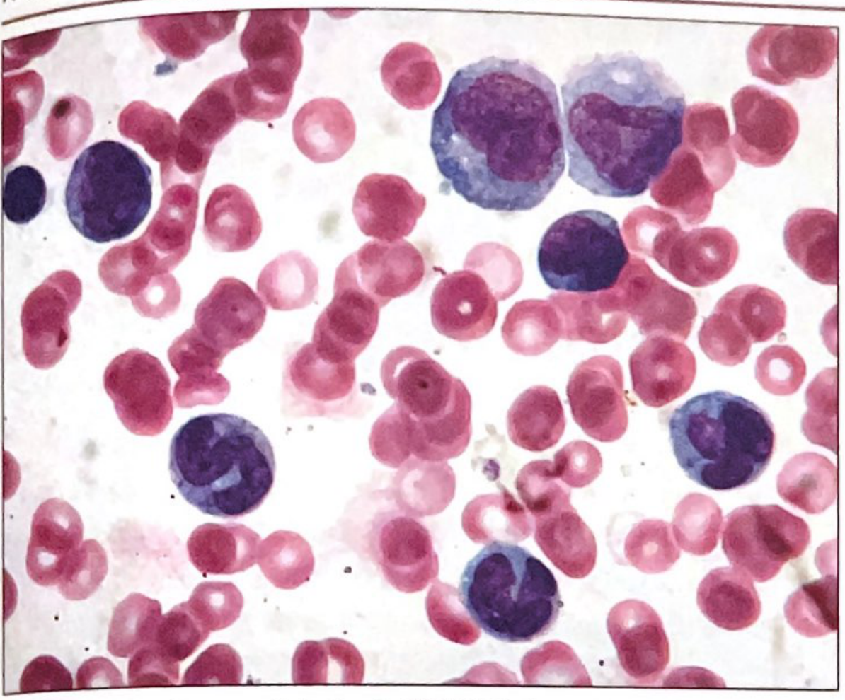
CMML - peripheral blood showing increased absolute monocytes with mild atypia
Immunophenotype
- CD13+, CD33+, variable CD14/CD68/CD64/CD4
Molecular Features
| Gene | Frequency | Notes |
|---|---|---|
| TET2 | 40-60% | Most common; epigenetic (DNA methylation) |
| SRSF2 | 40-50% | RNA splicing; co-mutation with TET2 highly specific for CMML phenotype |
| ASXL1 | 40-50% | Histone modification; adverse prognosis |
| RUNX1 | 15-20% | Transcription factor |
| SETBP1 | ~40% | Poor prognosis |
| NRAS/KRAS | 10-20% | RAS/MAPK signaling |
| EZH2 | 5-10% | Histone modification |
| JAK2 | 5-10% | |
| SF3B1, U2AF1 | 1-10% | Splicing |
| IDH1/IDH2, FLT3, TP53 | 1-10% | AML transformation risk |
Key diagnostic pearl: Co-mutation of TET2 + SRSF2 is highly characteristic of CMML and can support the diagnosis even when morphologic dysplasia is not definitive. TET2, SRSF2, or ASXL1 mutations occur in >80% of CMML cases.
Cytogenetics: Abnormal in 20-40% - most common: +8, -7/del(7q), del(12p).
Prognosis: Median survival 20-30 months; AML transformation in 15-30%.
2. Atypical CML, BCR-ABL1-Negative (aCML) / MDS/MPN with Neutrophilia
Rare entity with predominantly neutrophilic leukocytosis + dysgranulopoiesis, no Philadelphia chromosome.
Diagnostic Criteria
| Criterion | Value |
|---|---|
| Leukocytosis | WBC ≥13 × 10⁹/L |
| Neutrophil precursors (promyelocytes + myelocytes + metamyelocytes) | ≥10% of WBCs |
| Dysgranulopoiesis | Prominent (required) |
| Monocytes | <10% of WBCs (distinguishes from CMML) |
| Basophils | <2% (distinguishes from CML) |
| BCR-ABL1 / Ph chromosome | Absent |
| PDGFRA, PDGFRB, FGFR1, PCM1-JAK2 | Absent |
| Blasts | <20% |
Morphology
- Marked granulocytic hyperplasia with dysgranulopoiesis (abnormal granules, abnormal nuclear segmentation)
- Dysplasia in erythroid and megakaryocytic lineages may also be present
- Bone marrow: hypercellular with granulocytic proliferation
- Variant: "syndrome of abnormal chromatin clumping" - exaggerated chromatin condensation
Molecular Features
| Gene | Frequency | Notes |
|---|---|---|
| SETBP1 | ~25% | Most characteristic mutation; poor prognosis |
| ETNK1 | ~15% | Relatively specific, newly identified |
| JAK2 V617F | Some cases | |
| CSF3R | Controversial overlap with CNL | |
| ASXL1, TET2 | Variable |
Cytogenetics: Abnormal in up to 80% - +8, +13, del(20q), i(17q), del(12p).
Prognosis: Median survival ~20 months; AML transformation 25-40%.
3. MDS/MPN with SF3B1 Mutation and Thrombocytosis (MDS/MPN-SF3B1-T)
Formerly MDS/MPN with Ring Sideroblasts and Thrombocytosis (MDS/MPN-RS-T). WHO 2022 renamed this to reflect the molecular driver.
Diagnostic Criteria
| Criterion | Value |
|---|---|
| Ring sideroblasts | ≥15% of erythroid precursors (or ≥5% if SF3B1 mutated) |
| Thrombocytosis | Platelets ≥450 × 10⁹/L (persistent) |
| Anemia | Present |
| Blasts | <5% BM, <1% PB |
| SF3B1 mutation | Present in most cases (required for "SF3B1-T" designation) |
| No del(5q), -7/del(7q), or inv(3)/t(3;3) | Absent |
| BCR-ABL1, PDGFR rearrangements | Absent |
Molecular Features
| Gene | Notes |
|---|---|
| SF3B1 | Disease-defining in WHO 2022; ≥5% RS in SF3B1+ cases qualify |
| JAK2 V617F | Present in ~50%; contributes to the proliferative/thrombocytosis phenotype |
| CALR | Present in some SF3B1-negative cases |
| TET2, ASXL1 | Co-mutations common |
When SF3B1 is absent, WHO 2022 reclassifies these cases as MDS/MPN-RS-T-NOS (ICC retains MDS/MPN-RS-T-NOS as a separate subtype).
Morphology
- Ring sideroblasts on Prussian blue (iron encircling ≥1/3 of nucleus)
- Megakaryocytes: large with dense/hyperlobated nuclei (MPN-like, similar to ET/PV)
- Dyserythropoiesis prominent
- Dysgranulopoiesis mild or absent
4. Juvenile Myelomonocytic Leukemia (JMML)
(Moved to pediatric/germline-associated category in WHO 2022 and ICC 2022, but historically classified under MDS/MPN)
Clonal disorder of granulocytic and monocytic lineages in children under 6 years (most <3 years); male 2:1.
Diagnostic Criteria
| Feature | Finding |
|---|---|
| Monocytosis | >1 × 10⁹/L |
| No BCR-ABL1 | Absent |
| RAS/MAPK pathway mutation | Required (PTPN11, KRAS, NRAS, NF1, CBL) |
| Splenomegaly | Present (nearly always) |
| GM-CSF hypersensitivity | In vitro spontaneous CFU-GM formation = confirmatory |
Molecular Features (RAS pathway - one of these required)
| Gene | Frequency | Context |
|---|---|---|
| PTPN11 (SHP2) | ~35% | Usually somatic; if germline = Noonan syndrome |
| KRAS | ~20% | |
| NRAS | ~20% | |
| NF1 | ~10% | Neurofibromatosis type 1 (germline) |
| CBL | ~15% | Germline CBL mutations in some |
- Monosomy 7 in ~25%
- Elevated HbF in patients with normal karyotype
- Hypergammaglobulinemia and autoantibodies frequent
5. MDS/MPN, Not Otherwise Specified (MDS/MPN-NOS)
Diagnosed when:
- Clinical, morphologic, and/or laboratory features of both MDS and MPN are present
- Does not meet criteria for CMML, aCML, MDS/MPN-SF3B1-T, or JMML
- No prior MPN or MDS diagnosis that could explain overlap features
- No BCR-ABL1, no PDGFR/FGFR1/JAK2 rearrangements
- Blasts <20%
Molecular profile is heterogeneous (ASXL1, TET2, RUNX1, JAK2, RAS mutations).
Precursor Condition: Clonal Monocytosis of Undetermined Significance (CMUS)
New in ICC 2022: persistent monocytosis (≥0.5 × 10⁹/L, ≥10% WBC differential) with clonal mutation but insufficient for CMML diagnosis (no dysplasia, <10% blasts). Analogous to CHIP/CCUS in the MDS spectrum.
Key Diagnostic Pitfalls
| Pitfall | Resolution |
|---|---|
| CMML vs reactive monocytosis | Classical monocyte subset by flow (>94% of monocytes are "classical" CD14++CD16- in CMML) |
| CMML vs MDS with monocytosis | Monocyte count threshold: ≥1 × 10⁹/L AND ≥10% |
| aCML vs CML | BCR-ABL1/Ph chromosome absent in aCML |
| aCML vs CMML | Monocytes <10% in aCML; neutrophilia predominates |
| MDS/MPN-SF3B1-T vs ET with RS | SF3B1 mutation, ring sideroblasts, and thrombocytosis together; in pure ET, ring sideroblasts are absent |
| JMML vs CMML in children | Age <14 years; RAS pathway mutation; GM-CSF hypersensitivity |
Summary Table: Molecular Drivers by Entity
| Mutation | CMML | aCML | MDS/MPN-SF3B1-T | JMML | MDS/MPN-NOS |
|---|---|---|---|---|---|
| TET2 | +++++ | + | ++ | - | ++ |
| SRSF2 | +++++ | + | + | - | + |
| ASXL1 | +++++ | ++ | ++ | - | ++ |
| SETBP1 | ++ | +++ | - | - | + |
| ETNK1 | - | ++ | - | - | - |
| SF3B1 | + | - | +++++ | - | + |
| JAK2 V617F | + | + | ++++ | - | ++ |
| PTPN11/KRAS/NRAS | ++ (NRAS/KRAS) | + | - | +++++ | + |
| NF1/CBL | - | - | - | +++ | - |
Sources:
- Henry's Clinical Diagnosis and Management by Laboratory Methods, pp. 466-470 (CMML molecular) and pp. 3188-3235 (full MDS/MPN entities)
- Quick Compendium of Clinical Pathology 5th ed., pp. 11485-11527 (CMML, aCML, JMML criteria)
- Fontana et al. 2023, PMC10296742 - WHO/ICC 2022 MDS/MPN focused review
- Gerke et al. 2023, Cancers, PMID 37568631 - MDS/MPN definitions, biology, treatment landscape
This is a shared conversation. Sign in to Orris to start your own chat.