
retroperitoneum and intraperitoneum
Reading File
The retroperitoneum is the space behind the peritoneum containing structures like kidneys, aorta, and pancreas (partially), while the intraperitoneum (peritoneal cavity) is the space enclosed by peritoneum containing most of the bowel and solid organs. Let me pull detailed textbook content on both.
Searching sources...
Finding Sources
Finding Sources
Finding Sources
Reading File
Reading File
Finding Sources
Reading File
Finding Sources
Reading File
Reading File
Reading File
Reading File
Reading File
Reading File
Finding Sources
Reading File
Reading File
Reading File
Loading Image
Now I have comprehensive information from multiple textbooks. Here is a thorough, well-sourced answer.
The Peritoneum, Intraperitoneal Space, and Retroperitoneum
1. The Peritoneum - What It Is
The peritoneum is a thin serous membrane (surface area ~2 m² in adults - nearly equal to skin) that lines the walls of the abdominal cavity and reflects onto the abdominal viscera. It is composed of flat polyhedral mesothelial cells, one layer thick, resting on fibroelastic tissue with a rich subperitoneal capillary and lymphatic network.
It has two layers:
- Parietal peritoneum - lines the abdominal and pelvic walls, inferior surface of the diaphragm. Richly innervated by somatic afferents (spinal nerves T7-T12, L1). Pain is sharp and well-localized.
- Visceral peritoneum - covers the abdominal organs. Supplied by visceral (autonomic) afferents. Pain is vague, poorly localized, referred to the midline.
The peritoneal cavity is closed in men but has two openings in women (via the uterine tubes). In health, it contains <100 mL of sterile, pale-yellow, viscid fluid that lubricates the viscera and aids peristalsis. - Gray's Anatomy for Students, Yamada's Textbook of Gastroenterology
2. The Peritoneal (Intraperitoneal) Space
Definition
The peritoneal cavity is the potential space between parietal and visceral layers of peritoneum. Intraperitoneal organs are suspended within this cavity by peritoneal folds (mesenteries or ligaments), with visceral peritoneum lining their entire surface.
Intraperitoneal Organs
| Category | Organs |
|---|---|
| GI tract | Stomach, jejunum, ileum, transverse colon, sigmoid colon, cecum, appendix |
| Solid organs | Liver (except bare area), spleen, gallbladder |
| Female reproductive | Uterus (body), fallopian tubes, ovaries |
Key Peritoneal Folds
- Small bowel mesentery - suspends jejunum and ileum from the posterior abdominal wall; contains superior mesenteric vessels, lymph nodes, nerves, and fat
- Transverse mesocolon - suspends transverse colon; its root runs along the inferior pancreatic border
- Sigmoid mesocolon - inverted V-shape; apex at bifurcation of left common iliac artery
- Greater omentum - a well-vascularized double fold of peritoneum that "seals" perforations and delivers phagocytes
- Lesser omentum - from lesser curvature of stomach to liver (hepatoduodenal + gastrohepatic ligaments)
Physiology of the Peritoneal Cavity
- Acts as a bidirectional semipermeable biomembrane
- Fluid absorbs upward toward the diaphragm during expiration; bacteria and particulates are absorbed through diaphragmatic peritoneal "pores" within minutes
- Large surface area allows rapid spread of infection and malignant cells (peritoneal carcinomatosis)
- Used clinically for peritoneal dialysis and ventriculoperitoneal shunts
- Pathological fluid accumulation = ascites (seen in cirrhosis, nephrotic syndrome, heart failure, portal hypertension) - Bailey and Love's, Gray's Anatomy for Students
3. The Retroperitoneum
Definition and Boundaries
The retroperitoneum is the region behind (posterior to) the peritoneum. Its boundaries are:
-
Anterior: peritoneum
-
Posterior: iliopsoas and lumbar muscles
-
Superior: diaphragm
-
Inferior: levator ani muscles
-
Schwartz's Principles of Surgery, 11th Ed.
Three Spaces of the Retroperitoneum
The retroperitoneum is divided into three compartments (Schwartz):
| Space | Contents |
|---|---|
| Anterior pararenal space | Ascending colon, descending colon, duodenum (2nd-4th parts), pancreas |
| Perirenal space | Kidneys, adrenal glands, IVC, aorta |
| Posterior pararenal space | Largely fat; continuous with preperitoneal fat anteriorly |
Since the anterior boundary (peritoneum) is compliant and the other boundaries are rigid, pathological collections (tumors, hematomas, abscesses) tend to expand anteriorly into the peritoneal cavity.
Retroperitoneal Organs
| Category | Organs/Structures |
|---|---|
| GI | Duodenum (2nd-4th parts), ascending colon, descending colon, pancreas (body + tail) |
| Vascular | Aorta, IVC, iliac arteries, gonadal vessels |
| Renal | Kidneys, ureters, adrenal glands |
| Urogenital | Bladder, seminal vesicles, vas deferens, ovaries, vagina |
| Other | Nerves (lumbar plexus, sympathetic chains) |
- Yamada's Textbook of Gastroenterology, Table 105.1
Primary vs. Secondary Retroperitoneal Organs
- Primary retroperitoneal - were always behind the peritoneum (kidneys, adrenals, aorta, IVC)
- Secondary (secondarily retroperitoneal) - began intraperitoneally during development but fused to the posterior wall, losing their mesentery (pancreas, duodenum, ascending colon, descending colon) - Color Atlas of Human Anatomy
4. Side-by-Side Comparison
| Feature | Intraperitoneal | Retroperitoneal |
|---|---|---|
| Location | Within the peritoneal sac | Posterior to the peritoneum |
| Peritoneal covering | All surfaces (visceral peritoneum) | Only anterior surface (parietal peritoneum) |
| Mesentery | Yes - organs are "suspended" | No (or fused/fixed) |
| Mobility | Mobile (moves with peristalsis) | Fixed to posterior wall |
| Example GI organs | Stomach, jejunum, ileum, transverse colon | Duodenum D2-D4, pancreas, ascending/descending colon |
| Pain character | Visceral: poorly localized, referred | Can produce back/flank pain |
| Disease spread | Rapid peritoneal dissemination | Insidious; may not cause peritoneal signs |
5. Zones of the Retroperitoneum (Surgical/Trauma Relevance)
In vascular trauma, the retroperitoneum is divided into four zones (Mulholland and Greenfield):
- Zone I (central): Aorta, major branches, IVC - supramesocolic and inframesocolic subzones
- Zone II (bilateral): Kidneys and renal vessels
- Zone III: Pelvic retroperitoneum, iliac vessels
- Zone IV: Perihepatic - retrohepatic IVC and hepatic veins
Surgical rule: In penetrating trauma, all retroperitoneal hematomas are explored except Zone IV. In blunt trauma, only Zone I hematomas are opened unless expanding. - Mulholland & Greenfield's Surgery
6. Retroperitoneal Pathology
| Condition | Notes |
|---|---|
| Retroperitoneal abscess | From perforated retrocecal appendix, diverticulitis, pancreatitis, iatrogenic perforation; presents with back/flank/groin pain, fever; CT-guided drainage preferred |
| Retroperitoneal fibrosis (Ormond's disease) | Gray-white fibrous plaque in low lumbar region encasing ureters, aorta, common iliac vessels; mostly idiopathic; also from drugs (methysergide), lymphoma, aortic aneurysm |
| Retroperitoneal tumors | Primary arise from connective tissue (liposarcoma, leiomyosarcoma); secondary from kidney, ureter, adrenal; also lymphoma |
| Hematoma | Often from blunt trauma or ruptured aortic aneurysm |
| Retroperitoneoscopy | Minimally invasive access; lateral decubitus or prone position; used for adrenalectomy, nephrectomy - shorter hospital stay vs laparoscopic |
7. Peritoneal Pathology
| Condition | Notes |
|---|---|
| Peritonitis | Inflammation - bacterial (perforated viscus, PID), chemical (bile, barium), ischemic, allergic; localised or diffuse |
| Ascites | >100 mL peritoneal fluid; cirrhosis, heart failure, nephrotic syndrome, portal hypertension |
| Peritoneal carcinomatosis | Malignant spread via peritoneal fluid circulation; stomach, ovarian, colorectal cancers |
| Primary bacterial peritonitis (SBP) | Spontaneous infection in cirrhotic ascites (no perforation) |
The anatomy diagram below (from Schwartz's Principles of Surgery) shows the retroperitoneal anatomy with the aorta, IVC, kidneys, duodenum, and pancreas lying behind the parietal peritoneum, while the intraperitoneal organs (stomach, small bowel, transverse colon) are suspended by mesenteries in front.
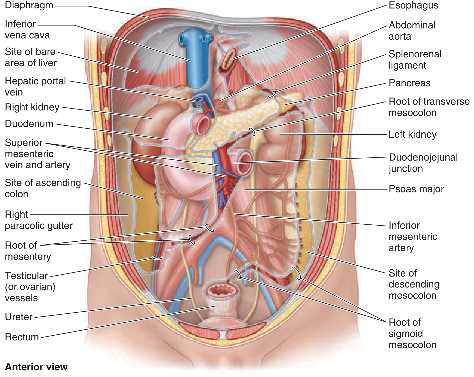
Anatomy of the retroperitoneum - anterior view showing retroperitoneal organs (kidneys, duodenum, IVC, aorta, ureters) after removal of intraperitoneal viscera. - Schwartz's Principles of Surgery, 11th Ed.
This is a shared conversation. Sign in to Orris to start your own chat.