
Glomus tumor in detail according to standard ENT textbook in simplified manner for long question answer
I'll start by reading the medical library skill, then search for comprehensive information on Glomus tumors in ENT textbooks.
Reading File
Now let me search for Glomus tumor content in the ENT textbooks in parallel:
Finding Sources
Finding Sources
Searching Images
glomus tumor jugulare tympanicum ear
Excellent! Found dedicated ENT sections in Cummings Otolaryngology and Shambaugh. Let me read them all in parallel:
Reading File
Reading File
Finding Sources
Found great content. Let me read the main Cummings block for glomus tympanicum/jugulare sections and the Shambaugh reference:
Reading File
Reading File
Excellent content found. Let me now get the remaining sections on treatment, and also the Cummings block 25 (Nomenclature section) for more detail:
Reading File
Reading File
Loading Image
Now let me get the treatment sections and Shambaugh content for surgical approaches:
Reading File
Reading File
I now have comprehensive content from Cummings Otolaryngology. Let me get one more section - the treatment/management section for glomus tumors from the block40 file, and also read the Shambaugh section for additional ENT detail:
Reading File
Finding Sources
Excellent! Found K.J. Lee's Essential Otolaryngology content. Let me get that and also the surgical approach section from Cummings:
Reading File
Reading File
I now have comprehensive information. Let me also fetch the clinical image of glomus tumor through the tympanic membrane:
Loading Image
Loading Image
I now have all the necessary information. Let me compile a comprehensive, well-structured answer on Glomus Tumors in ENT.
GLOMUS TUMOR (Paraganglioma of the Temporal Bone / Neck)
Based on: Cummings Otolaryngology Head and Neck Surgery, K.J. Lee's Essential Otolaryngology, Shambaugh Surgery of the Ear
1. INTRODUCTION AND NOMENCLATURE
Glomus tumors are the most common benign vascular neoplasms of the middle ear and one of the most common primary tumors of the temporal bone. They belong to a broader family called paragangliomas (PGLs) - neoplasms arising from extra-adrenal paraganglionic cells derived from the neural crest (neuroectoderm).
Confusing names - simplified:
| Old/Popular Name | Modern Preferred Term | Meaning |
|---|---|---|
| Glomus jugulare | Jugulotympanic paraganglioma | Arises from jugular bulb |
| Glomus tympanicum | Jugulotympanic paraganglioma | Arises from middle ear glomus bodies |
| Glomus vagale | Vagal paraganglioma | Arises from vagus body in neck |
| Carotid body tumor | Carotid paraganglioma | Arises at carotid bifurcation |
| Chemodectoma | Not recommended - avoid | Inaccurate older term |
The terms glomus tympanicum and glomus jugulare persist in clinical practice despite "paraganglioma" being the preferred scientific term. - Cummings Otolaryngology, p. 3404
2. ORIGIN / ANATOMY
Where do glomus bodies come from?
Temporal bone paraganglia are small clusters of neuroendocrine tissue found:
- Along Jacobson's nerve (branch of CN IX - tympanic branch) - in the middle ear
- Along Arnold's nerve (auricular branch of CN X) - in the middle ear
- In the adventitia of the jugular bulb - at the jugular foramen
- Along the vagus nerve in the neck
Tumors arising from the middle ear glomus bodies = glomus tympanicum
Tumors arising from the jugular bulb = glomus jugulare
3. HISTOLOGY / PATHOLOGY
The paraganglion system contains two cell types (same in all PGLs):
Type I - Chief cells (granular cells / APUD cells)
- Predominate in the tumor
- Contain dense-core granules filled with catecholamines
- Part of the amine precursor and uptake decarboxylase (APUD) system
- Polygonal, with abundant granular eosinophilic cytoplasm
- Stain positively for: neuron-specific enolase (NSE), chromogranin A, synaptophysin
- Arranged in a characteristic nested pattern = Zellballen
Type II - Sustentacular cells (supporting cells)
- Elongated, closely resemble Schwann cells
- Peripherally surround the Type I cells
- Difficult to see on light microscopy (spindle-shaped, basophilic)
- Stain with S-100 and focally with GFAP
Important: Nuclear pleomorphism and cellular hyperchromatism are common in PGLs and should NOT be taken as evidence of malignancy. - Cummings Otolaryngology
Malignancy criteria: Only ~3-5% are malignant. Malignancy is defined by distant metastasis (to lymph nodes, bone, liver, lungs), NOT by histological appearance alone.
4. EPIDEMIOLOGY
- Most common benign neoplasm of the middle ear
- Predominantly affects women (F:M ratio ~6:1 for jugulotympanic)
- Peak incidence: 5th and 6th decades
- Can be sporadic or familial
- Rule of 10s (K.J. Lee's Essential Otolaryngology):
- 10% bilateral
- 10% familial
- 10% malignant
Familial type:
- Autosomal dominant inheritance
- Associated with mutations in succinate dehydrogenase (SDH) subunit genes (SDHB, SDHC, SDHD)
- Higher rates of bilateral and multicentric tumors in familial cases
5. VASOACTIVE PROPERTIES
Glomus tumors can secrete vasoactive substances, including:
- Catecholamines
- Norepinephrine, dopamine
- Somatostatin
- Vasoactive intestinal polypeptide (VIP)
- Calcitonin
Only 1-3% of glomus jugulare tumors are functionally secreting, but all patients should be screened before surgery.
If patient has headache, palpitations, flushing, diarrhea, or hypertension:
- Order 24-hour urine VMA (vanillylmandelic acid) and serum catecholamine levels
- If elevated, get abdominal CT to rule out pheochromocytoma
- Treat adrenergic symptoms before surgery
6. CLINICAL FEATURES
A. Glomus Tympanicum
| Feature | Details |
|---|---|
| Origin | Glomus bodies along Jacobson's nerve in middle ear |
| Most common symptom | Pulsatile tinnitus (synchronous with pulse) |
| Hearing loss | Conductive hearing loss |
| Otoscopy | Red/bluish pulsatile mass behind tympanic membrane |
| Size at presentation | Small (causes symptoms early due to proximity to TM and ossicles) |
| Incidental finding | Possible on routine exam |
Brown's sign: When pneumatic otoscopy (Siegle speculum) is used, the mass blanches with pressure and returns to its original color when pressure is released. This is due to the highly vascular nature.
Aquino's sign: Pulsation of the mass disappears when the ipsilateral carotid artery is compressed.
B. Glomus Jugulare
| Feature | Details |
|---|---|
| Origin | Dome of jugular bulb |
| Presentation | Often late after significant growth and bony destruction |
| Most common early symptom | Pulsatile tinnitus |
| Hearing loss | Conductive (from middle ear involvement) or SNHL (from labyrinthine erosion) |
| Cranial nerve involvement | CN IX, X, XI (at jugular foramen) |
| CN XII | Involved in large tumors |
| Facial nerve | Paresis if tumor extends into mastoid |
| Otoscopy | Large tumors extend into middle ear - appear as pulsatile mass behind/through TM |
| Extreme cases | Erodes TM and presents as bleeding mass in EAC |
C. Glomus Vagale
- Arises from vagus body high in neck at C1-C2 level
- May extend superiorly to mimic glomus jugulare
- Anterior displacement of the high cervical ICA on imaging
7. OTOSCOPIC / CLINICAL SIGNS
Classic otoscopic appearance: Reddish-blue pulsatile mass visible through (or bulging the) tympanic membrane.
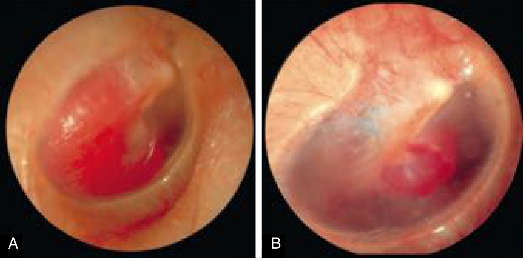
Key clinical distinction:
- If all borders of the mass are visible through the TM → Glomus tympanicum (small, contained)
- If borders are not all visible → could be large glomus tympanicum or glomus jugulare extending into middle ear
- If cranial nerve deficits are present → more likely glomus jugulare
8. INVESTIGATIONS
Radiology
CT Scan (best for bone details):
- Glomus jugulare: destruction of the bony septum between ICA and IJV (highly specific finding)
- Irregular permeative ("moth-eaten") pattern of bone destruction
- Erosion of the jugular foramen
- Separates from aberrant ICA (which lacks this bony destruction)
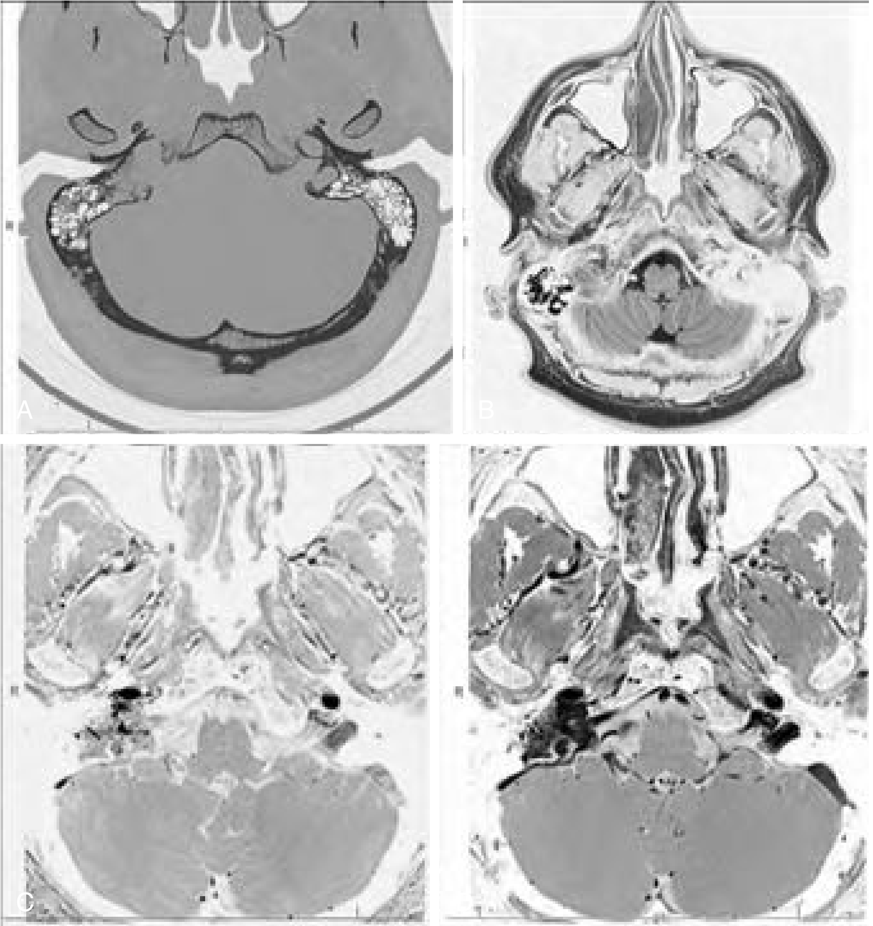
MRI (best for soft tissue extent):
- Classic "salt and pepper" appearance on T1/T2
- "Pepper" (black) = flow voids from enlarged vessels (high vascular flow)
- "Salt" (white) = areas of slow flow or subacute hemorrhage
- Intense post-gadolinium enhancement
- Defines intracranial extension
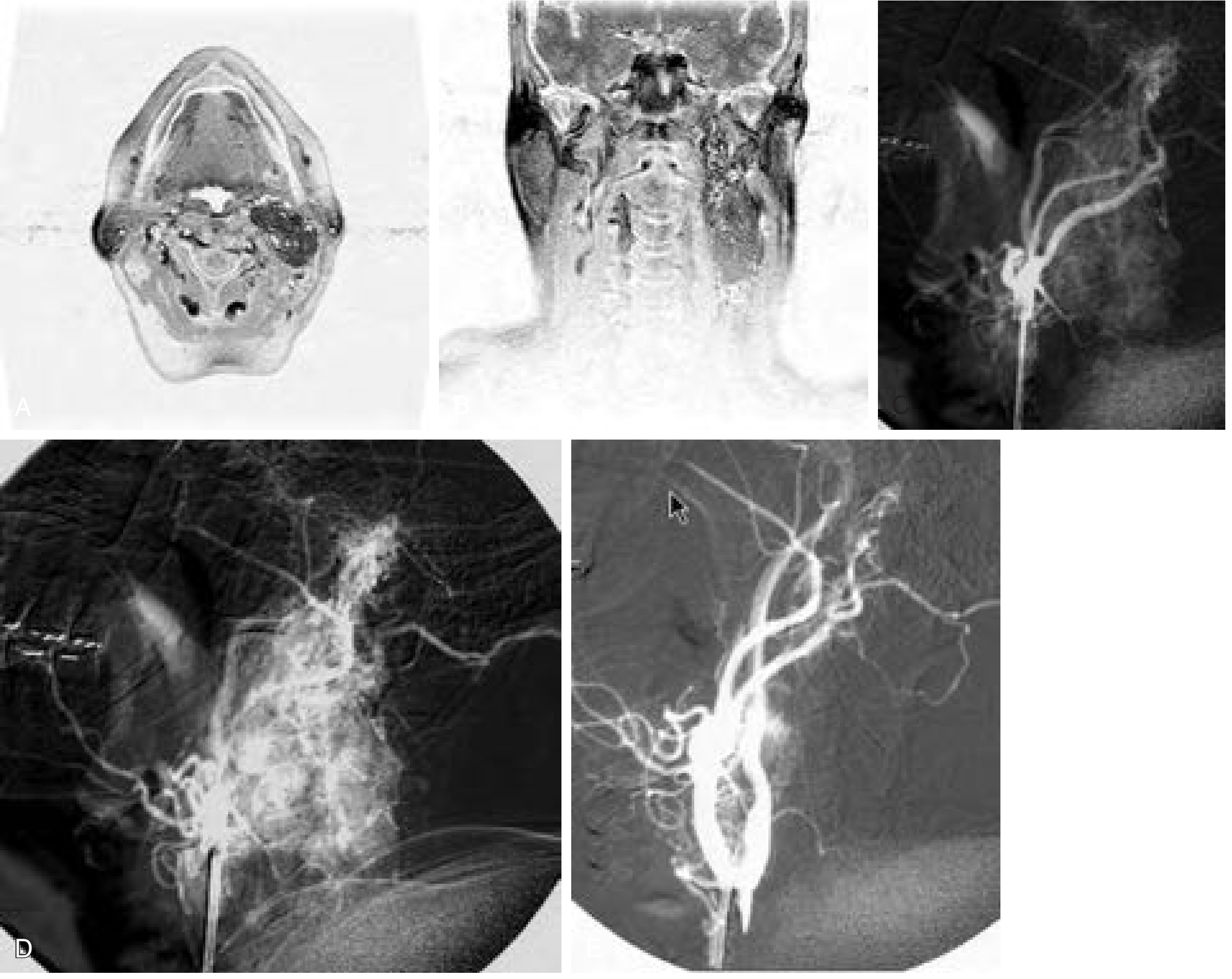
Angiography (Digital Subtraction Angiography - DSA):
- Gold standard for vascular mapping
- Identifies feeding vessels for preoperative embolization
- Shows characteristic intense tumor blush
- Main feeders: ascending pharyngeal artery, posterior division of middle meningeal artery
- Less common: stylomastoid branch of occipital artery, posterior auricular artery
MRI with salt-and-pepper appearance of a glomus jugulare/vagale tumor (axial enhanced MRI showing flow voids and intense enhancement):
Pre-operative workup:
- 24-hour urine metanephrines / VMA
- Serum catecholamines (if secreting suspected)
- Abdominal CT (to rule out pheochromocytoma)
- Preoperative angiography with embolization
9. STAGING / CLASSIFICATION
Two major classification systems exist:
Fisch Classification (1978)
| Stage | Description |
|---|---|
| A | Limited to the middle ear cleft |
| B | Limited to the tympanomastoid area, with or without erosion of the jugular bulb |
| C | Involvement and/or destruction of infralabyrinthine and apical compartments of petrous bone |
| D1 | Intracranial extension <2 cm in greatest diameter |
| D2 | Intracranial extension >2 cm in greatest diameter |
| D3 | Inoperable intracranial invasion |
Glasscock-Jackson Classification (1982)
| Stage | Description |
|---|---|
| I | Small tumor involving jugular bulb, middle ear, and mastoid |
| II | Tumor extending under the internal auditory canal; may have intracranial extension |
| III | Tumor extending into the petrous apex; may have intracranial extension |
| IV | Tumor extending beyond the petrous apex into the clivus and infratemporal fossa; may have intracranial extension |
Source: Cummings Otolaryngology, Table 178.1
10. DIFFERENTIAL DIAGNOSIS
| Condition | Key Distinguishing Feature |
|---|---|
| Aberrant ICA | No bony septum between ICA and middle ear; pulsatile reddish mass; CT shows absent posterior carotid canal wall |
| High-riding jugular bulb | Blue mass inferiorly in middle ear; no pulsation with otoscopy |
| Schwannoma (CN IX, X, XI) | Destroys pars nervosa (medial part) of jugular foramen; no tumor blush |
| Cholesteatoma | White mass; no pulsation; CT shows erosion of ossicles |
| Hemangioma | Different CT/MRI characteristics |
| Meningioma | Different signal characteristics on MRI |
Important: Never biopsy a suspected glomus tympanicum without proper imaging, to exclude aberrant ICA or high-riding jugular bulb.
11. MANAGEMENT
Preoperative Preparation
- Screen for secreting tumor (24-hr urine metanephrines)
- Preoperative embolization 24-48 hours before surgery for sizeable tumors:
- Significantly reduces intraoperative blood loss and operative time
- Uses particles like Embospheres (100-500 μm) or PVA
- Avoid embolizing stylomastoid artery (CN VII risk) - use lidocaine provocative test
- Anesthesiology team coordination if secreting tumor
Surgical Treatment
Glomus Tympanicum:
| Approach | Indication |
|---|---|
| Transcanal approach | Small tumor with all borders visible through TM |
| Endaural / postauricular approach | If transcanal doesn't give enough access |
| Mastoidectomy with extended facial recess approach | If borders are not entirely visible on CT-proven glomus tympanicum |
Glomus Jugulare:
- Most commonly approached by the Fisch Type A infratemporal fossa approach
- This involves: transposition of the facial nerve, closure of EAC, and wide exposure of the jugular foramen
- Staging may be necessary for large tumors with extensive intracranial extension
- Cranial nerves VII-XII may be involved at the skull base
- Superficial involvement: segmental removal of epineurium
- Frank neural invasion: may require segmental neurectomy (with caution)
- Priority: nerve preservation due to significant morbidity of lower cranial nerve resection (may need tracheostomy + gastrostomy)
- ICA involvement: Preoperative balloon occlusion test or stenting if tumor infiltrates horizontal petrous ICA
Key intraoperative principle:
- Intraoperative ligation of feeding vessels (ascending pharyngeal, stylomastoid, caroticotympanic, internal maxillary, superior tympanic arteries) before tumor removal to reduce bleeding.
Radiotherapy
Historically not preferred for resectable tumors; however now well-established as primary or adjunct treatment:
| Type | Outcome |
|---|---|
| Conventional RT / IMRT | Tumor control rates 90-100% in recent series |
| Stereotactic radiosurgery (Gamma Knife / CyberKnife) | 90-100% tumor control in most series; some report 25% failure |
| Long-term follow-up series | Complete control in 93% at >20 years |
Indications for radiotherapy:
- Elderly or medically unfit patients
- Bilateral tumors (to spare the contralateral side)
- Residual tumor after surgery
- Unresectable disease
- Patient preference (avoiding surgical morbidity)
Observation (Watch and Wait)
- Small, incidentally found, asymptomatic tumors in elderly patients
- Slow-growing nature justifies close surveillance with MRI
12. COMPLICATIONS
Surgical complications:
- CN VII palsy (facial nerve injury)
- CN IX, X, XI, XII deficits (dysphagia, hoarseness, aspiration, shoulder weakness)
- Cerebrospinal fluid leak
- Significant intraoperative hemorrhage
- Sensorineural hearing loss
- Need for tracheostomy / gastrostomy (in severe lower CN palsy)
Radiotherapy complications:
- Worsening facial nerve function
- Hearing loss
- Hoarseness
- Ataxia (rare)
13. MALIGNANCY
- Rare: ~3-5% of paragangliomas are malignant
- Malignancy is defined by presence of distant metastases (lymph nodes, bone, lung, liver) - NOT histological appearance
- Local invasion alone does not mean malignancy
- SDHB mutations are associated with higher risk of malignancy
14. QUICK MEMORY AID
GLOMUS TUMOR - KEY POINTS

Origin:      Neural crest → paraganglia cells
Location:    Jugular bulb (jugulare) / Middle ear (tympanicum)
Type:        Most common benign tumor of middle ear
Histology:   Zellballen pattern; Chief cells + Sustentacular cells
Symptoms:    Pulsatile tinnitus + Conductive hearing loss
Sign:        Reddish-blue mass behind TM (pulsatile)
Brown's sign: Blanching on pneumatic otoscopy
MRI:         "Salt and Pepper" appearance
CT:          Permeative bone erosion; absent bony septum (jugulare)
Angio:       Gold standard; intense tumor blush
Rule of 10s: 10% bilateral, 10% familial, 10% malignant
Surgery:     Fisch Type A (jugulare); transcanal (small tympanicum)
Pre-op:      Embolization 24-48h before; urine VMA screening
Radiotherapy: 90-100% tumor control rates
REFERENCES
- Cummings Otolaryngology Head and Neck Surgery, 7th Ed - Chapters 136 & 178 (Paragangliomas of Temporal Bone)
- K.J. Lee's Essential Otolaryngology, 11th Ed - Chapter: Paragangliomas
- Shambaugh Surgery of the Ear - Glomus Tumor Section
This is a shared conversation. Sign in to Orris to start your own chat.