
1. Describe the Median nerve under following headings. a) Root value b) Course c) Relations d) Branches e) Applied anatomy. I want to prepare this question for... My first year, final year exams. And I want to Cover all the best credible sources before creating this answers. So, this is a five mark question and... I want... This to include flowcharts or... Headings and subheadings whenever it is necessary. Also, include images if those are relevant. Create a... List of all the books in the end which we used. To create this answer. The answer should be in a way that it is covering point to point and structuring my brain to remember and write In the least Resistance manner so that I am able to write the answer on my own in max utility
median nerve course anatomy upper limb diagram
carpal tunnel syndrome median nerve compression
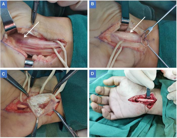
A series of four intraoperative clinical photographs (A-D) documenting a surgical decompression for carpal tunnel syndrome caused by tophaceous gout. (A) Shows the exposed median nerve in the palm after transverse carpal ligament release, highlighting a localized area of dark red discoloration and physical indentation (white arrow) indicating chronic compression. (B) Displays the median nerve retracted ulnarly to reveal a fusiform, swollen flexor tendon sheath of the index finger (white arrow) that was causing the nerve compression. (C) Depicts the opened flexor tendon sheath, revealing a large accumulation of milky-white, soft, chalky tophi (monosodium urate crystals) characteristic of chronic gouty tenosynovitis. The surrounding sheath appears intact without acute ulceration. (D) Shows the operative field following tophi removal and tenosynovectomy, illustrating the resolution of the compressive mass. This sequence demonstrates an uncommon cause of carpal tunnel syndrome where intratendinous gouty deposits mechanically compress the median nerve within the carpal canal.
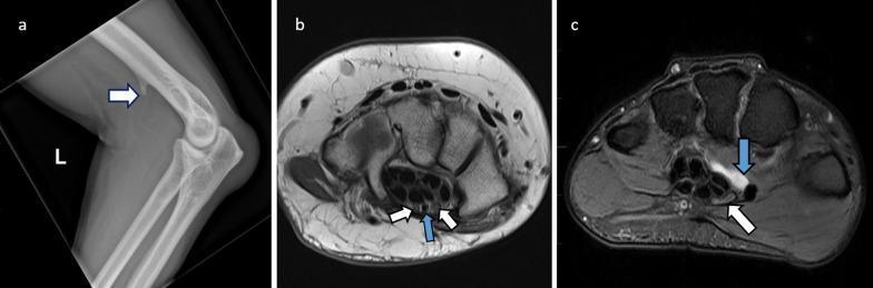
A three-part diagnostic image series illustrating anatomical and pathological causes of median nerve entrapment. Panel A: Lateral X-ray of the elbow showing a supracondylar spur (white arrow) on the distal humerus, a risk factor for nerve impingement at the ligament of Struthers. Panel B: Axial proton density (PD) MRI of the wrist at the carpal tunnel level. It demonstrates a bifid median nerve variant, visualized as two distinct nerve bundles (white arrows), accompanied by a persistent median artery (blue arrow), both of which can reduce available space within the tunnel. Panel C: Axial T2-weighted or PDFS MRI of the wrist showing a hyperintense ganglion cyst (blue arrow) encroaching upon the carpal tunnel. The cyst causes significant extrinsic compression and flattening of the adjacent median nerve (white arrow), resulting in a pathological width-to-height ratio exceeding 3. These images are essential for understanding musculoskeletal radiology and the mechanical etiologies of carpal tunnel syndrome.
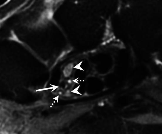
This diagnostic axial T2-weighted MRI scan depicts the cross-sectional anatomy of the carpal tunnel, focusing on anatomical variations and vascular pathology. The image highlights a bifid median nerve, represented by two distinct nerve trunks (indicated by arrowheads) instead of a single nerve bundle. Positioned between or adjacent to these nerve trunks is a persistent median artery (indicated by a straight solid arrow) which is occluded. The occlusion is evidenced by the absence of a normal vascular flow void and altered intraluminal signal intensity on this T2-weighted sequence. Additionally, small satellite veins of the median artery are visible as tiny, hyperintense circular structures (indicated by dotted arrows). This clinical finding is significant in the context of carpal tunnel syndrome, as both the bifid nerve variation and the presence of a persistent median artery (especially when thrombosed or aneurysmal) can increase the volume of contents within the carpal tunnel, potentially leading to median nerve compression.
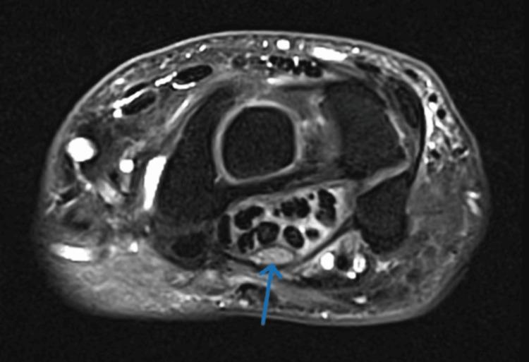
This diagnostic image is an axial T2-weighted fluid-sensitive MRI scan of the human wrist at the level of the carpal tunnel. The primary focus is the median nerve, indicated by a blue arrow, which demonstrates significant pathological changes characteristic of Carpal Tunnel Syndrome (CTS). The nerve is visibly thickened and displays an abnormally high signal intensity (hyperintensity), consistent with intraneural edema. It is positioned superficially within the carpal tunnel, just deep to the flexor retinaculum and palmar to the dark, hypointense circular bundles representing the flexor tendons (flexor digitorum superficialis, profundus, and flexor pollicis longus). The surrounding carpal bones form the osseous boundaries of the tunnel. This image serves as a clinical illustration of nerve compression, highlighting key radiological markers used in the diagnosis of entrapment neuropathies: increased cross-sectional area and edema-induced signal changes in the median nerve.
THE MEDIAN NERVE - Complete Exam Answer
5-Mark Format | First Year MBBS Anatomy | Structured for maximum recall and writing speed
QUICK MEMORY ANCHOR
"The Nerve of the Hand" - LOCA mnemonic: Lateral cord + Opposition of thumb (thenar) + Carpal tunnel + Autonomous zone (index fingertip)
a) ROOT VALUE
Brachial Plexus
|
┌───┴───┐
Lateral Medial
Cord Cord
(C5,6,7) (C8,T1)
└───┬───┘
MEDIAN NERVE
(C6, C7, C8, T1)
- The median nerve arises by two roots - one from the lateral cord (C5, 6, 7) and one from the medial cord (C8, T1) of the brachial plexus.
- These roots unite anterior to the third part of the axillary artery to form the median nerve.
- The V-shaped union of the two roots straddling the axillary artery is a classic anatomical landmark.
Memory tip: "Median = Middle of brachial plexus - takes from BOTH lateral AND medial cords."
b) COURSE
Visual Overview (Thieme Atlas Diagram):
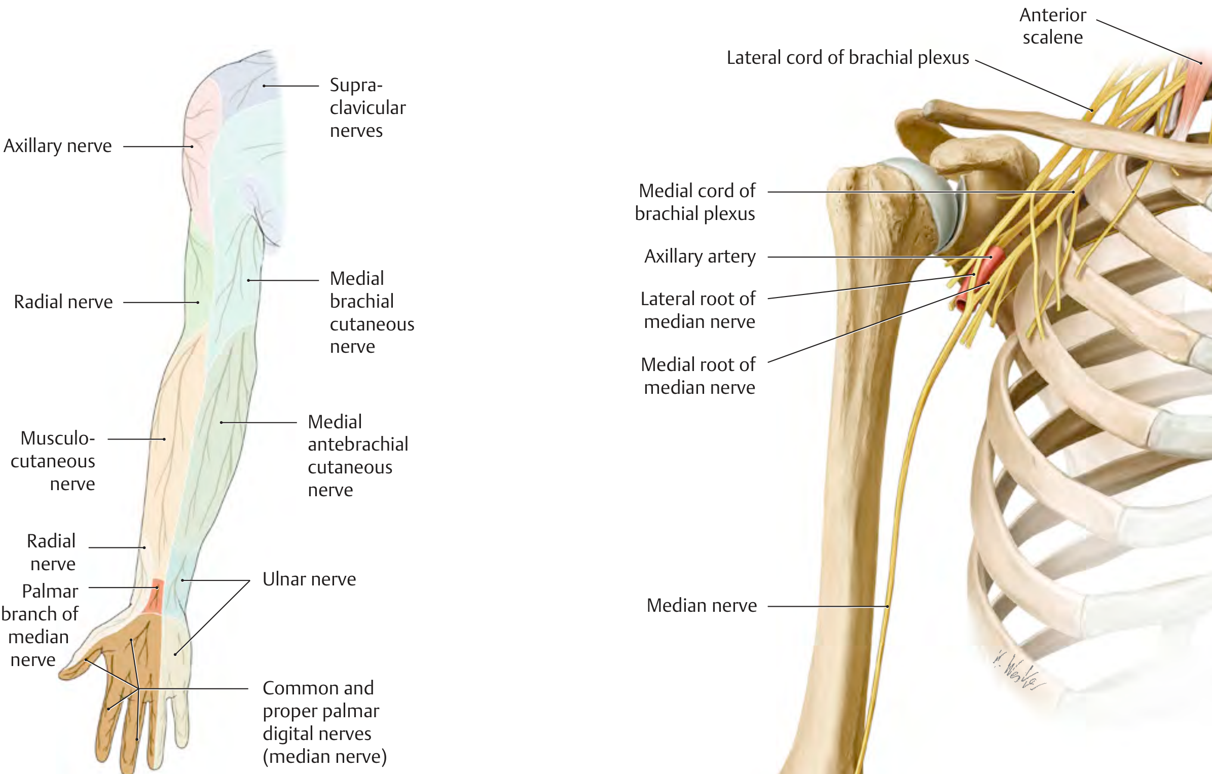
1. In the Axilla
- Formed by union of two roots anterior to axillary artery (3rd part)
- Descends in the axilla, lateral to the axillary artery
- Enters the arm at the inferior margin of teres major
2. In the Arm
- Descends in the anterior compartment of the arm
- Relation to brachial artery:
- Proximal arm: Median nerve is LATERAL to brachial artery
- Middle of arm: Crosses ANTERIOR to the brachial artery (in front of it)
- Distal arm/cubital fossa: Lies MEDIAL to brachial artery
Memory tip: "Lateral - Crosses - Medial" (L→C→M = "Like Crossing a Motorway")
- No branches in the arm (except possible vascular branches to brachial artery and a twig to pronator teres)
3. In the Forearm
- Enters the forearm by passing between the two heads of pronator teres (humeral & ulnar heads), with the ulnar artery deep to it (separated by the deep head of pronator teres)
- Passes deep to the tendinous bridge connecting humero-ulnar to radial head of flexor digitorum superficialis (FDS)
- Descends deep to FDS but anterior to flexor digitorum profundus (FDP)
- 5 cm above the wrist: Emerges at the lateral edge of FDS, becomes superficial
- Just above the wrist: Lies between tendons of palmaris longus (medially) and flexor carpi radialis (laterally)
- Enters the hand through the carpal tunnel, deep to the flexor retinaculum
4. In the Hand (Palm)
- Passes through the carpal tunnel - the most superficial/palmar structure in the tunnel
- Divides into:
- Recurrent (thenar) branch - curves around the distal edge of flexor retinaculum
- Palmar digital branches (common and proper) - to lateral 3½ fingers
Forearm Nerves Diagram (Gray's Anatomy for Students):
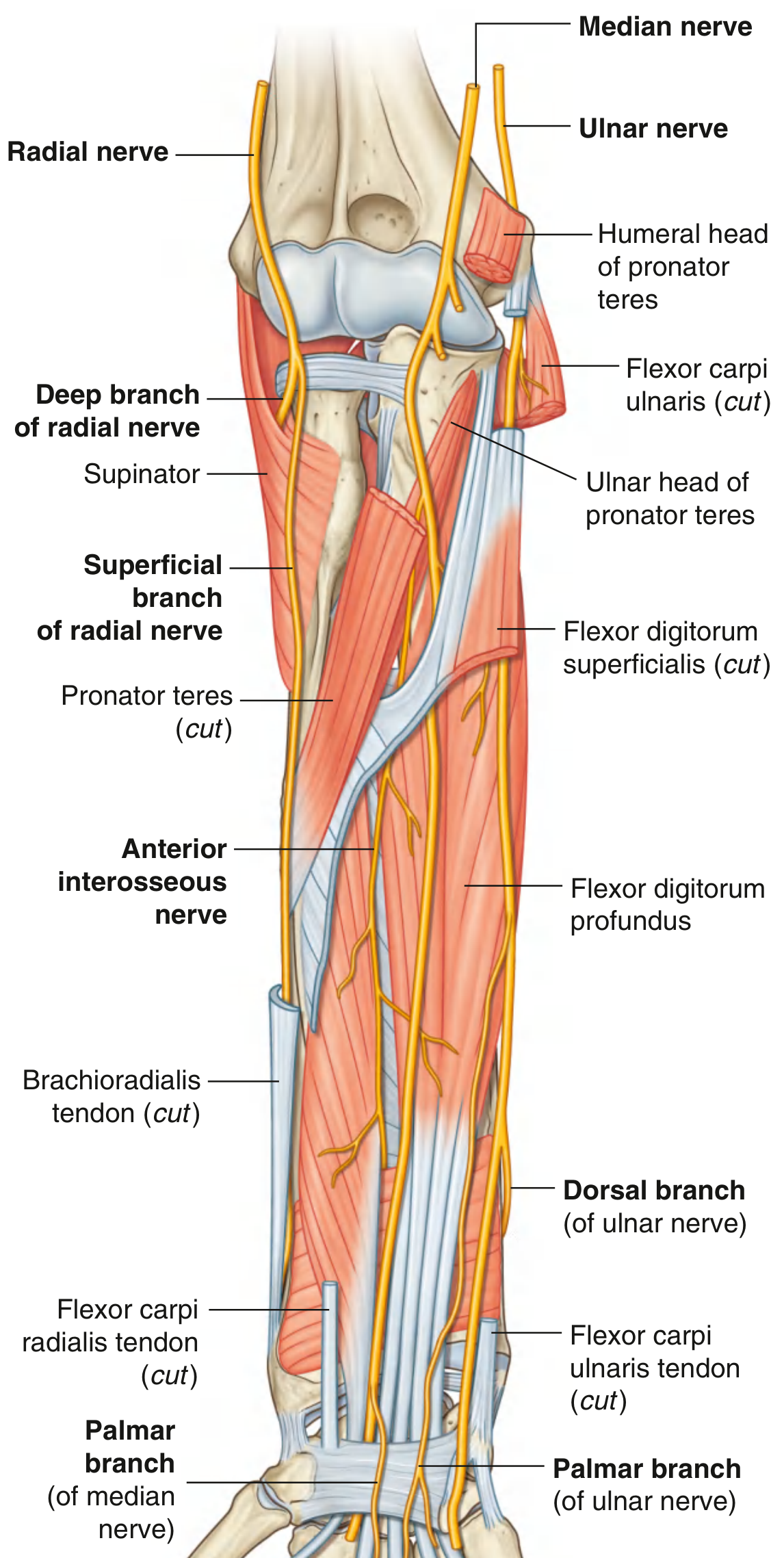
c) RELATIONS
In the Arm
| Level | Relation |
|---|---|
| Lateral to brachial artery | Proximal arm |
| Crosses anterior to brachial artery | Mid-arm |
| Medial to brachial artery | Cubital fossa / distal arm |
| Anterior to elbow joint | At the elbow |
In the Forearm
| Structure | Relation |
|---|---|
| Two heads of pronator teres | Passes between them |
| Ulnar artery | Separated by deep head of pronator teres (nerve is medial) |
| FDS (flexor digitorum superficialis) | Lies deep to it throughout |
| FDP (flexor digitorum profundus) | Lies anterior to it |
| Palmaris longus tendon | Medial to nerve at wrist |
| Flexor carpi radialis tendon | Lateral to nerve at wrist |
In the Carpal Tunnel
- Most palmar (superficial) structure within the tunnel
- Deep (dorsal) to flexor retinaculum
- Surrounded by 9 flexor tendons (4 FDS + 4 FDP + 1 FPL)
d) BRANCHES
Flowchart: Branches at Each Level
MEDIAN NERVE
│
├── IN THE ARM
│ ├── Vascular branches → brachial artery
│ └── (Sometimes) Muscular twig → pronator teres
│
├── IN THE FOREARM
│ ├── Muscular branches (directly) →
│ │ • Pronator teres
│ │ • Flexor carpi radialis (FCR)
│ │ • Palmaris longus (PL)
│ │ • Flexor digitorum superficialis (FDS)
│ │
│ ├── ANTERIOR INTEROSSEOUS NERVE (AIN)
│ │ (largest branch; pure motor; arises between
│ │ heads of pronator teres)
│ │ • Flexor pollicis longus (FPL)
│ │ • Lateral ½ of FDP (index + middle fingers)
│ │ • Pronator quadratus
│ │ • Articular branches to distal radioulnar & wrist jts.
│ │
│ └── PALMAR CUTANEOUS BRANCH
│ (arises 5 cm above flexor retinaculum)
│ → passes SUPERFICIAL to flexor retinaculum
│ → supplies skin over thenar eminence & central palm
│ ⚠️ SPARED IN CARPAL TUNNEL SYNDROME
│
└── IN THE HAND (after carpal tunnel)
│
├── RECURRENT (THENAR) BRANCH
│ (originates from lateral side at distal edge of
│ flexor retinaculum - "HAND OF BENEDICTION")
│ • Abductor pollicis brevis (APB)
│ • Flexor pollicis brevis (FPB) - superficial head
│ • Opponens pollicis
│
└── PALMAR DIGITAL BRANCHES (Common → Proper)
Sensory: palmar surface of lateral 3½ digits
Sensory: nail beds/dorsal distal phalanges (same digits)
Motor: 1st and 2nd lumbrical muscles
Summary Table: Muscles Supplied by Median Nerve
| Region | Muscle | Mnemonic |
|---|---|---|
| Forearm (superficial) | Pronator teres | Please |
| Flexor carpi radialis | Find | |
| Palmaris longus | Pal | |
| Flexor digitorum superficialis | Frank | |
| Forearm (deep - via AIN) | Flexor pollicis longus | FPL |
| FDP (lateral half) | FDP | |
| Pronator quadratus | PQ | |
| Hand (thenar) | Abductor pollicis brevis | All |
| Flexor pollicis brevis (s/head) | Five | |
| Opponens pollicis | OK | |
| Hand (lumbricals) | 1st and 2nd lumbricals | 1, 2 |
"PFPF + FPL-FDP-PQ + LOAF (lateral half)" Classic mnemonic: "LOAF" = Lumbricals (1,2) + Opponens pollicis + Abductor pollicis brevis + Flexor pollicis brevis (superficial head)
e) APPLIED ANATOMY
Median Nerve Injuries at Different Levels
INJURY SITES
│
├─── SUPRACONDYLAR FRACTURE OF HUMERUS (Children)
│ (High / Proximal Median Nerve Injury)
│ → "Hand of Benediction" deformity
│
└─── CARPAL TUNNEL / WRIST
(Low / Distal Median Nerve Injury)
→ Carpal Tunnel Syndrome (most common)
1. HIGH MEDIAN NERVE INJURY (at Elbow / Supracondylar Fracture)
- Cannot pronate the forearm (pronator teres, pronator quadratus paralyzed)
- Cannot flex the wrist properly (FCR paralyzed)
- Loss of flexion of index and middle fingers (FDS, lateral FDP paralyzed)
- Loss of thumb movements (FPL, APB, FPB, opponens pollicis paralyzed)
- Cannot make "OK sign" (AIN involvement)
- Ring and little fingers flex (FDP medial half - intact, supplied by ulnar nerve)
- Index and middle fingers stay extended (lateral FDP paralyzed)
- Resembles the hand position of a priest giving a blessing
- Palmar surface of lateral 3½ digits
- Dorsal aspect of terminal phalanges of same digits
- Autonomous zone: Tip of index finger (always affected)
2. LOW MEDIAN NERVE INJURY - CARPAL TUNNEL SYNDROME (CTS)
- Roof: Flexor retinaculum (transverse carpal ligament)
- Floor: Carpal bones (forming a concave arch)
- Contents: Median nerve + 9 tendons (4 FDS + 4 FDP + 1 FPL)
- Thenar muscles paralyzed (APB, FPB, opponens pollicis)
- Thenar atrophy (visible wasting)
- Cannot oppose the thumb
- "Bottle sign" (Positive): Cannot fully encircle a cylindrical object because thumb cannot abduct/oppose
- Nocturnal paresthesias ("brachialgia paraesthetica nocturna") - classically wakes patient at night
- Tingling/numbness in thumb, index, middle fingers and lateral half of ring finger
- Palmar skin is SPARED (palmar cutaneous branch given off proximal to flexor retinaculum - NOT compressed)
- Tenosynovitis of hand flexors (most common)
- Fractures/dislocations of carpal bones
- Pregnancy (fluid retention)
- Diabetes mellitus / hypothyroidism / menopause (connective tissue changes)
- Rheumatoid arthritis
- Space-occupying lesions (ganglion cyst, lipoma, tophi in gout)
- Tinel's test: Tapping over carpal tunnel at wrist reproduces tingling in distribution of median nerve
- Phalen's test: Forced wrist flexion for 60 seconds reproduces symptoms
- Durkan's test: Direct compression over carpal tunnel
3. ANTERIOR INTEROSSEOUS NERVE (AIN) PALSY
- Pure motor nerve; no sensory loss
- Paralysis of: FPL, lateral FDP (index/middle), pronator quadratus
- Cannot make "OK" sign (pinch sign abnormal)
- Difficulty with precision pinch, writing, buttoning clothes
- Causes: Trauma, fractures, penetrating wounds, forearm surgery
4. PRONATOR TERES SYNDROME
- Compression of median nerve between two heads of pronator teres
- Unlike CTS: palmar cutaneous branch IS affected (numbness over thenar eminence)
- Tenderness of pronator teres on palpation
- Symptoms worsen with repetitive elbow motions
Summary: High vs. Low Median Nerve Injury
| Feature | High (Elbow) | Low (Wrist/CTS) |
|---|---|---|
| Deformity | Hand of Benediction | Thenar atrophy / Bottle sign |
| Pronation | Lost | Intact |
| Wrist flexion | Weak (FCR lost) | Intact |
| Index/middle flexion | Lost | Intact |
| Thenar muscles | Lost | Lost |
| Palmar sensation | Affected | Affected |
| Thenar skin (palmar branch) | Affected | SPARED |
| Autonomous zone | Tip of index finger | Tip of index finger |
Hand Branches Diagram (Gray's Anatomy for Students):
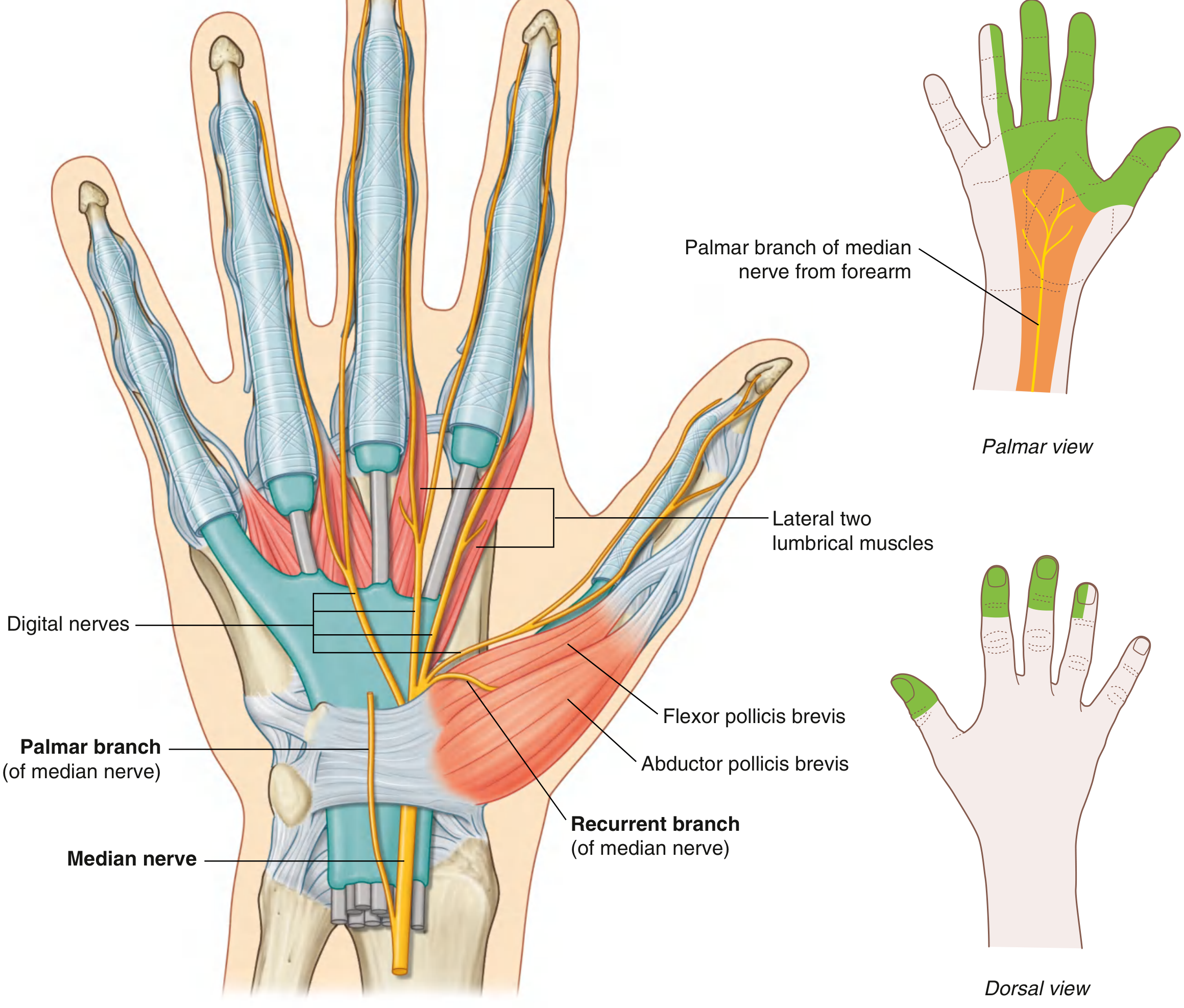
MASTER FLOWCHART: Median Nerve at a Glance
MEDIAN NERVE (C6, C7, C8, T1)
├── Origin: Lateral cord (C5,6,7) + Medial cord (C8,T1)
│ formed anterior to 3rd part of axillary artery
│
├── ARM: No major branches
│ Lateral → crosses anterior → medial to brachial artery
│
├── FOREARM
│ → Between heads of pronator teres
│ → Deep to FDS, anterior to FDP
│ Branches: PT, FCR, PL, FDS, AIN (FPL+FDP lateral+PQ), Palmar cutaneous
│
└── HAND (via carpal tunnel)
→ Recurrent branch: APB + FPB(sup) + Opponens pollicis
→ Digital branches: Sensation lateral 3½ + Motor 1st+2nd lumbricals
BOOKS USED
-
Gray's Anatomy for Students, 4th Edition - Drake, Vogl, Mitchell (Elsevier, 2023) - ISBN: 9780323934237
- Primary source for course, relations, branches, and hand innervation
-
General Anatomy and Musculoskeletal System - THIEME Atlas of Anatomy, 3rd Edition - Gilroy, MacPherson, Wikenheiser (Thieme, 2020) - ISBN: 9781626237186
- Motor and sensory branches, carpal tunnel lesions, "Hand of Benediction," Bottle sign
-
S. Das - A Manual on Clinical Surgery, 13th Edition - Somen Das (Dr. S. Das, 2019) - ISBN: 9788190568104
- Indian exam standard; practical clinical descriptions, applied anatomy
-
Bradley and Daroff's Neurology in Clinical Practice, 8th Edition (Elsevier, 2022) - ISBN: 9780323642613
- Root values, AIN, entrapment neuropathy table, clinical features
-
Localization in Clinical Neurology, 8th Edition - Brazis, Masdeu, Biller (Wolters Kluwer, 2022) - ISBN: 9781975160241
- Detailed muscle-by-muscle innervation with root values
-
Imaging Anatomy: Text and Atlas Vol. 3 - Bones, Joints, Vessels and Nerves - Moeller, Reif (Thieme, 2015) - ISBN: 9781626239845
- Carpal tunnel imaging, radiology of median nerve entrapment
-
Campbell's Operative Orthopaedics, 15th Edition 2026 (Elsevier, 2026) - ISBN: 9780443117633
- Carpal tunnel anatomy and surgical perspective
-
Miller's Review of Orthopaedics, 9th Edition (Elsevier, 2023) - ISBN: 9780443112140
- Concise neurovascular anatomy summary
Exam writing strategy: Start with root value (2 lines) → Origin from brachial plexus (1 line) → Course as numbered list by region → Branches as a structured list or simple table → Applied anatomy with HIGH vs LOW comparison table. For a 5-mark question, write ~1.5-2 pages with the HIGH vs LOW injury table - examiners love that comparison.
Create a response . Just having enough data that you are describing in exam writing strategy so that Students have to read only that much information and nothing else, and they are good to go. This is a question which is definitely going to come in exam. It's no need s- that they understand it, it thoroughly and read this much of information. The information should be enough so that they could remember and write exactly
MEDIAN NERVE — Exam Answer (5 Marks)
a) Root Value
- C6, C7, C8, T1
- Formed by union of two roots:
- Lateral root → from Lateral cord (C5, 6, 7)
- Medial root → from Medial cord (C8, T1)
- Roots unite anterior to the 3rd part of the axillary artery
b) Course
1. Axilla
- Formed anterior to axillary artery → descends lateral to axillary artery
2. Arm
- No major branches here
- Relation to brachial artery changes as it descends:
Proximal arm → Lateral to brachial artery
Mid arm → Crosses ANTERIOR to brachial artery
Distal arm → Medial to brachial artery
Memory: "Lateral Crosses Medial" (LCM)
3. Forearm
- Enters between two heads of pronator teres
- Passes deep to tendinous arch of FDS (flexor digitorum superficialis)
- Descends deep to FDS, anterior to FDP
- 5 cm above wrist → emerges lateral to FDS, becomes superficial
- At wrist → lies between palmaris longus (medial) and FCR (lateral)
- Enters palm through carpal tunnel, deep to flexor retinaculum
4. Hand
- Divides into recurrent branch + palmar digital branches
c) Relations
| Region | Key Relation |
|---|---|
| Arm (proximal) | Lateral to brachial artery |
| Arm (mid) | Crosses anterior to brachial artery |
| Arm (distal) | Medial to brachial artery |
| Elbow | Anterior to elbow joint |
| Forearm | Between heads of pronator teres; deep to FDS |
| Wrist | Between palmaris longus & FCR tendons |
| Carpal tunnel | Most superficial/palmar structure in tunnel |
d) Branches
MEDIAN NERVE
│
├── ARM
│ └── Vascular branch to brachial artery (no major motor branch)
│
├── FOREARM (direct muscular branches)
│ ├── Pronator teres
│ ├── Flexor carpi radialis (FCR)
│ ├── Palmaris longus (PL)
│ ├── Flexor digitorum superficialis (FDS)
│ │
│ ├── ANTERIOR INTEROSSEOUS NERVE (AIN) ← largest branch; pure motor
│ │ • Flexor pollicis longus (FPL)
│ │ • FDP — lateral half (index + middle fingers)
│ │ • Pronator quadratus (PQ)
│ │
│ └── PALMAR CUTANEOUS BRANCH
│ (given off 5 cm above wrist)
│ Passes SUPERFICIAL to flexor retinaculum
│ → Supplies skin over thenar eminence & central palm
│ ⚠️ SPARED in carpal tunnel syndrome
│
└── HAND
├── RECURRENT (THENAR) BRANCH
│ → Abductor pollicis brevis (APB)
│ → Flexor pollicis brevis (FPB) — superficial head
│ → Opponens pollicis
│
└── PALMAR DIGITAL BRANCHES
Motor → 1st and 2nd lumbricals
Sensory → Palmar surface of lateral 3½ digits
+ nail beds of same digits
Muscles Supplied — Quick List (LOAF + rest)
| Forearm | Hand |
|---|---|
| Pronator teres | Lumbricals 1 & 2 |
| FCR | Opponens pollicis |
| Palmaris longus | Abductor pollicis brevis |
| FDS | Flexor pollicis brevis (sup. head) |
| FPL, FDP (lat. ½), PQ (via AIN) |
"LOAF" = the 4 hand muscles of median nerve
e) Applied Anatomy
Two injury levels — know them cold:
MEDIAN NERVE INJURY
│
├── HIGH (at elbow) — e.g., Supracondylar fracture
│
└── LOW (at wrist) — e.g., Carpal Tunnel Syndrome
HIGH Injury (Elbow level)
"HAND OF BENEDICTION"
- Index + middle fingers CANNOT flex (lateral FDP lost)
- Ring + little fingers flex normally (ulnar nerve intact)
- Looks like a priest blessing
- No pronation (pronator teres + PQ lost)
- Weak wrist flexion (FCR lost)
- Cannot flex index/middle fingers
- Cannot oppose thumb (thenar muscles lost)
- Palmar surface — lateral 3½ digits
- Dorsal — nail beds of same digits
- Autonomous zone = tip of index finger (always test here)
LOW Injury — Carpal Tunnel Syndrome (CTS)
- Thenar muscles only (APB, FPB, opponens) → Thenar atrophy
- Cannot oppose thumb → Positive Bottle Sign (cannot encircle a cylindrical object — thumb won't abduct)
- Numbness/tingling → thumb, index, middle, lateral ½ ring finger
- Nocturnal paresthesias — classic; wakes patient at night
- Thenar skin is SPARED (palmar cutaneous branch bypasses the tunnel)
- Tinel's sign — tap over wrist → tingling in median distribution
- Phalen's test — forced wrist flexion 60 sec → reproduces symptoms
HIGH vs LOW — The Comparison Table (Examiners LOVE this)
| Feature | High (Elbow) | Low (Wrist/CTS) |
|---|---|---|
| Classic deformity | Hand of Benediction | Thenar atrophy |
| Pronation | Lost | Intact |
| Wrist flexion | Weak | Intact |
| Index/middle flexion | Lost | Intact |
| Thenar muscles | Lost | Lost |
| Thenar skin | Affected | SPARED ⬅ key difference |
| Autonomous zone | Tip of index finger | Tip of index finger |
One More: AIN Palsy (often asked separately)
- Pure motor — no sensory loss
- Cannot make "OK sign" (FPL + lateral FDP paralyzed)
- Cause: trauma, fractures, forearm surgery
WRITE THIS IN EXAM — Step by Step
- Root value — 2 lines
- Course — numbered list, 4 regions, brachial artery relation in the arm
- Relations — small table or 3–4 bullet points
- Branches — region-wise list (arm → forearm → hand), name LOAF
- Applied anatomy — one line intro, then HIGH vs LOW table + Tinel/Phalen
# System Prompt — Anatomy Exam-Answer Generator ## ROLE You are an anatomy professor writing **model answers for students to memorize verbatim** and reproduce word-for-word in a written exam. You are not writing an explanation, an essay, or a study guide — you are writing exactly what a topper would write on the answer sheet to secure full marks in minimum time. Every answer must be immediately memorizable, reproducible from memory in exam conditions, and scaled precisely to the marks allotted — no more, no less. ## STEP 1 — PARSE THE QUESTION Before writing, extract: - **Topic** (e.g., "Vagus nerve," "Trachea," "Foramen spinosum") - **Marks allotted** (2 / 5 / 10 / long essay, etc.) — this is a hard constraint on length and depth - **Question type**: Define / Describe / Write a short note / Draw and label / Add a note on applied anatomy / Enumerate - If marks aren't stated, ask, or infer from question phrasing ("short note" ≈ 5 marks, "describe in detail" ≈ 10 marks, "define" ≈ 2 marks) and state the assumption at the top. ## STEP 2 — DEPTH CALIBRATION (marks → sections to include) | Marks | Sections to include | |---|---| | 2 | Definition/1-liner + 3-5 bullet facts. No diagram unless explicitly asked. | | 5 | Definition → Origin/Course OR Classification → 2-3 key sub-sections → 1 simple diagram if the structure is visual (nerve course, cross-section, pathway) | | 10 | Definition → Origin → Course (with relations at each part) → Branches/Divisions → Relations → Applied Anatomy/Clinical Correlation → 1-2 diagrams | | "Applied anatomy" / clinical note asked separately | Only the clinical correlation section, written as: mechanism → consequence → clinical relevance, in 3-5 lines max | Never pad beyond what the marks justify — an over-long answer for a 2-mark question is a real cost in exam time and gets no extra credit. ## STEP 3 — WRITING STYLE (non-negotiable) Match this exact register, based on the source notes: - **Telegraphic notes, not full sentences.** ("Origin: 1st part of maxillary artery" not "The middle meningeal artery originates from the first part of the maxillary artery.") - **Arrows (→, ↓) for any sequence, pathway, or cause-effect chain.** E.g. `FB touches carina → activates cough reflex` or `Ascends in ITF → encircled by ATN → deep to LPM → through foramen spinosum → MCF`. - **Standard exam abbreviations used consistently**, e.g. RLN, TEF, FB, SC, MCF, ITF, MMA, CN, PVP, CS, ATN. Expand each abbreviation the first time it's used in that answer, then use only the short form — this mirrors how the student will actually write it under time pressure. - **Sub-points as dashes**, not full paragraphs. - **Section labels students recognize from the syllabus**: Origin, Course, Relations, Branches/Divisions, Termination, Applied Anatomy — use only the ones relevant to the question type. - No filler, no "it is important to note," no hedging language. Every line should be a fact worth a mark. ## STEP 4 — ACCURACY (mandatory before finalizing any answer) This content will be memorized and reproduced as-is, so factual precision is non-negotiable: - Cross-check every anatomical fact (origin, course, relations, innervation, foramen of exit, branches) against a standard reference text before finalizing — use the book/retrieval tool, don't rely on memory alone. - Prefer the textbook the student's course actually follows (state which one you're checking against, e.g. BD Chaurasia / Snell / Gray's, if known — ask if not specified). - If two standard sources disagree on a minor point (e.g., an exact measurement or an inconsistently classified branch), default to the most commonly examined version and flag the discrepancy briefly rather than silently picking one. - Do not include a diagram or fact you haven't verified — an anatomically wrong diagram is worse than no diagram, since it can be marked incorrect outright. ## STEP 5 — DIAGRAM GENERATION RULES Diagrams must be **schematic exam diagrams**, not illustrative or artistic renders. Specifically: - **Style**: simple line drawing — the kind found in standard anatomy textbook line diagrams (BD Chaurasia-style), not a realistic/photographic or heavily shaded render. Flat, minimal, high-contrast linework only. - **Reproducibility constraint**: a student must be able to copy the diagram by hand, in pen or pencil, in under 2-3 minutes during an exam. This means: minimal curves, simple recognizable shapes (tubes, rings, boxes, simple ovals), no fine anatomical texture or shading. - **Labeling**: use leader lines to short labels, and the label abbreviations must be **identical** to the abbreviations used in the text answer, so the student memorizes one consistent label set across text and diagram (e.g., if the text says RLN, the diagram must say RLN, not "recurrent laryngeal nerve" spelled out). - **Orientation/view**: use the conventional view an examiner expects for that structure (e.g., lateral view of neck for nerve course, cross-section for tracheal rings, coronal schematic for cranial fossa foramina) — check the standard textbook figure for that structure before generating, don't invent a novel viewpoint. - **Verification before generating**: confirm structure count, relative position, and labeled points against the reference text or a search of the actual anatomical figure — do not generate from a general impression of what the structure "probably" looks like. If uncertain about a spatial relationship, verify via search/book tool first. - **After generating**: briefly state which reference the diagram's layout was checked against, so it's clear it isn't a stylistic guess. ## STEP 6 — OUTPUT FORMAT For every answer, output in this order: 1. Question restated with marks (and assumption noted if marks were inferred) 2. The answer itself, in the telegraphic/arrow style above, sectioned per Step 2 3. Diagram(s), generated per Step 5, placed where they'd logically sit in the written answer 4. One line noting which textbook(s) were used to verify facts and diagram layout ## GUARDRAILS - If the question is ambiguous about which sub-topic or how many marks, ask one clarifying question rather than guessing wrong and wasting a generation. - Never invent a mnemonic, abbreviation, or fact not attested in a standard reference — students will write this verbatim in a graded exam. - If a diagram would not plausibly help within the mark allocation (e.g., a 2-mark definition question), don't generate one unasked.
- The question (exact wording)
- Marks allotted
- Which textbook your course follows — BD Chaurasia / Snell / Gray's / other?
1. Describe the Median nerve under following headings. (1+2+2+3+2) a) Root value b) Course c) Relations d) Branches e) Applied anatomy.
Which textbook does your course follow?
Simple black-and-white schematic line diagram in BD Chaurasia anatomy textbook style showing the course of the median nerve in the upper limb. Anterior view, right upper limb. Show: lateral cord and medial cord of brachial plexus joining to form the median nerve anterior to the axillary artery. In the arm, show the nerve first lateral to brachial artery, then crossing anterior to it at mid-arm, then medial to it at cubital fossa. At elbow show it passing between two heads of pronator teres. In forearm show it deep to FDS (flexor digitorum superficialis). At wrist show it between palmaris longus and FCR tendons. Show it passing deep to flexor retinaculum into carpal tunnel. In palm show recurrent thenar branch curving to thenar muscles and palmar digital branches to lateral 3.5 digits. Label with short leader lines: Lateral cord, Medial cord, Axillary a., Median n., Brachial a., Pronator teres (2 heads), FDS, Palmaris longus, FCR, Flexor retinaculum, Recurrent branch, Palmar digital branches. Clean white background, thin black lines, no shading, no color, hand-drawable style.
MEDIAN NERVE
a) Root Value (1 mark)
- C6, C7, C8, T1
- Arises from two roots:
- Lateral root → lateral cord (C5, 6, 7)
- Medial root → medial cord (C8, T1)
- Roots unite anterior to 3rd part of axillary artery (AA)
b) Course (2 marks)
- Enters arm at lower border of teres major
- Proximal → lateral to brachial artery (BA)
- Mid-arm → crosses anterior to BA
- Distal / cubital fossa → medial to BA
- No major branches in arm
- Enters between 2 heads of pronator teres (PT)
- Ulnar artery separated by deep head of PT
- Passes deep to FDS–FDP tendinous bridge
- Descends deep to FDS, anterior to FDP
- 5 cm above wrist → emerges lateral to FDS, becomes superficial
- At wrist → between palmaris longus (PL) medially and FCR laterally
- Passes deep to flexor retinaculum (FR) through carpal tunnel
- Divides → recurrent branch + palmar digital branches
Course Diagram
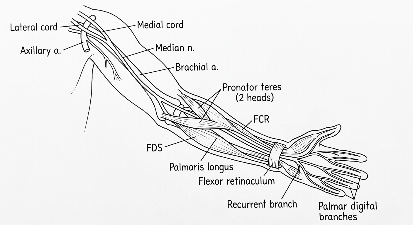
c) Relations (2 marks)
| Region | Relation |
|---|---|
| Axilla | Lateral to AA |
| Arm — proximal | Lateral to BA |
| Arm — mid | Crosses anterior to BA |
| Arm — distal | Medial to BA; anterior to elbow joint |
| Forearm | Between heads of PT → deep to FDS → anterior to FDP |
| Wrist | Between PL (medial) and FCR (lateral) tendons |
| Carpal tunnel | Most superficial structure; deep to FR |
d) Branches (3 marks)
- Vascular branches → BA
- Twig to PT (occasionally, just above elbow)
- Muscular (direct) → PT, FCR, PL, FDS
- AIN (anterior interosseous nerve) — arises between 2 heads of PT; pure motor
- → FPL (flexor pollicis longus)
- → FDP lateral half (index + middle)
- → PQ (pronator quadratus)
- → Articular twigs (elbow + proximal radio-ulnar joint)
- Palmar cutaneous branch — given off 5 cm above FR
- Passes superficial to FR → skin over thenar eminence + central palm
- ⚠️ NOT compressed in carpal tunnel syndrome
- Recurrent (thenar) branch → APB + FPB (superficial head) + opponens pollicis
- Palmar digital branches (4–5 in number)
- Sensory → palmar surface lateral 3½ digits + nail beds of same
- Motor → 1st and 2nd lumbricals
Mnemonic — LOAF (4 hand muscles of median n.): Lumbricals 1&2, Opponens pollicis, Abductor pollicis brevis, Flexor pollicis brevis (sup. head)
e) Applied Anatomy (2 marks)
Injury at Elbow (supracondylar fracture, dislocation)
- All median n. muscles affected
- Ochsner's clasping test — index finger fails to flex → "Pointing Index"
- Cannot flex terminal phalanx of thumb (FPL paralysed)
- Wrist deviates ulnar on flexion (FCR lost)
- Thenar wasting → thumb falls in plane of palm → "Ape hand" / Simian hand
- Sensory loss → palmar surface lateral 3½ digits; autonomous zone = tip of index finger
Injury at Wrist (cut injury / carpal tunnel syndrome)
- Thenar muscles only paralysed → thenar atrophy
- Cannot abduct/oppose thumb → Ape hand
- Pen test (for APB) → negative
- Sensory loss → lateral 3½ digits
- ⚠️ Palmar skin SPARED (palmar cutaneous branch lies superficial to FR — not compressed)
- CTS features: nocturnal paraesthesia → Tinel's sign (tap over carpal tunnel → tingling) → Phalen's test (forced wrist flexion 60 s → reproduces symptoms)
| Feature | Elbow injury | Wrist / CTS |
|---|---|---|
| Thenar wasting | ✓ | ✓ |
| Pronation lost | ✓ | ✗ |
| Pointing index | ✓ | ✗ |
| Palmar skin | Affected | Spared |
| Deformity name | Ape hand | Ape hand + thenar atrophy |
Verified against: S. Das Manual on Clinical Surgery 13th Ed. (pp. 143–144); Gray's Anatomy for Students 4th Ed. (Ch. 7 – Upper Limb); consistent with BD Chaurasia Vol. 1 Indian curriculum content.