
Give me the types of nerve injury classification of nerve injury and walllerian degeneration and regeneration and picture of nerve
Reading File
Now let me search the medical library for nerve injury classification, Wallerian degeneration, and nerve images simultaneously.
Finding Sources
Finding Sources
Reading File
Finding Sources
Finding Sources
Excellent! I have great source material. Now let me read the key sections and fetch the nerve images simultaneously.
Reading File
Reading File
Reading File
Loading Image
Now let me get the nerve regeneration image from Junqueira's and the detailed Wallerian + Sunderland classification content.
Loading Image
Reading File
I have excellent content. Let me fetch the nerve classification diagram and get the Wallerian degeneration details.
Loading Image
Reading File
I now have all the key images and content. Let me fetch the Wallerian degeneration figure and read the remaining regeneration text.
Loading Image
Reading File
I now have everything needed for a comprehensive, well-illustrated answer. Let me compile it.
Peripheral Nerve Injury: Classification, Wallerian Degeneration & Regeneration
1. Types of Nerve Injury
There are two main classification systems that are used together.
Seddon's Classification (3 types)
| Seddon | Structure damaged | Recovery |
|---|---|---|
| Neurapraxia | Myelin sheath only - axon intact | Complete, spontaneous (days to ~6 weeks) |
| Axonotmesis | Axon disrupted; endoneurium ± perineurium intact | Wallerian degeneration occurs; regeneration possible |
| Neurotmesis | Complete nerve division including connective tissue | Unlikely without surgical repair |
Sunderland's Classification (5 grades) + MacKinnon's 6th
Based on which connective tissue layers are disrupted:
| Grade | Seddon Equivalent | What is Damaged | Prognosis |
|---|---|---|---|
| I | Neurapraxia | Myelin only - focal conduction block, axon intact | Full recovery, no Wallerian degeneration |
| II | Axonotmesis | Axon + myelin; endoneurium intact | Good - regeneration guided by intact tubes |
| III | Axonotmesis | Axon + endoneurium disrupted; perineurium intact | Incomplete - misdirection may occur |
| IV | Neurotmesis | Axon + endoneurium + perineurium disrupted; epineurium intact | Poor - neuroma in continuity |
| V | Neurotmesis | Complete nerve trunk transection (all layers) | Needs surgical repair |
| VI | - | Mixed combination of I-V in same nerve | Variable - partial/complex injury |
(MacKinnon and Dellon, Surgery of the Peripheral Nerve, 1988)
Nerve Structure Diagram (showing all 5 Sunderland grades)
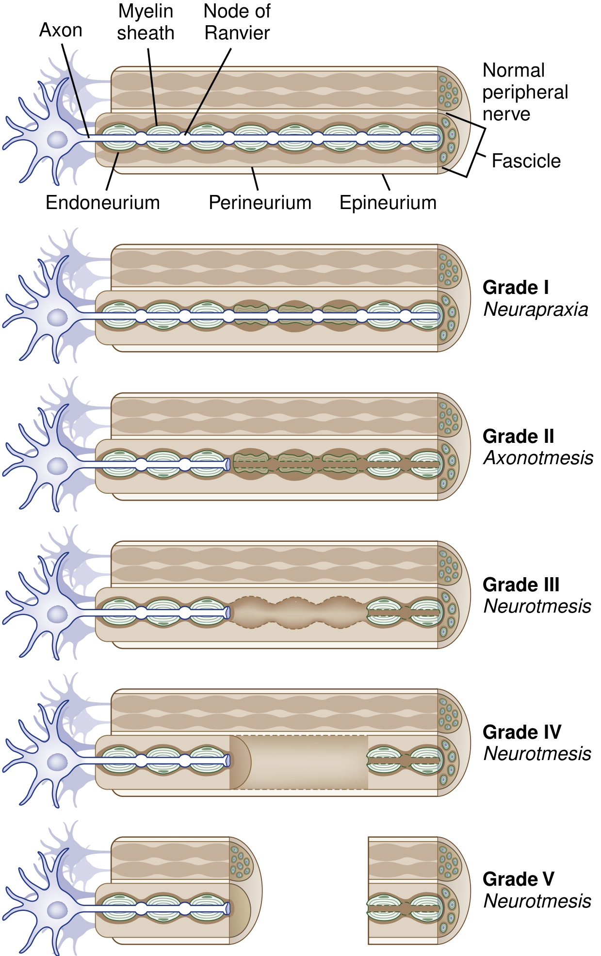
Fig. Classification of Peripheral Nerve Trauma. Grades I (Neurapraxia) through V (complete Neurotmesis). From Bradley and Daroff's Neurology in Clinical Practice, courtesy of Cleveland Clinic / David Schumick, CMI.
2. Wallerian Degeneration
Wallerian degeneration follows Grade II to Grade V injuries (any injury with axonal disruption). It is a programmed, sequential breakdown of the axon and its myelin distal to the injury site.
Sequence of Events
Distal to injury:
- Hours 0-3: Rapid influx of extracellular Ca²+ and Na⁺ through disrupted axonal membrane activates a cascade resembling apoptosis
- Day 1-3: Axonal cytoskeleton fragments; Schwann cells retract from nodes of Ranvier; leukocytes are recruited; cytokine signaling begins
- Day 3-7: Schwann cells dedifferentiate (upregulate c-Jun protein) and switch from myelin-manufacturing to "repair cells"; they begin digesting myelin debris together with recruited macrophages
- ~1 week: The entire axonal process of Wallerian degeneration is complete
- Weeks following: Schwann cells proliferate to form Bungner bands (cords) - tubes that guide regenerating axons
Proximal to injury (cell body):
- Chromatolysis occurs - the rough endoplasmic reticulum (Nissl substance) breaks up and disperses, the nucleus shifts eccentrically
- Gene expression switches from axon maintenance to protein synthesis (regeneration mode)
- Very proximal injuries (e.g., near spinal cord) may trigger cell body apoptosis
Target organ:
- Denervated muscle fibers undergo progressive atrophy
- If reinnervation does not occur, permanent degeneration of the target organ results
Wallerian Degeneration Diagram
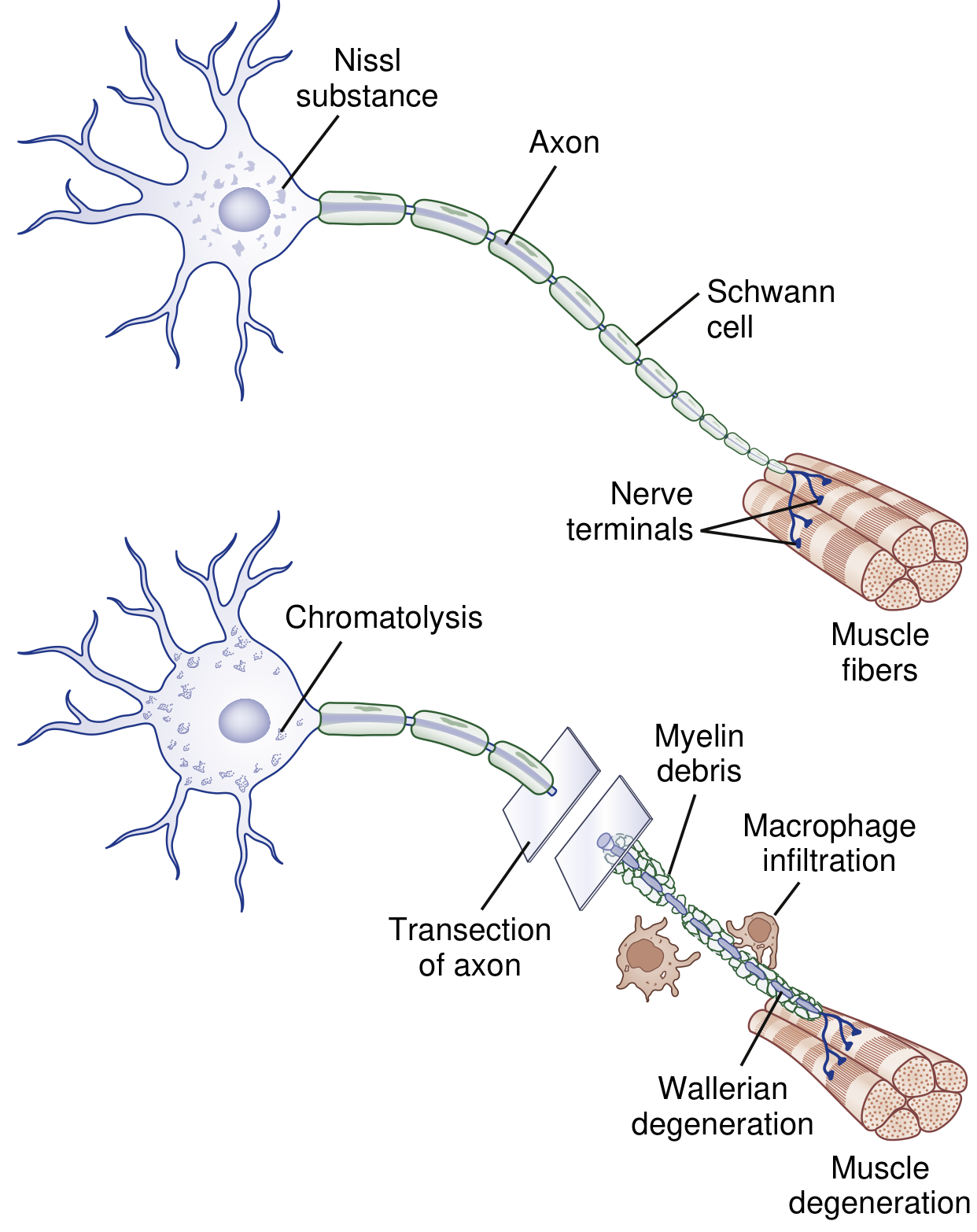
Fig. Wallerian Degeneration. After axotomy, the distal axon and myelin sheath degenerate; macrophages clear debris; the cell body undergoes chromatolysis - switching to regeneration mode. From Bradley and Daroff's Neurology in Clinical Practice.
3. Nerve Regeneration
The method depends on injury grade:
Grade I (Neurapraxia) - Remyelination
- Schwann cells divide and remyelinate the intact axon
- New myelin sheath is thinner with shorter internodes than original
- Recovery within weeks to a few months - no Wallerian degeneration needed
Grade II-V - Axonal Regrowth
Collateral sprouting (partial injuries):
- Intact neighboring axons sprout new branches from nodes of Ranvier (nodal sprouts) or nerve terminals (terminal sprouts)
- Begins as early as 4 days post-injury
- Adopts denervated muscle fibers, enlarging surviving motor units
- Clinical recovery takes 3-6 months
Proximal-to-distal regeneration (severe/complete injuries):
- Begins only after Wallerian degeneration is complete
- Schwann cells dedifferentiate, proliferate, and form Bungner bands - physical tubes that guide the regrowing axon
- Regenerating sprouts grow at 0.5-3 mm/day (approximately 1-4 mm/day clinically)
- Motor axons preferentially reinnervate distal motor branches when possible
- Tinel's sign (tingling with tapping along the nerve) is a clinical indicator of advancing axon regeneration
Key factors for successful regeneration:
- Cell body viability (intact perikaryon)
- A permissive scaffold (intact endoneurial tubes)
- Guidance cues (neurotrophins, extracellular matrix molecules)
Nerve Regeneration Sequence (Histology)
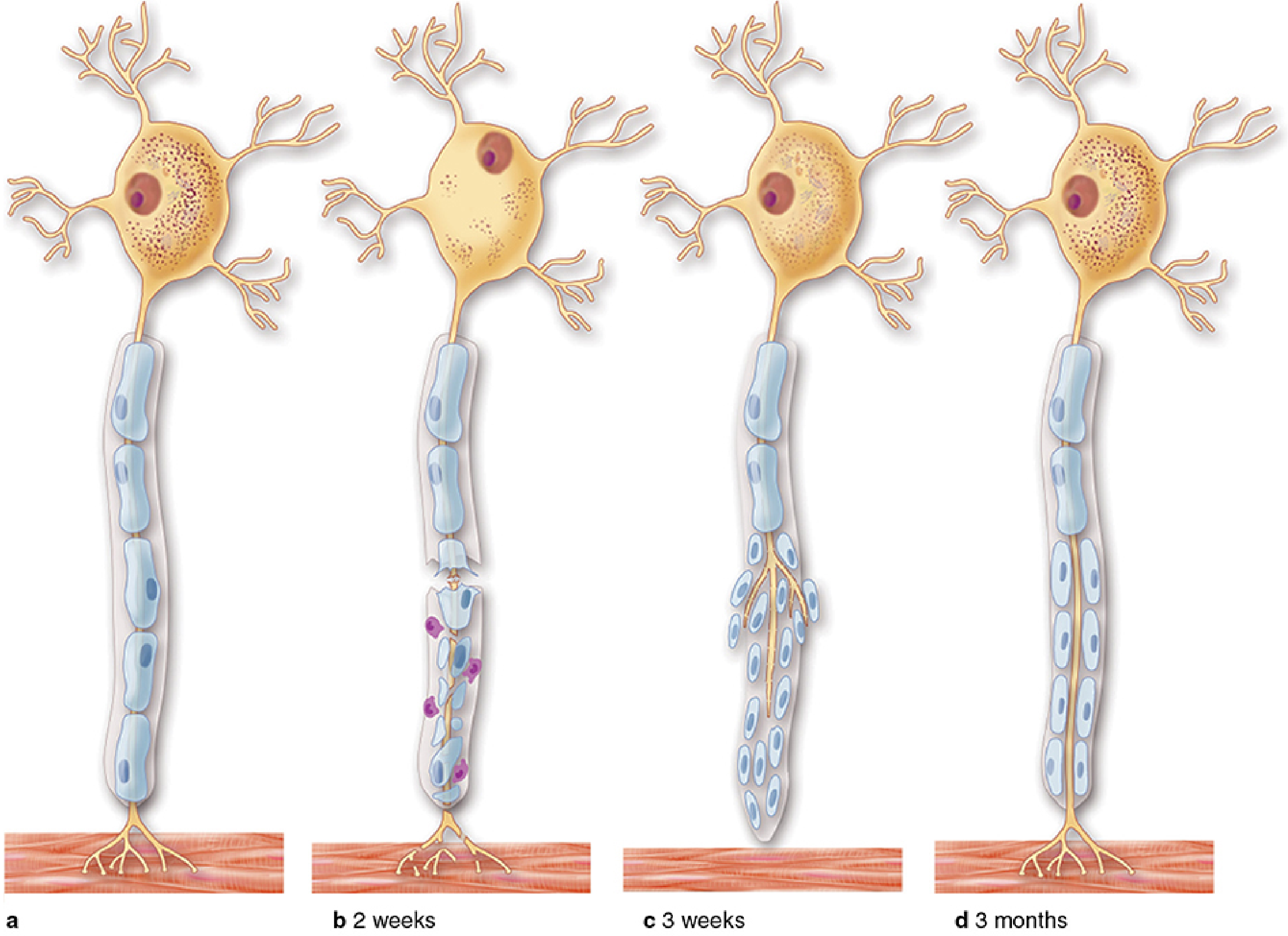
Fig. Peripheral nerve regeneration at (a) baseline, (b) 2 weeks, (c) 3 weeks, and (d) 3 months after injury. From Junqueira's Basic Histology, 17e.
Patterns of Axonal Degeneration vs. Demyelination vs. Regeneration
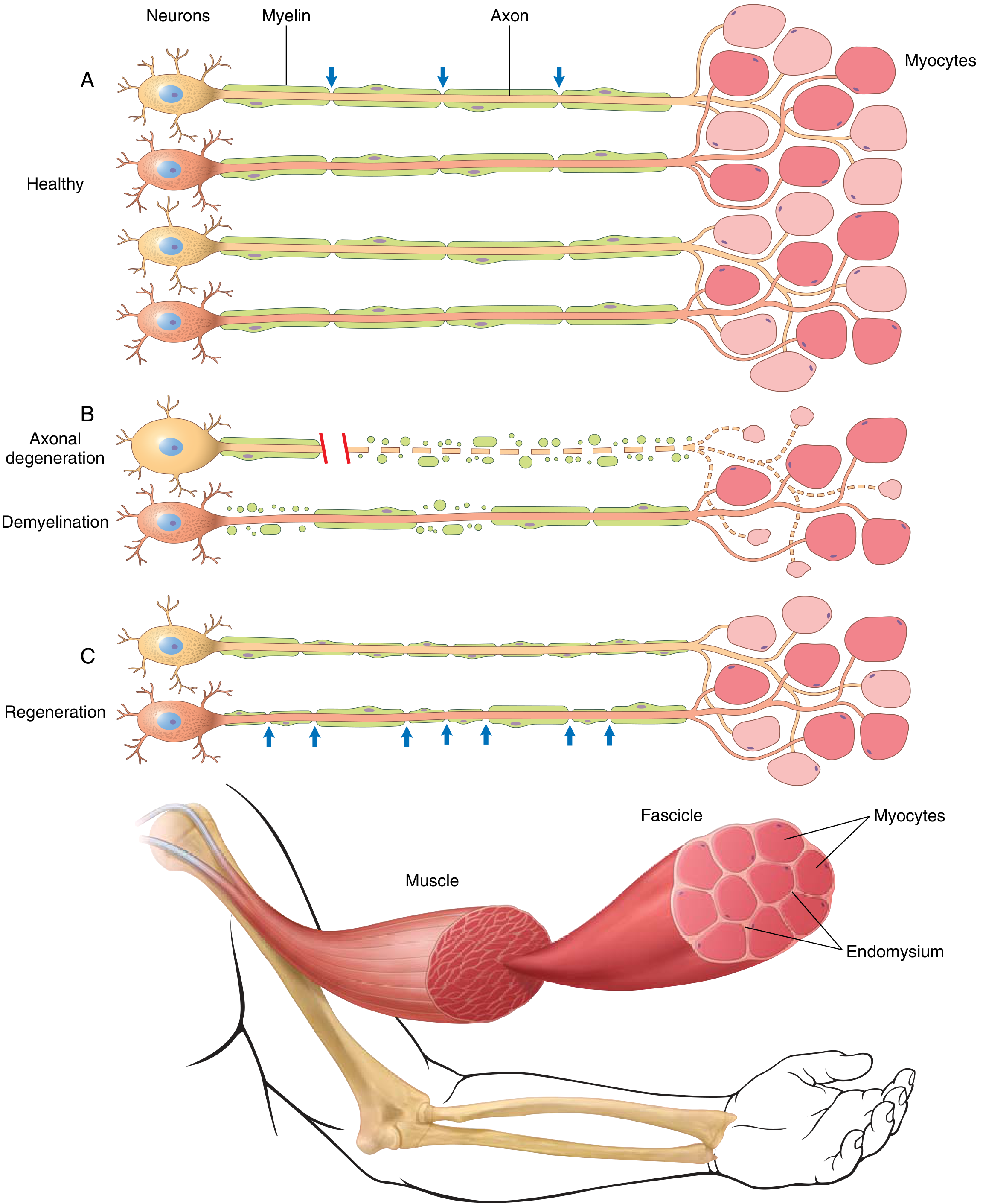
Fig. Patterns of peripheral nerve damage. (A) Healthy. (B) Axonal injury (top) causes distal Wallerian degeneration + myofiber atrophy; demyelination (bottom) spares the axon. (C) Regenerated axon has thinner, shorter internodes; remyelinated segments also have thinner sheaths. From Robbins & Kumar Basic Pathology.
Quick Summary Table
| Feature | Neurapraxia (I) | Axonotmesis (II-IV) | Neurotmesis (V) |
|---|---|---|---|
| Axon | Intact | Disrupted | Transected |
| Wallerian degeneration | No | Yes | Yes |
| Endoneurium | Intact | II: intact; III/IV: disrupted | Disrupted |
| Spontaneous recovery | Yes - complete | Yes - partial/variable | No - needs surgery |
| Rate of recovery | Weeks | 1-4 mm/day | Depends on repair |
| Tinel's sign | No | Yes | Yes (advancing) |
Sources: Bradley and Daroff's Neurology in Clinical Practice | Campbell's Operative Orthopaedics 15th Ed | Robbins & Kumar Basic Pathology | Junqueira's Basic Histology 17e
This is a shared conversation. Sign in to Orris to start your own chat.