
analyse_media
Reading File
Finding Sources
Finding Sources
Reading File
Reading File
Reading File
Loading Image
Based on the image analysis and textbook evidence, here is a thorough assessment:
What This Likely Is: Tinea Cruris ("Jock Itch")
The lesion in your photo is a large, hyperpigmented, dry, scaly plaque on the upper inner thigh at the inguinal fold (groin crease). This is a classic location and appearance for tinea cruris - a superficial fungal infection of the groin caused by dermatophytes (most commonly Trichophyton rubrum or Epidermophyton floccosum).
Key Features Matching the Photo
| Feature | Seen in Your Photo |
|---|---|
| Location: inner thigh / inguinal fold | ✓ |
| Large oval-to-round plaque | ✓ (~5-8 cm) |
| Well-defined, raised, scaly border | ✓ |
| Hyperpigmented (dark brown) | ✓ |
| Dry, thickened (lichenified) surface | ✓ |
| Some central clearing / ring-like pattern | ✓ |
As Fitzpatrick's Dermatology describes: "Tinea cruris presents classically as a well-marginated annular plaque with a scaly raised border that extends from the inguinal fold to the inner thigh." The hyperpigmentation and lichenification you see are signs of a chronic or partially treated infection.
Here is how classic tinea cruris looks in more acute/inflammatory form (textbook reference image):
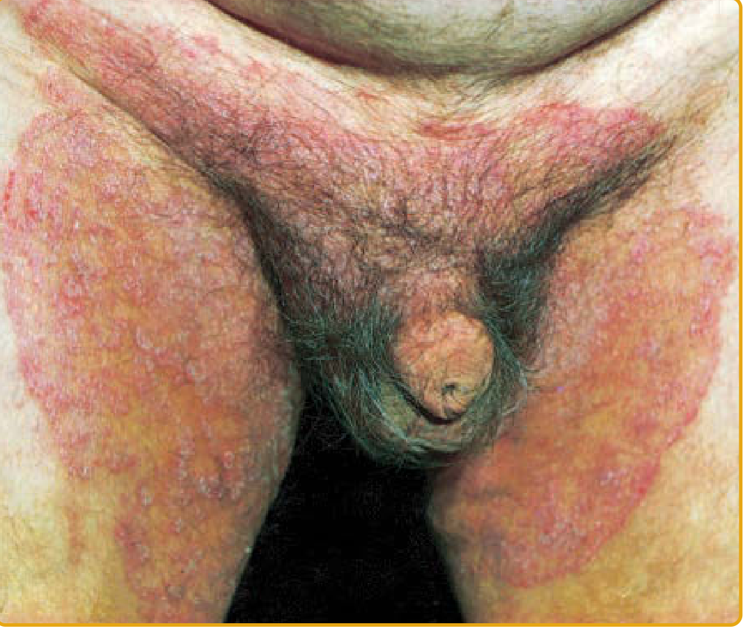
Differential Diagnosis to Consider
These conditions can look similar and should be considered by a doctor:
- Erythrasma - bacterial infection (Corynebacterium minutissimum) that gives a copper-brown color; fluoresces coral-red under Wood's lamp
- Cutaneous candidiasis - usually moister, more inflamed, may involve satellite pustules, often involves scrotum
- Inverse psoriasis - shiny red plaques, often bilateral, associated with psoriasis elsewhere
- Intertrigo - friction/moisture rash, no raised active border
- Seborrheic dermatitis - usually also involves chest and axillae
- Contact dermatitis - history of irritant/allergen exposure
What You Should Do
See a doctor or dermatologist. While this strongly resembles tinea cruris, confirmation requires:
- KOH (potassium hydroxide) examination of a skin scraping - shows fungal hyphae
- Fungal culture if KOH is negative
- Wood's lamp to rule out erythrasma
Treatment (if confirmed as tinea cruris)
Topical antifungals are first-line (per Andrews' Diseases of the Skin and Fitzpatrick's):
| Agent | Regimen |
|---|---|
| Terbinafine 1% cream | Once daily for 1-2 weeks |
| Clotrimazole 1% cream | Twice daily for 2-4 weeks |
| Miconazole 2% cream | Twice daily for 2-4 weeks |
| Econazole 1% cream | Once daily for 2-4 weeks |
Oral therapy (for extensive, chronic, or recurrent cases):
- Terbinafine 250 mg/day for 2-4 weeks
- Itraconazole 200 mg/day for 1-2 weeks
Supportive measures (reduce recurrence):
- Keep the area dry - use loose-fitting cotton underwear and clothing
- Apply antifungal or plain talcum powder to absorb moisture
- If you also have athlete's foot (tinea pedis), treat it simultaneously (it is a common reservoir for reinfection)
- Put on socks before underwear to avoid spreading fungus from feet to groin
- Avoid tight, synthetic clothing
Important disclaimer: This is an informational assessment only, not a medical diagnosis. Please consult a healthcare professional or dermatologist for an accurate diagnosis and a treatment plan tailored to your situation.
This is a shared conversation. Sign in to Orris to start your own chat.