
I just completed sarcoidosis from robbins
Reading File
That's great! Sarcoidosis is a high-yield topic. Let me pull the content from Robbins to build a solid revision set for you.
Finding Sources
Finding Sources
Finding Sources
Both Robbins books have sarcoidosis content. Let me read both simultaneously.
Reading File
Reading File
Reading File
I went too far in the file. Let me get the sarcoidosis clinical features and images from the correct section.
Reading File
Reading File
Reading File
I now have all the Robbins sarcoidosis content I need. Let me also fetch the histology image and check the clinical features section (lines 740-800) that got cut off.
Reading File
I now have everything I need from Robbins to build a thorough revision note. Here it is:
Sarcoidosis - Robbins Revision Notes
Quick Recall Box
| Feature | Key Fact |
|---|---|
| Etiology | Unknown (multisystem granulomatous disease) |
| Histology | Non-caseating epithelioid granuloma |
| Key cells | CD4+ Th1 cells (driver); epithelioid macrophages |
| Most common presentation | Bilateral hilar lymphadenopathy on CXR |
| Lung involvement | 90% of patients |
| Unique epidemiology | Higher prevalence in nonsmokers (unique among lung diseases) |
Epidemiology
- Adults < 40 years predominantly affected
- Higher incidence in Danish/Swedish populations and African Americans (2-3x more than European descent)
- Unique feature: higher prevalence among nonsmokers - virtually unique to sarcoidosis among pulmonary diseases
Pathogenesis
Disordered immune regulation in genetically predisposed individuals exposed to undefined environmental agents.
Immunologic clues (all CD4+ Th1 driven):
- Intraalveolar and interstitial accumulation of CD4+ Th1 cells
- Oligoclonal expansion of CD4+ Th1 T cells within the lung
- Increased Th1 cytokines: IL-2 (T-cell proliferation) and IFN-γ (macrophage activation)
- Increased IL-8, TNF, MIP-1α → recruit more T cells and monocytes → granuloma formation
Paradox: Blood CD4+ T cells are often LOW → associated with anergy to common skin test antigens (Candida, PPD)
After lung transplantation, sarcoidosis recurs in the new lungs in at least one-third of patients.
Morphology (MORPHOLOGY box - HIGH YIELD)
Cardinal feature: Non-necrotizing (non-caseating) epithelioid granuloma
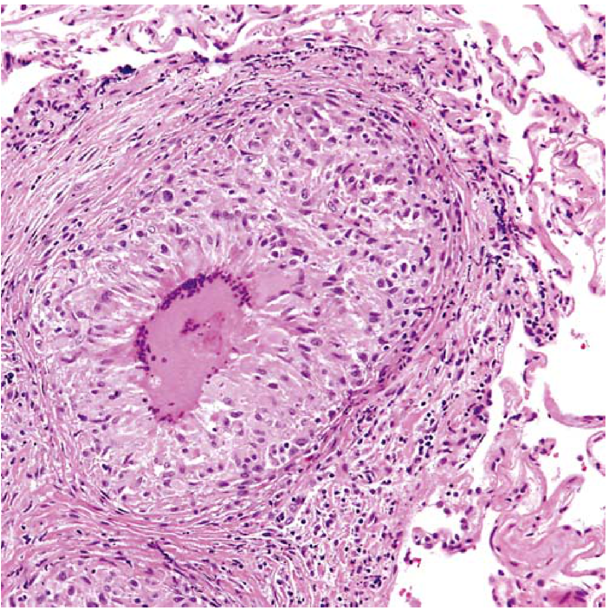
Characteristic noncaseating granuloma with a large central multinucleated giant cell
Structure of granuloma:
- Discrete compact collection of epithelioid macrophages
- Rimmed by an outer zone rich in CD4+ T cells
- Intermixed multinucleate giant cells (from fused macrophages)
- Thin layer of laminated fibroblasts peripherally → over time → hyalinized scar
Special bodies (not required for diagnosis, not specific):
- Schaumann bodies - laminated concretions of calcium and proteins
- Asteroid bodies - stellate inclusions within giant cells
Note: Rarely, foci of central necrosis may appear (nodular form) - but caseation is NOT a feature.
Organ Involvement
Lungs (90%)
- Granulomas in the interstitium (not air spaces)
- "Lymphangitic" distribution - around bronchioles, pulmonary venules, and pleura
- BAL: abundant CD4+ T cells
- 5-15% progress to diffuse interstitial fibrosis → honeycomb lung
Lymph Nodes (75-90%)
- Intrathoracic hilar and paratracheal enlargement
- 1/3 have peripheral lymphadenopathy
- Nodes are painless, firm, rubbery
- Key distinction from TB: "nonmatted" (non-adherent), no necrosis
Skin (~25%)
- Erythema nodosum - hallmark of acute sarcoidosis; bilateral raised, red, tender nodules on anterior legs; it's a panniculitis - classic sarcoid granulomas are actually uncommon in these lesions
- Discrete painless subcutaneous nodules - these DO contain typical noncaseating granulomas
Eyes (20-50%)
- Iritis or iridocyclitis (uveitis) - unilateral or bilateral
- Complications: corneal opacities, glaucoma, vision loss
- Posterior uveal tract: choroiditis, retinitis, optic nerve involvement
- Lacrimal gland involvement → sicca syndrome (dry eyes)
Parotid glands (<10%)
- Painful parotid enlargement, xerostomia (dry mouth)
- Mikulicz syndrome = combined uveoparotid involvement
Spleen
- Granulomas in ~75% microscopically
- Clinical splenomegaly in ~10%
Liver
- Granulomas (usually in portal triads) in ~75%
- Hepatomegaly or abnormal LFTs in ~1/3
Bone Marrow
- Granulomas in up to 40%; rarely causes severe manifestations
Diagnosis
No definitive test exists. Diagnosis requires:
- Clinical + radiologic findings consistent with sarcoidosis
- Exclusion of other disorders (especially TB must be excluded)
- Identification of noncaseating granulomas in involved tissues
Clinical Course & Prognosis
- Many cases: asymptomatic, discovered incidentally on CXR (bilateral hilar adenopathy) or at autopsy
- Course is unpredictable - chronic progressive OR remissions (spontaneous or steroid-induced)
- Prognosis:
- 65-70% recover with minimal/no residual manifestations
- 20% develop permanent lung dysfunction or visual impairment
- 10-15% succumb to progressive pulmonary fibrosis and cor pulmonale
High-Yield Exam Traps
| Trap | Correct Answer |
|---|---|
| Granuloma type in sarcoidosis | Non-caseating (caseation = TB) |
| Low blood CD4+ cells means immunodeficiency? | No - it's just skin test anergy; lung CD4+ cells are HIGH |
| Erythema nodosum granulomas | Classic sarcoidal granulomas are UNCOMMON in EN lesions |
| Sarcoid lymph nodes vs TB nodes | Sarcoid = non-matted, non-necrotic; TB = matted, caseating |
| Schaumann/asteroid bodies - specific to sarcoid? | NO - also seen in other granulomatous diseases |
| Smoking and sarcoidosis | Sarcoidosis is MORE common in nonsmokers |
| After lung transplant | Sarcoidosis recurs in ≥1/3 of patients |
Summary Mnemonic - SARCOID
- S - Schaumann/asteroid bodies (inclusions)
- A - Anergy to PPD/Candida skin tests (blood CD4 low)
- R - Restrictive lung disease (interstitial fibrosis)
- C - CD4+ Th1 cells drive it; Cor pulmonale (end-stage)
- O - Ocular (iritis/iridocyclitis), unknown etiology
- I - IL-2, IFN-γ (Th1 cytokines); Incidence in African Americans
- D - Diagnosis of exclusion; Diffuse hilar adenopathy on CXR
Source: Robbins & Kumar Basic Pathology, pp. 461-463; Robbins, Cotran & Kumar Pathologic Basis of Disease
Everything about thyroid neoplasms
Finding Sources
Finding Sources
Finding Sources
Reading File
Reading File
Excellent content. Now let me get the rest: the full basic pathology neoplasm section (adenoma, follicular, anaplastic, medullary) and the big Robbins PBD section with NIFTP and molecular genetics.
Reading File
Reading File
Reading File
Reading File
Reading File
I now have comprehensive content from both Robbins books. Let me compile the full revision notes.
Thyroid Neoplasms - Complete Robbins Revision Notes
Overview
Thyroid tumors range from benign follicular adenomas to highly aggressive anaplastic carcinomas.
Clinical clues suggesting malignancy in a thyroid nodule:
- Solitary nodule (vs. multiple)
- Age < 30 years or male gender
- History of radiation exposure
- Cold nodule on radionuclide scan (does not take up radioactive iodine)
- Majority of solitary nodules are benign; carcinomas account for < 1% of solitary thyroid nodules
Important: Ultrasound + fine-needle aspiration (FNA) biopsy are essential for preoperative evaluation.
1. Thyroid Adenoma (Follicular Adenoma)
Key Features
- Typically solitary, spherical lesion derived from follicular epithelium
- Most are nonfunctional; small proportion are toxic adenomas (produce thyroid hormone → thyrotoxicosis)
- Follicular adenomas are generally NOT forerunners of carcinoma, though shared genetics (RAS, PTEN) raise the possibility that some follicular carcinomas arise in preexisting adenomas
Genetics
| Adenoma Type | Mutation |
|---|---|
| Toxic adenoma (functional) | Gain-of-function TSHR mutation (most common); less commonly GNAS (Gsα subunit) |
| Nonfunctional adenoma | RAS mutations (<20%), PTEN mutations |
Morphology
- Gross: Solitary, spherical, compresses adjacent thyroid
- Micro: Uniform follicles with colloid; cells uniform in size, shape, nuclear morphology; mitotic figures rare
- Key hallmark: Intact capsule encircling the ENTIRE tumor - this is what distinguishes adenoma from follicular carcinoma
- Occasional oxyphil (Hürthle cell) change - brightly eosinophilic granular cytoplasm
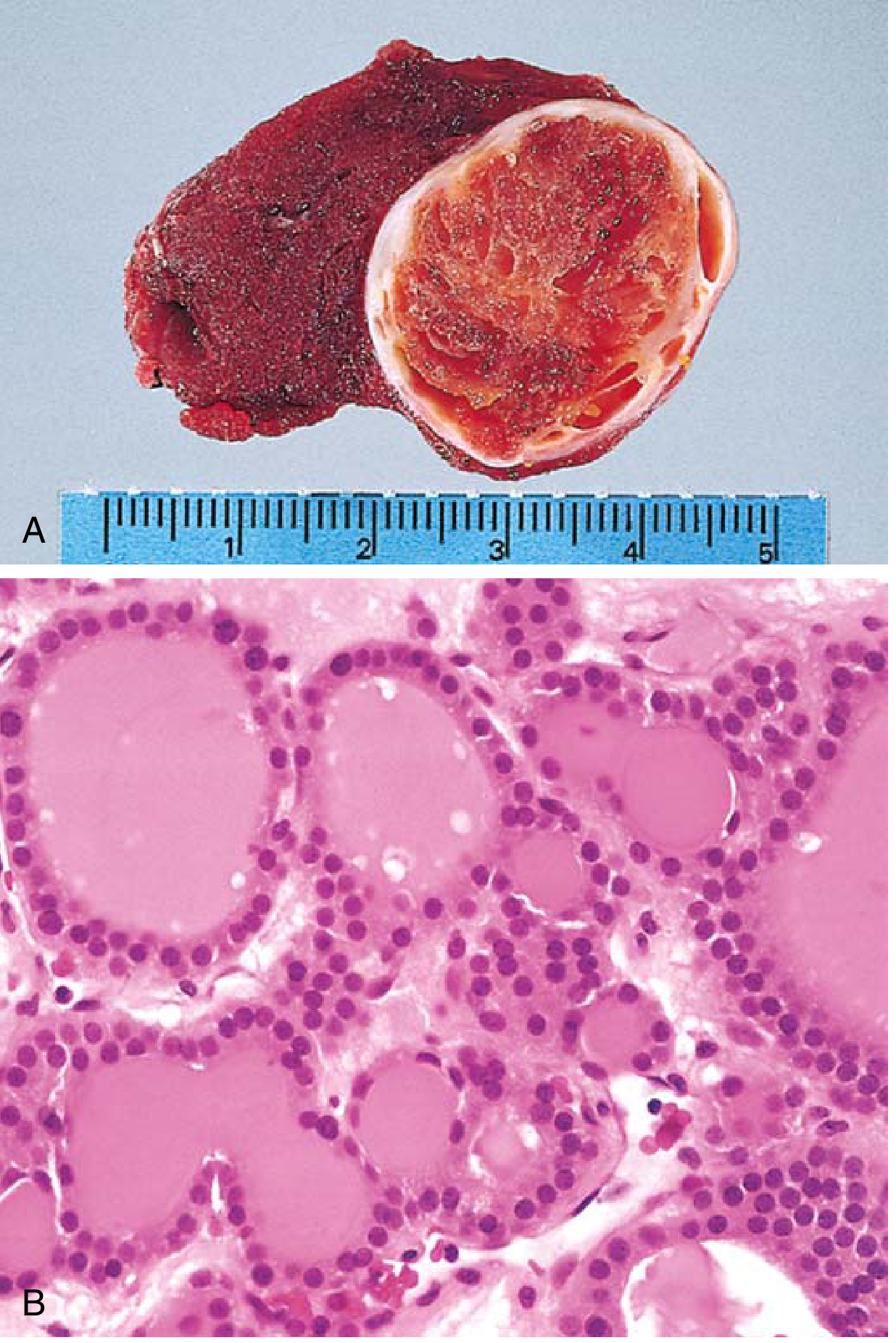
Radionuclide Scan
- Nonfunctional (cold nodule): Takes up less iodine than normal thyroid - up to 10% of cold nodules prove malignant
- Toxic adenoma (hot/warm nodule): Malignancy is uncommon in hot nodules
Prognosis
Excellent - do not recur after surgical removal.
2. Thyroid Carcinomas - Overview
| Type | Frequency | Origin | Key Feature |
|---|---|---|---|
| Papillary thyroid carcinoma (PTC) | ~85% | Follicular epithelium | Orphan Annie nuclei, psammoma bodies |
| Follicular thyroid carcinoma (FTC) | 5-15% | Follicular epithelium | Vascular/capsular invasion (NO lymph spread) |
| Medullary thyroid carcinoma (MTC) | 5% | Parafollicular C cells | Calcitonin, amyloid, RET mutation |
| Anaplastic (undifferentiated) | <5% | Follicular epithelium | Most aggressive, worst prognosis |
3. Papillary Thyroid Carcinoma (PTC)
Epidemiology
- Most common thyroid malignancy (~85% of cases in the US)
- Most often between 25-50 years of age
- Most thyroid carcinomas associated with prior ionizing radiation are papillary
Genetics (HIGH YIELD)
- BRAF V600E mutation - most common; associated with more aggressive behavior
- RET/PTC rearrangements - especially in radiation-induced PTC
- RAS mutations - seen in follicular variant of PTC
Morphology
Gross:
- May be solitary or multifocal
- Some well-circumscribed, others infiltrate adjacent parenchyma (ill-defined margins)
- May contain fibrosis, calcification; often cystic
Micro (HALLMARKS - HIGH YIELD):
-
Branching papillae with fibrovascular stalk covered by cuboidal epithelial cells (single to multiple layers)
-
Orphan Annie eye nuclei (ground-glass nuclei) - finely dispersed chromatin giving optically clear/empty appearance
- Plus: nuclear membrane irregularities → nuclear grooves (coffee-bean shapes) and pseudoinclusions (cytoplasm invaginating into nucleus)
-
Psammoma bodies - concentrically calcified structures within papillary cores or adjacent stroma
- Almost NEVER found in follicular or medullary carcinomas
- Strong indicator of PTC in FNA material
-
Lymphatic invasion commonly present; blood vessel involvement uncommon (contrast with follicular carcinoma)
-
Cervical lymph node metastases in up to 50% of cases
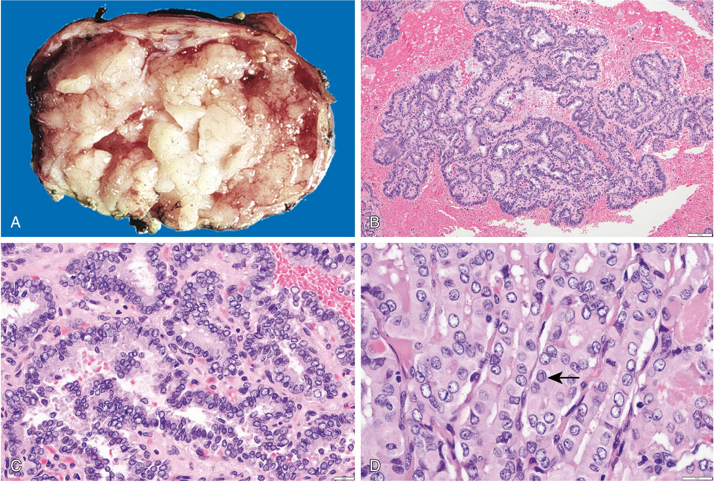
Clinical Features
- Usually presents as asymptomatic thyroid nodule; first manifestation may be enlarged cervical lymph node
- Nodules move freely with thyroid during swallowing
- Hoarseness, dysphagia, cough, dyspnea = advanced disease
- Cold mass on scintigraphy
- "Hot" nodules = almost never malignant (only do scintigraphy if TSH is low)
Prognosis
- Excellent - 10-year survival > 95%
- 5-20% have local/regional recurrences; 10-15% have distant metastases
- Prognosis worse with: age > 55, extrathyroidal extension, distant metastases
NIFTP (HIGH YIELD - new entity)
Noninvasive Follicular Thyroid Neoplasm with Papillary-like Nuclear Features (NIFTP)
- Encapsulated follicular variant of PTC with NO invasion
- Reclassified from malignant to a low-risk neoplasm (no longer called carcinoma)
- Distinguished from invasive encapsulated follicular variant of PTC by absence of capsular/vascular invasion
4. Follicular Thyroid Carcinoma (FTC)
Epidemiology
- 5-15% of primary thyroid cancers (US/iodine-sufficient areas)
- In iodine-deficient areas: rises to 25-40% of thyroid cancers
- Presents in older patients than PTC - peak incidence 40-60 years
Genetics
- RAS mutations (most common)
- PAX8-PPARG rearrangement (translocation) - characteristic of FTC
Morphology
Gross:
- Usually solitary, circumscribed or widely infiltrative
- Sharply demarcated lesions can be indistinguishable from follicular adenomas grossly
- Gray to tan to pink; may be translucent (large colloid-filled follicles)
- Degenerative changes: central fibrosis, coarse calcification
Micro (KEY DISTINCTION from adenoma):
- Uniform cells forming small follicles with colloid - resembles normal thyroid
- Nuclei LACK papillary carcinoma features; NO psammoma bodies
- Occasional oncocytic (Hürthle cell) variant - abundant granular eosinophilic cytoplasm
- Adenoma vs. Carcinoma: NO reliable cytologic difference on FNA - requires extensive histologic sampling of tumor capsule to identify:
- Capsular invasion and/or
- Vascular invasion (venous vessels within/just beyond capsule)
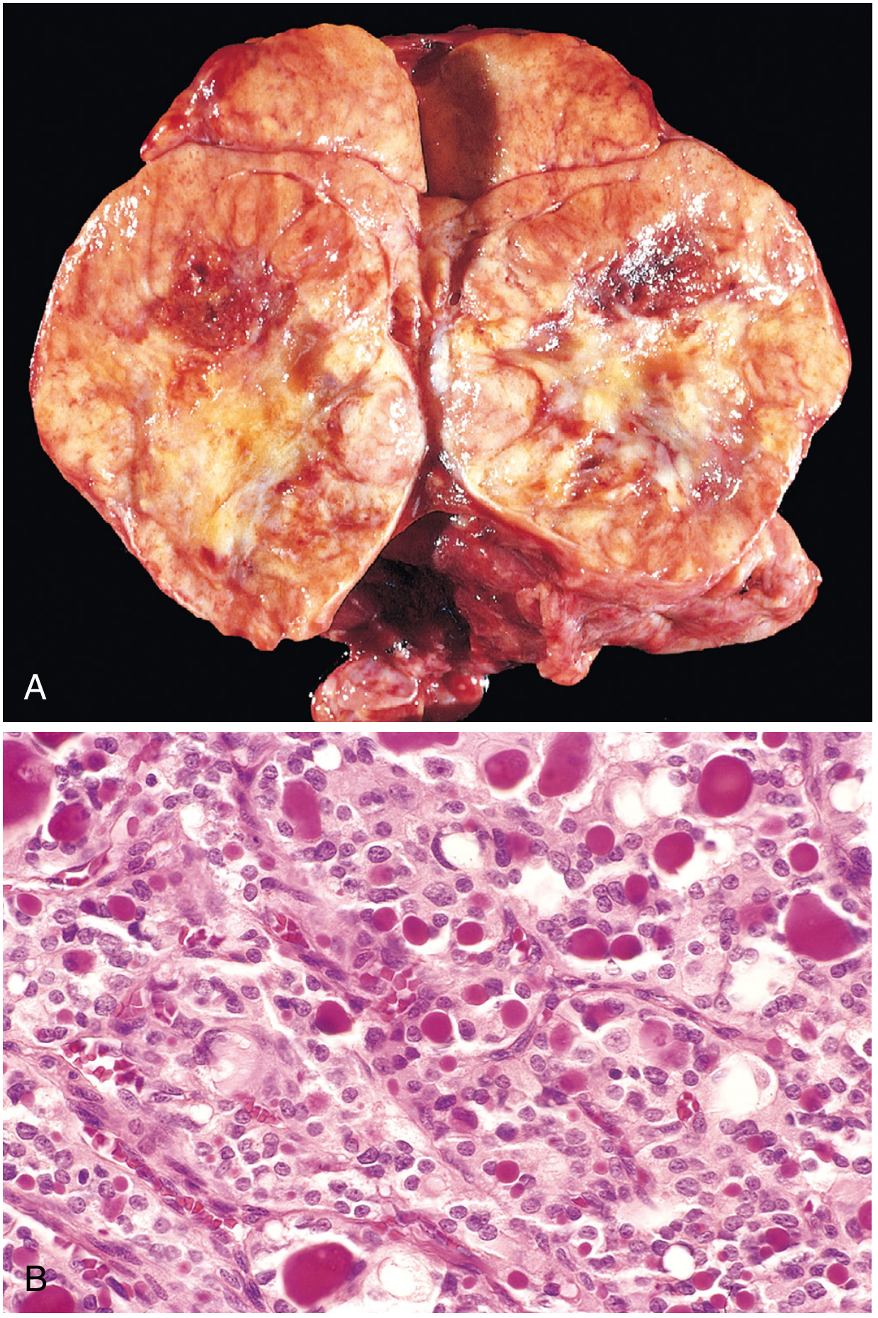
Spread Pattern (KEY CONTRAST WITH PTC)
- Hematogenous spread (not lymphatic) → distant metastases
- Lymph node involvement uncommon (opposite of PTC)
- Hematogenous mets most commonly to lung (and bone)
Prognosis
- Worse than PTC
- Minimally invasive FTC (capsular invasion only): excellent prognosis
- Widely invasive FTC: significantly worse
5. Medullary Thyroid Carcinoma (MTC)
Origin
- Arises from parafollicular C cells (NOT follicular epithelium)
- C cells secrete calcitonin
Epidemiology & Association
- 5% of thyroid carcinomas
- Sporadic (70%): usually presents in 5th-6th decade, typically solitary
- Familial (30%): associated with RET proto-oncogene mutations
- MEN-2A (Sipple syndrome): MTC + pheochromocytoma + parathyroid hyperplasia
- MEN-2B: MTC + pheochromocytoma + mucosal neuromas + marfanoid habitus
- Familial MTC (without MEN features)
- Familial cases: often bilateral and multicentric
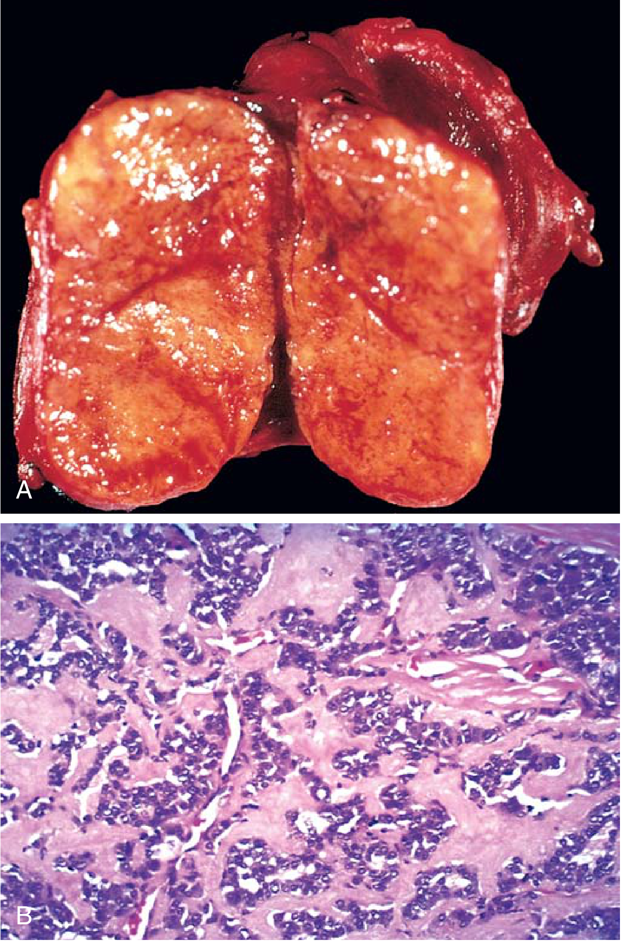
Morphology
- Polygonal to spindle-shaped cells in nests, trabeculae, or follicles
- Amyloid deposits in stroma (derived from calcitonin molecules) - distinctive feature
- C-cell hyperplasia precedes familial MTC (identified in prophylactic thyroidectomies)
- Bilateral/multicentric in familial cases
Clinical Features
- Sporadic: presents as neck mass ± compression effects (dysphagia, hoarseness)
- May secrete other peptide hormones → diarrhea (vasoactive intestinal peptide), flushing (calcitonin, serotonin)
- Screening: serum calcitonin levels or RET mutation testing for family members
- Members of MEN-2 kindreds with RET mutations → prophylactic thyroidectomy offered
Key Markers
- Serum calcitonin (elevated) - diagnostic marker and screening tool
- Amyloid in stroma (calcitonin-derived)
6. Anaplastic (Undifferentiated) Thyroid Carcinoma
Key Features
- Represents <5% of thyroid tumors but is the most aggressive
- Usually occurs in patients >65 years of age
- May arise from dedifferentiation of pre-existing differentiated carcinoma (PTC or FTC) or de novo
- Near-100% mortality; most patients die within 1 year of diagnosis
Genetics
- TP53 mutations (most common) - often with co-existing RAS or BRAF mutations (suggesting progression from differentiated carcinoma)
- TERT promoter mutations
Morphology
- Highly pleomorphic, undifferentiated cells - may be:
- Spindle cells
- Giant cells
- Mixed
- Extensive necrosis and mitotic figures
- Invades adjacent structures extensively
Clinical Features
- Rapidly enlarging, hard, fixed neck mass
- Often presents with airway obstruction, dysphagia, hoarseness at time of diagnosis
- Distant metastases common at presentation
High-Yield Comparison Table
| Feature | Papillary | Follicular | Medullary | Anaplastic |
|---|---|---|---|---|
| Frequency | 85% | 5-15% | 5% | <5% |
| Origin | Follicular cells | Follicular cells | C cells (parafollicular) | Follicular cells |
| Age | 25-50 | 40-60 | Variable | >65 |
| Spread | Lymphatic (LN mets in 50%) | Hematogenous (lung, bone) | Both | Direct invasion |
| Psammoma bodies | YES | No | No | No |
| Orphan Annie nuclei | YES | No | No | No |
| Amyloid stroma | No | No | YES | No |
| Calcitonin marker | No | No | YES | No |
| RET mutation | RET/PTC rearrangement | No | RET point mutation (MEN2) | TP53 |
| BRAF V600E | YES (most common) | No | No | Sometimes |
| RAS | Yes (follicular variant) | YES | No | Sometimes |
| PAX8-PPARG | No | YES | No | No |
| FNA diagnosis | Yes (reliable) | No - can't distinguish from adenoma | Yes | Yes |
| Prognosis | Excellent (>95% 10yr) | Good (minimally invasive) | Intermediate | Dismal (<5% 5yr) |
Nodule Evaluation: Hot vs. Cold (HIGH YIELD)
| Nodule Type | Radioiodine Uptake | Malignancy Risk |
|---|---|---|
| Cold (nonfunctional) | Less than normal thyroid | Up to 10% malignant |
| Warm/Hot (functional) | Equal or greater than normal | Malignancy very rare |
Rule: Always do scintigraphy only if TSH is low (otherwise not clinically indicated).
Key Exam Traps
| Trap | Correct Fact |
|---|---|
| FNA can distinguish follicular adenoma from carcinoma | WRONG - capsular/vascular invasion can only be assessed on surgical histology |
| Psammoma bodies in medullary or follicular carcinoma | Almost NEVER - strongly indicates papillary carcinoma |
| PTC spreads via blood | No - PTC spreads via lymphatics; FTC spreads via blood |
| MTC is derived from follicular cells | No - from C cells (parafollicular) |
| All hot nodules need biopsy | No - hot nodules are almost never malignant |
| Familial MTC = always bilateral | YES - bilateral/multicentric in familial cases |
| Anaplastic carcinoma - any chance of cure | Essentially no - near 100% fatality |
| NIFTP is a carcinoma | No - it was reclassified as a low-risk neoplasm |
Mnemonic: PFMA (types in frequency order)
- Papillary - 85% - Psammoma, Papillae, Paediatric/young adults
- Follicular - 5-15% - Fibrovascular invasion needed, Far (haematogenous) spread
- Medullary - 5% - MEN-2, Marker calcitonin, aMyloid
- Anaplastic - <5% - Awful prognosis, Aged patients, Aggressive
Sources: Robbins & Kumar Basic Pathology, pp. 735-740; Robbins, Cotran & Kumar Pathologic Basis of Disease, pp. 1005-1012
This is a shared conversation. Sign in to Orris to start your own chat.