
From Guyton and Hall Textbook of Medical Physiology Fourth, South Asia edition, writer, 10 - long mark answer on Genesis of Action Potential-describe ; Properties of AP-enumerate
Reading File
Finding Sources
Finding Sources
Searching PubMed
"action potentials" AND physiology
Searching the Web
action potential physiology generation refractory period potassium sodium conductance
Finding Sources
Finding Sources
Reading File
Reading File
Reading File
Reading File
Searching Images
diagram neuronal action potential phases depolarization repolarization hyperpolarization sodium potassium conductance
Reading File
Genesis of Action Potential and Properties of Action Potential
Definition
An action potential (AP) is a rapid, transient, and propagated reversal of membrane potential in an excitable tissue such as a nerve fiber. The membrane potential changes from its resting negative value to a positive value and then rapidly returns to the resting level.
In a typical large nerve fiber:
- Resting membrane potential: about -70 mV
- Threshold potential: about -55 mV
- Peak or overshoot: about +30 to +35 mV
- Total duration: a few milliseconds
Guyton and Hall Textbook of Medical Physiology, p. 80.
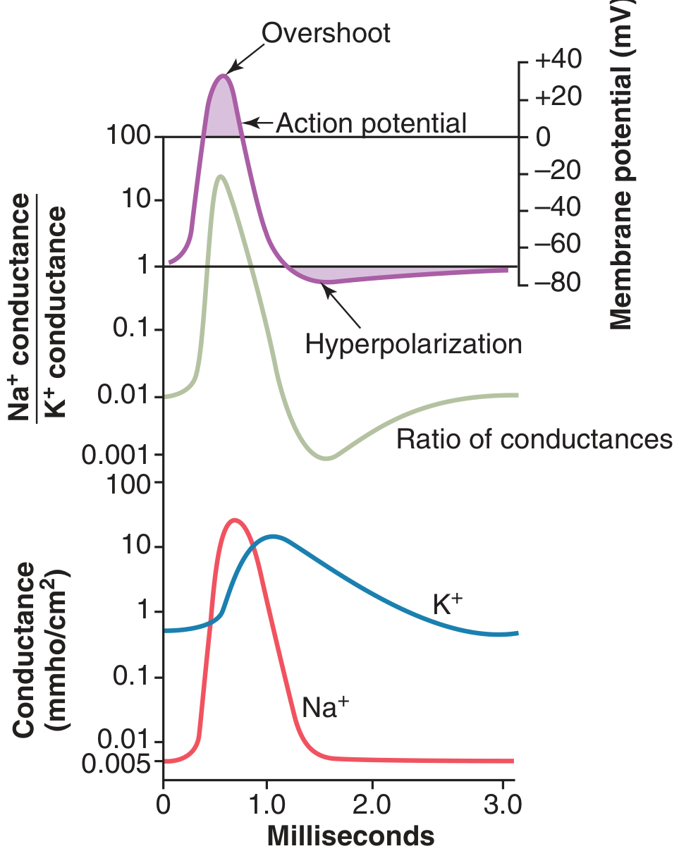
Genesis of Action Potential
The action potential is generated by sequential changes in the permeability of the nerve membrane to sodium (Na⁺) and potassium (K⁺) ions through voltage-gated ion channels.
1. Resting stage or polarized state
- Before stimulation, the nerve membrane is polarized.
- The inside of the cell is negative relative to the outside, usually about -70 mV.
- At rest, potassium conductance is much greater than sodium conductance because of potassium leak channels.
- Voltage-gated Na⁺ channels are closed. Their activation gate is closed and inactivation gate is open.
- Voltage-gated K⁺ channels are also closed.
The Na⁺-K⁺ pump maintains the ionic gradients by pumping 3 Na⁺ out and 2 K⁺ in.
2. Threshold and initiation of AP
A suitable stimulus produces local depolarization. When membrane potential reaches the threshold, usually about -55 mV, voltage-gated Na⁺ channels open.
At threshold, Na⁺ influx becomes greater than K⁺ efflux. This initiates a regenerative positive-feedback cycle:
- Initial depolarization opens some voltage-gated Na⁺ channels.
- Na⁺ enters the cell rapidly.
- The membrane becomes still more depolarized.
- More Na⁺ channels open.
- Further Na⁺ influx occurs, producing an explosive depolarization.
Thus, an action potential develops only after threshold is reached. A subthreshold stimulus produces only a local response and does not cause an AP.
Guyton and Hall Textbook of Medical Physiology, p. 83.
3. Depolarization phase
- Once threshold is reached, the Na⁺ activation gates open rapidly.
- Membrane permeability to Na⁺ rises very markedly, approximately 500 to 5000 times.
- Na⁺ diffuses rapidly into the axon down its electrochemical gradient.
- The interior of the cell becomes progressively less negative and then positive.
This produces the steep ascending limb of the action potential.
4. Overshoot
- Continued Na⁺ influx causes the membrane potential to cross 0 mV.
- The inside of the nerve fiber becomes positive relative to the outside.
- This positive part of the AP is called the overshoot.
In large nerve fibers, the potential may rise to about +35 mV. In small fibers and many CNS neurons, it may only approach zero without a clear overshoot.
5. Repolarization phase
Repolarization begins within a fraction of a millisecond because of two simultaneous events:
-
Inactivation of voltage-gated Na⁺ channels
- The Na⁺ channel inactivation gate closes more slowly than the activation gate.
- Therefore Na⁺ influx stops.
-
Opening of voltage-gated K⁺ channels
- K⁺ channels open with a slight delay.
- K⁺ diffuses out of the cell.
- Loss of positive charge makes the interior negative again.
Thus, decreased Na⁺ entry plus increased K⁺ exit restores the negative membrane potential.
Guyton and Hall Textbook of Medical Physiology, pp. 80-81.
6. After-hyperpolarization or negative after-potential
- K⁺ channels close slowly.
- K⁺ continues to leave the cell for a short time even after normal resting potential is restored.
- Consequently, the membrane becomes more negative than the resting level.
This phase is called after-hyperpolarization, undershoot, or negative after-potential. When K⁺ channels finally close, the membrane returns to its resting potential.
Ionic basis in brief
| Phase | Main channel event | Main ionic movement |
|---|---|---|
| Resting state | K⁺ leak conductance predominates | K⁺ tends to diffuse outward |
| Threshold | Na⁺ activation gates open | Initial Na⁺ influx |
| Depolarization | Marked increase in Na⁺ conductance | Rapid Na⁺ influx |
| Overshoot | Continued Na⁺ influx | Cell interior becomes positive |
| Repolarization | Na⁺ channel inactivation and delayed K⁺ channel opening | Na⁺ influx stops; K⁺ efflux rises |
| Hyperpolarization | Delayed closure of K⁺ channels | Continued K⁺ efflux |
During the early AP, the ratio of Na⁺ to K⁺ conductance rises more than 1000-fold. Later, Na⁺ conductance falls while K⁺ conductance predominates, producing rapid repolarization. Guyton and Hall Textbook of Medical Physiology, pp. 82-83.
Properties of Action Potential
1. All-or-none law
- An AP occurs only when the stimulus reaches threshold.
- A subthreshold stimulus does not produce an AP.
- Once threshold is reached, the AP is generated with full amplitude.
- Increasing stimulus strength above threshold does not increase the amplitude of AP in a single nerve fiber.
A stronger stimulus increases the frequency of action potentials or recruits more nerve fibers, rather than increasing AP size.
2. Stereotyped response
- Each action potential in a particular nerve fiber has approximately the same:
- amplitude
- shape
- duration
- rate of rise
- Therefore, AP is a uniform or stereotyped event.
3. Non-decremental propagation
- AP is conducted along the nerve fiber without loss of amplitude.
- Each excited segment produces local circuit currents that depolarize the next resting segment to threshold.
- Therefore, the AP is regenerated continuously along the membrane.
This differs from an electrotonic or graded potential, which decreases in amplitude with distance.
4. Self-propagating nature
- Once initiated, the AP spreads automatically into adjacent resting membrane.
- Local circuit current from an active area depolarizes the next area to threshold.
- The newly excited area then generates a fresh AP.
Thus, conduction is regenerative and self-propagated.
5. Refractory period
After an AP, the nerve cannot immediately generate another normal AP.
a. Absolute refractory period
- No second AP can be produced, irrespective of stimulus strength.
- It occurs during depolarization and early repolarization.
- The cause is inactivation of voltage-gated Na⁺ channels.
- Na⁺ channels cannot reopen until the membrane has repolarized sufficiently.
b. Relative refractory period
- A second AP can occur, but only with a stronger-than-normal stimulus.
- It occurs during late repolarization and after-hyperpolarization.
- The cause is persistence of increased K⁺ conductance and incomplete recovery of Na⁺ channels.
The refractory period limits the maximum frequency of nerve discharge.
Guyton and Hall Textbook of Medical Physiology, p. 83.
6. Physiological unidirectional conduction
- If a nerve is stimulated in the middle experimentally, APs can travel in both directions.
- Under normal physiological conditions, transmission is functionally unidirectional because the segment behind the advancing AP is refractory.
- Hence, an AP normally proceeds from the site of initiation toward the nerve terminal.
7. No summation
- Individual action potentials do not summate.
- This is because an AP is an all-or-none event and is followed by refractoriness.
- In contrast, graded potentials can undergo temporal and spatial summation.
8. No fatigue in nerve fibers
- Nerve fibers can conduct repeated action potentials for long periods with little fatigue.
- Fatigue is more characteristic of synapses, especially neuromuscular and central synapses, than of nerve conduction itself.
Conclusion
Action potential is generated when threshold depolarization opens voltage-gated Na⁺ channels, causing rapid Na⁺ influx and depolarization. Subsequent Na⁺ channel inactivation and delayed opening of K⁺ channels cause K⁺ efflux, repolarization, and a brief after-hyperpolarization. Its main properties are all-or-none response, stereotyped form, self-propagation, non-decremental conduction, refractoriness, physiological unidirectionality, absence of summation, and relative resistance to fatigue.
Reference: Guyton and Hall Textbook of Medical Physiology, “Membrane Potentials and Action Potentials,” pp. 80-84.
This is a shared conversation. Sign in to Orris to start your own chat.