
Headache management
Reading File
Now let me search the medical library and PubMed in parallel for headache management.
Finding Sources
Searching PubMed
"headache" AND "management"
Good. Now let me read the key Harrison's section and also search for more specific headache topics in parallel.
Reading File
Finding Sources
Finding Sources
Now let me read the key sections on migraine treatment and cluster headache in parallel.
Reading File
Reading File
Finding Sources
Finding Sources
Let me now read the migraine prophylaxis section, cluster headache treatment, and also the Harrison's migraine management chapter for a complete picture.
Reading File
Reading File
Reading File
I now have excellent data. Let me read the prophylaxis/prevention section from Harrison's and tension headache treatment quickly.
Reading File
Reading File
Let me now get the migraine prophylaxis data from Harrison's.
Reading File
Let me get the migraine prophylaxis table from Harrison's quickly.
Finding Sources
Finding Sources
Reading File
I now have comprehensive data from multiple authoritative sources. Let me compile the full answer.
Headache Management
A structured, evidence-based overview covering classification, acute treatment, prophylaxis, and special headache types.
1. Classification - Red Flags First
Before managing headache, exclude secondary (dangerous) causes. The key warning signs are:
- "Thunderclap" - sudden, worst-ever headache (subarachnoid hemorrhage until proven otherwise)
- New headache in age >50 (giant cell arteritis, mass lesion)
- Fever + neck stiffness (meningitis)
- Headache with focal neurologic signs or papilloedema
- Progressive worsening over days/weeks
- Headache in a cancer or immunocompromised patient
90% of patients presenting to primary care with headache have migraine. - Harrison's Principles of Internal Medicine 22E, 2025
2. Primary Headache Types & Their Management
A. Migraine
Acute (Abortive) Treatment
Step up based on attack severity:
Step 1 - Mild attacks:
- Aspirin, acetaminophen, or NSAIDs (ibuprofen, naproxen)
- Combination: acetaminophen + aspirin + caffeine (FDA-approved)
- Add a dopamine antagonist (metoclopramide 10 mg, prochlorperazine 10 mg) if nausea is present - also enhances GI absorption
Step 2 - Moderate-to-severe attacks (after NSAID failure):
| Drug | Route | Dose |
|---|---|---|
| Sumatriptan | PO | 50-100 mg |
| Rizatriptan | PO | 10 mg |
| Eletriptan | PO | 40 mg |
| Almotriptan | PO | 12.5 mg |
| Zolmitriptan | PO/nasal | 2.5 mg / 5 mg nasal |
| Sumatriptan | SC | 6 mg (fastest onset) |
| Sumatriptan | Nasal | 20 mg |
| Rimegepant (CGRP antagonist) | PO | 75 mg |
| Ubrogepant (CGRP antagonist) | PO | 50-100 mg |
| Lasmiditan (5-HT1F agonist) | PO | 50-200 mg |
Triptans are selective 5-HT1B/1D receptor agonists. Rizatriptan and eletriptan are the most efficacious on a population basis. Sumatriptan and zolmitriptan offer multiple formulations. - Harrison's
Triptan timing is key: Give oral/nasal forms at aura onset; SC at headache onset. Do NOT give during aura to prevent headache - they are ineffective in the aura phase but safe if given then.
Triptan contraindications: Ischemic heart disease, uncontrolled hypertension, basilar or hemiplegic migraine, concurrent MAOIs.
Special clinical situations (from Harrison's treatment table):
| Situation | Preferred Option |
|---|---|
| Early vomiting | Zolmitriptan 5 mg nasal / Sumatriptan 6 mg SC |
| Rapid onset headache | SC or nasal triptans / DHE IM |
| Headache recurrence | Ergotamine 2 mg PR / Naratriptan 2.5 mg / Eletriptan 40 mg |
| Poor tolerance of acute therapy | Naratriptan, Almotriptan, Rimegepant, neuromodulation |
| Menstrual migraine prevention | Frovatriptan, Rimegepant, or estrogen patches during menses |
Dihydroergotamine (DHE): 1 mg IV (over 3 min) or IM, or 2 mg nasal spray. Pretreat with antiemetic. Contraindicated in pregnancy, ischemic heart disease, within 24h of triptan use.
ED/Parenteral setting:
- IV prochlorperazine 5-10 mg (superior to opioids in RCTs)
- IV metoclopramide 10 mg
- IV ketorolac 15-30 mg
- IV mixture: prochlorperazine 5 mg + DHE 0.5 mg over 2 min (common protocol)
- Dexamethasone 6-10 mg IV as adjunct to reduce recurrence
- Magnesium sulfate 2 g IV over 30 min (nonvalidated)
- Opioids: Only for severe infrequent headache unresponsive to other therapies; avoid routine use as they worsen long-term outcomes and decrease future triptan responsiveness
Tintinalli's Emergency Medicine, Table 165-7
Neuromodulation (non-pharmacologic acute options):
- Single-pulse transcranial magnetic stimulation (sTMS) - FDA cleared
- Noninvasive vagus nerve stimulator (nVNS) - FDA cleared
- Remote electrical neuromodulation (arm device)
- Transcutaneous supraorbital nerve stimulation
Migraine Prophylaxis (Preventive Treatment)
Indicated when: ≥4 headache days/month, attacks significantly impairing function, failure/contraindication to acute therapies, medication overuse headache risk, or patient preference.
Established preventive agents:
| Drug Class | Agents | Notes |
|---|---|---|
| Beta-blockers | Propranolol, metoprolol, timolol | First-line; avoid in asthma, depression |
| Anticonvulsants | Topiramate, valproate | FDA-approved; valproate teratogenic |
| TCAs | Amitriptyline | Also treats comorbid depression/sleep |
| Calcium channel blockers | Verapamil | More evidence for cluster headache |
| CGRP monoclonal antibodies | Erenumab, fremanezumab, galcanezumab, eptinezumab | New class; SC/IV monthly; very effective |
| CGRP receptor antagonists | Rimegepant (also used acutely) | Oral, twice-weekly for prevention |
| Supplements | Magnesium, riboflavin (B2), CoQ10, feverfew | Modest evidence, good safety |
B. Tension-Type Headache (TTH)
Characteristics: Bilateral, pressing/tightening (non-pulsating), mild-to-moderate, no vomiting, no significant nausea, no aggravation by activity.
Acute treatment:
- Simple analgesics: aspirin, acetaminophen, NSAIDs (first-line for episodic TTH)
- More severe headaches may require prescription analgesics, but no specific preparation has proven superior
- Avoid opioids and barbiturate-containing combinations due to dependence and medication overuse risk
Prevention/chronic TTH:
- Amitriptyline (single bedtime dose) - treatment of choice when anxiety/depression comorbid
- Some evidence for calcium channel blockers, phenelzine, cyproheptadine
- Propranolol and ergotamine are ineffective unless migraine features co-exist
- Biofeedback, relaxation techniques, massage for anxious/stressed patients
- Medication overuse is a key concern - gradual analgesic withdrawal is essential in chronic daily headache
Adams and Victor's Principles of Neurology, 12th Ed.
C. Cluster Headache
Characteristics: Unilateral periorbital/temporal, excruciating (15-180 min), autonomic features (lacrimation, conjunctival injection, nasal congestion, ptosis), episodic or chronic, M > F.
Acute treatment:
- 100% oxygen via mask for 10-15 min at attack onset (highly effective, no side effects)
- Sumatriptan 6 mg SC or zolmitriptan 5 mg nasal spray
- Intranasal lidocaine (adjunct)
- Ergotamine 2 mg orally at bedtime (for predictable nocturnal attacks)
Preventive/transitional treatment:
- Verapamil (up to 480 mg/d) - first-line; requires ECG monitoring in older patients
- Prednisone 75 mg/d tapering over ~3 weeks (short-term bridge)
- Lithium 600-900 mg/d (chronic cluster; monitor for toxicity)
- Galcanezumab (anti-CGRP monoclonal antibody) - halved weekly cluster frequency in clinical trials
- Topiramate, melatonin as alternatives
Adams and Victor's Principles of Neurology, 12th Ed.; Harrison's 22E
D. Trigeminal Autonomic Cephalalgias (TACs) Comparison
| Feature | Cluster | Paroxysmal Hemicrania | SUNCT/SUNA |
|---|---|---|---|
| Duration | 15-180 min | 2-30 min | 5-240 sec |
| Frequency | 1-8/day | 1-20/day | 3-200/day |
| Abortive Rx | O2, sumatriptan SC | Indomethacin (diagnostic) | IV lidocaine |
| Prevention | Verapamil, galcanezumab | Indomethacin | Lamotrigine |
Paroxysmal hemicrania: Complete, absolute response to indomethacin is pathognomonic - start at 25 mg TID, titrate to 75 mg TID.
SUNCT/SUNA: Best prevented with lamotrigine 200-400 mg/d. Topiramate and gabapentin are alternatives.
Hemicrania continua: Unilateral continuous pain + autonomic features; complete response to indomethacin is diagnostic and therapeutic.
E. Medication Overuse Headache (MOH)
- Occurs when acute medications are used >10-15 days/month
- Highest risk: opioids and barbiturates > triptans > NSAIDs
- Treatment: gradual withdrawal of the offending medication (can be done abruptly for triptans/NSAIDs; slower for opioids/barbiturates)
- Preventive therapy should be started during withdrawal
- Multidisciplinary support improves outcomes
3. When to Refer
Per Harrison's 22E: Refer to a headache specialist when:
- No clear diagnosis
- Primary headache other than migraine or TTH
- Unresponsive to two or more standard therapies for the diagnosed type
- Any suspected secondary headache requiring specialist workup
Recent Evidence Note
A 2024 BMJ network meta-analysis (PMID 39293828) on acute migraine management confirmed triptans as effective first-line agents and provided comparative efficacy data across drug classes - consistent with current guideline recommendations.
Abdominal koch’s pain abdomen mana
Abdominal tuberculosis (Koch's) causing pain abdomen - the management involves anti-TB therapy as the cornerstone, with surgical intervention for complications like obstruction, perforation, or abscess. Let me pull the full textbook content.
Searching sources...
Reading File
Finding Sources
Searching PubMed
"abdominal tuberculosis" AND management
Finding Sources
Finding Sources
Good - several rich sources found. Let me read all the key sections in parallel.
Reading File
Reading File
Finding Sources
Excellent - rich data. Now read the treatment details, peritonitis section, and anti-TB drug regimen in parallel.
Reading File
Reading File
Finding Sources
Reading File
Finding Sources
Finding Sources
Finding Sources
Reading File
Reading File
I now have comprehensive data. Let me also quickly fetch the barium meal image and compile the full answer.
Loading Image
Now I have all the data needed. Here is the comprehensive answer:
Abdominal Tuberculosis (Koch's) - Pain Abdomen Management
Overview
Abdominal TB (Koch's abdomen) refers to TB involvement of the gastrointestinal tract, peritoneum, mesenteric lymph nodes, and/or solid organs. The abdomen is involved in ~11% of extrapulmonary TB cases. 50-80% of abdominal TB patients have peritoneal involvement. - Bailey and Love's Short Practice of Surgery, 28th Ed.
Forms of Abdominal TB
| Form | Features |
|---|---|
| Intestinal TB | Terminal ileum/ileocaecal region most common (75%), ulcerative/hypertrophic/ulcerohypertrophic |
| Tuberculous peritonitis | Wet ascitic type (90%), dry fibrotic type (rare) |
| Mesenteric lymphadenitis | Enlarged nodes, central caseation - can be seen on CT |
| Solid organ | Liver, spleen involvement less common |
Clinical Presentation - Pain Abdomen in Abdominal TB
Chronic/Subacute:
- Abdominal pain - nonspecific, chronic; most common complaint (80-90% of patients)
- Weight loss, evening fever, malaise, night sweats
- Alternating diarrhoea and constipation
- Palpable RIF mass in 25-50% (ileocaecal involvement)
- Ascites and abdominal distension (peritoneal TB)
- "Doughy feel" of abdomen on palpation - a classical sign
Acute (Emergency presentation):
- Features of acute distal small bowel obstruction - colicky pain, abdominal distension, bilious/faeculent vomiting, absolute constipation
- Rarely: peritonitis from perforation of a tuberculous ulcer
- Acute-on-chronic: superimposed obstruction on longstanding disease
These patients are often extremely ill - dehydrated, malnourished, anaemic, often with active pulmonary TB. - Bailey & Love
Investigations
Bloods:
- FBC - mild anaemia, normal or mildly elevated WBC
- Raised ESR/CRP
- LFTs (especially with hepatic involvement)
- Urea and electrolytes (dehydration in obstruction)
Microbiological:
- Sputum AFB smear and culture (even without pulmonary symptoms, CXR is essential)
- IGRA (Interferon-Gamma Release Assay) - Quantiferon or T-SPOT
- Ascitic fluid analysis: straw-coloured exudate, protein >25-30 g/L, WBC >500/mL with lymphocytes >40%, AFB smear often negative, culture takes 4-8 weeks
- Adenosine deaminase (ADA) in ascitic fluid - high sensitivity and specificity for peritoneal TB
- Xpert MTB/RIF assay (on tissue, stool, or fluid) - more sensitive than AFB smear (~65%)
- Tissue biopsy: granulomas with caseation on histology (seen in 50-80% of cases)
Imaging:
- Plain AXR in obstruction: dilated small bowel loops, valvulae conniventes (concertina effect), fluid levels, featureless ileum
- CXR: pulmonary infiltrates/TB lesions (may be normal)
- USS abdomen: ascites (may be loculated), lymphadenopathy, bowel wall thickening
- CT abdomen: ileocaecal valve thickening, asymmetric bowel wall thickening, massive lymphadenopathy with central necrosis (characteristic), cecal contraction, Stierlin sign
- Barium meal/small bowel follow-through: multiple ileal strictures, subhepatic caecum (caecum pulled up by fibrosis - pathognomonic), ileum entering caecum in a straight line from below (Figure 6.38)
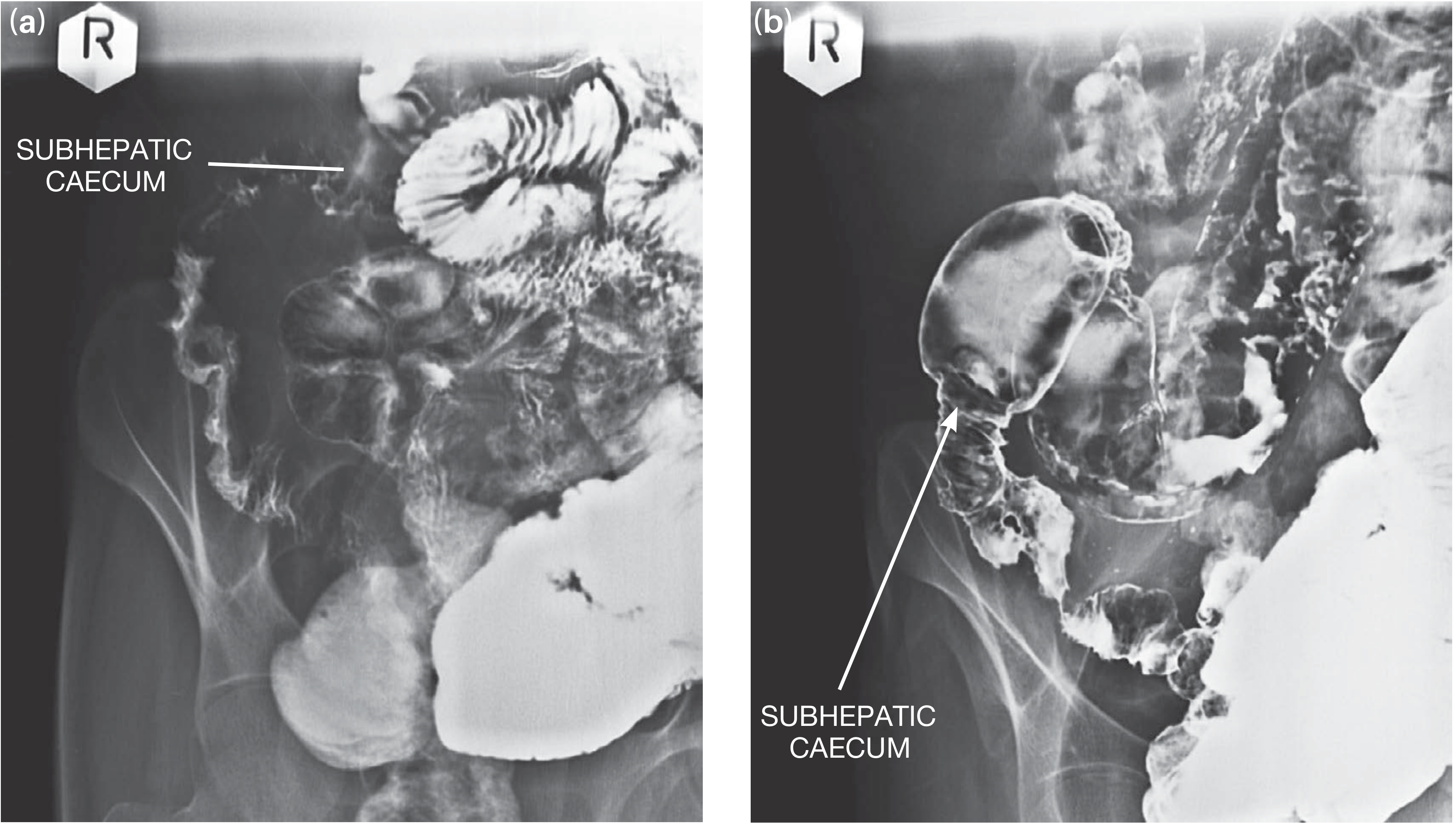
Barium follow-through showing ileal strictures with the caecum in a subhepatic position - Bailey & Love, 28th Ed.
Endoscopy:
- Colonoscopy with biopsy from ileocaecal region - most useful diagnostic procedure
- AFB staining, PCR, and culture on biopsy tissue (with drug sensitivity testing)
- Wear isolation masks when performing
Laparoscopy: Tubercles on bowel serosa, multiple strictures, high caecum, caseous lymph nodes, ascites - useful when biopsy via colonoscopy is inconclusive
Management
A. General Supportive Measures (All Patients)
- Nutrition - correct malnutrition; nasogastric or parenteral nutrition if required
- Hydration/fluid resuscitation - IV fluids for dehydration, especially in obstruction
- NGT decompression - in obstruction/ileus
- Anaemia correction - transfusion if Hb critically low pre-operatively
- Notify public health authorities - TB is a reportable disease; contact tracing
B. Medical Management - Anti-TB Therapy (MAINSTAY)
Abdominal TB is treated identically to pulmonary TB with standard short-course chemotherapy. A high index of suspicion warrants starting empirical therapy even before culture results - clinical response often occurs within 2 weeks. However, masses, hyperplasia, and strictures respond more slowly.
Standard Regimen (Drug-Sensitive TB):
| Phase | Duration | Drugs | Abbreviation |
|---|---|---|---|
| Intensive phase | 2 months | Isoniazid (H) + Rifampicin (R) + Pyrazinamide (Z) + Ethambutol (E) | 2HRZE |
| Continuation phase | 4 months | Isoniazid (H) + Rifampicin (R) | 4HR |
| Total | 6 months |
For extrapulmonary TB including abdominal TB, a total duration of 12-18 months may be preferred in some guidelines/settings due to the depth of tissue penetration and risk of relapse. - Bailey & Love
Important notes:
- Send drug sensitivity testing (DST) from cultures - MDR-TB may be more prevalent in abdominal than pulmonary TB
- Add pyridoxine (Vitamin B6) 10-25 mg/day with isoniazid (prevents peripheral neuropathy)
- In HIV co-infection, ART should be initiated; standard TB regimens are used but with close monitoring for drug interactions (rifampicin and antiretrovirals)
- Adjunctive corticosteroids (prednisolone) may be considered in peritoneal TB with ascites to reduce inflammation and adhesion formation (though evidence is limited)
- Engage a TB specialist for management guidance
C. Surgical Management
Surgery is reserved for complications - it is NOT the primary treatment.
Indications for Surgery:
| Indication | Notes |
|---|---|
| Intestinal obstruction not responding to medical therapy | Most common surgical indication |
| Intestinal perforation | Emergency - peritoneal contamination |
| Intestinal hemorrhage | Uncontrolled bleeding |
| Abdominal abscess | Drainage required |
| Diagnostic uncertainty | Biopsy when endoscopic diagnosis not possible |
| Fistula formation | Entero-enteric or entero-cutaneous |
Key principle: Many cases of intestinal obstruction from TB will respond to anti-TB treatment without surgery. Always give adequate medical therapy before deciding on surgical intervention. - Bailey & Love, Summary Box 65.6
Surgical Options (Elective / Post-Medical Treatment):
After completion of medical treatment, re-image the small bowel. If significant strictures remain causing subacute obstruction:
| Surgical Procedure | Indication |
|---|---|
| Limited ileocolic resection + anastomosis | Terminal ileal/ileocolic hyperplastic disease |
| Strictureplasty | Single ileal stricture (bowel-conserving) |
| Bowel resection | Multiple closely placed strictures |
| Right hemicolectomy | Extensive ileocolic disease precluding limited resection |
Surgical principles are similar to Crohn's disease - resections should be kept as conservative as possible to prevent short bowel syndrome. - Bailey & Love, 28th Ed.
Emergency Surgery (Acute Obstruction/Perforation):
These patients present in extremis - dehydrated, malnourished, anaemic, often with active pulmonary TB.
Pre-operative resuscitation is mandatory:
- IV fluids - vigorous fluid resuscitation
- NGT decompression
- Correct electrolytes
- Blood transfusion if severely anaemic
- Broad-spectrum antibiotics (for perforation/secondary peritonitis)
At laparotomy - minimum life-saving procedure:
- Resection of the diseased segment + proximal ileostomy + distal ileal or colonic mucus fistula (preferred)
- This avoids primary anastomosis, which has a high leak rate in the presence of active infection and poor general condition
- One-stage resection and anastomosis is only performed if the patient's general condition permits (rare in the emergency setting)
Peritoneal TB - Specific Management
- Primarily medical (anti-TB chemotherapy as above)
- Therapeutic paracentesis for tense ascites causing respiratory compromise
- Laparoscopy + peritoneal biopsy if diagnosis in doubt
- Distinguish from peritoneal metastases (CT-guided biopsy or laparoscopy)
- Intestinal obstruction in peritoneal TB (fibrous "dry" type - matted loops) may respond to anti-TB therapy + corticosteroids alone before considering surgery
TB vs Crohn's Disease - Differential Diagnosis Points
| Feature | Intestinal TB | Crohn's Disease |
|---|---|---|
| Ileocaecal valve | Incompetent (both sides involved) | Usually competent |
| Ulcer orientation | Transverse, circumferential | Longitudinal (along bowel axis) |
| Lymph nodes | Large (>1 cm), hypodense (caseous) | Small, non-necrotic |
| Stierlin sign | Present | Absent |
| Granulomas | Caseating | Non-caseating |
| AFB/PCR | Positive | Negative |
Summary Algorithm
Suspected Abdominal Koch's (Pain abdomen)
        ↓
Red flags? (Obstruction / Perforation / Haemorrhage)
        ↓                        ↓
      YES                       NO
        ↓                        ↓
Emergency resuscitation    Confirm diagnosis
  + surgery if required    (Colonoscopy + biopsy, CT, ADA)
                                  ↓
                        START ANTI-TB THERAPY
                        (2HRZE → 4HR, 6-18 months)
                                  ↓
                     Clinical response within 2 weeks?
                        ↓                    ↓
                       YES                  NO
                        ↓                    ↓
                  Continue ATT          Re-evaluate diagnosis
                        ↓             Consider MDR-TB / biopsy
              Post-treatment imaging
                        ↓
              Residual stricture/obstruction?
                        ↓
              ELECTIVE SURGERY
              (Strictureplasty / Limited resection)
Sources: Sleisenger & Fordtran's Gastrointestinal and Liver Disease; Bailey and Love's Short Practice of Surgery, 28th Ed.; Goldman-Cecil Medicine
Recent evidence note: A 2025 systematic review on abdominal TB in children (PMID 40533676) and a 2025 diagnostic evaluation and management review (PMID 40023548) confirm that standard 6-month HRZE/HR regimens remain the backbone of treatment, with surgical intervention reserved for complications unresponsive to medical therapy.
U/l limb swelling differentials
Upper limb swelling differentials span vascular, lymphatic, inflammatory, and neoplastic causes - DVT, lymphedema, cellulitis, and axillary pathology being the most common. Let me pull the full textbook content.
Searching sources...
Reading File
Finding Sources
Finding Sources
Finding Sources
Finding Sources
Finding Sources
Reading File
Finding Sources
Finding Sources
Excellent - the "Symptom to Diagnosis" book has a dedicated differential diagnosis ranking for upper extremity swelling. Let me read that directly.
Reading File
Finding Sources
Reading File
Reading File
Reading File
Finding Sources
Reading File
Reading File
I now have comprehensive, high-quality data from multiple authoritative sources. Here is the complete structured answer:
Upper Limb (U/L) Swelling - Differential Diagnoses
A systematic approach organized by mechanism, from common to rare and must-not-miss.
Framework for Approaching Upper Limb Swelling
Ask these key questions first:
| Question | Narrows Towards |
|---|---|
| Unilateral or bilateral? | Unilateral = local/structural; Bilateral = systemic |
| Pitting or non-pitting? | Pitting = venous/cardiac/hypoalbuminaemia; Non-pitting = lymphedema/myxoedema |
| Acute or chronic onset? | Acute = DVT, cellulitis, trauma; Chronic = lymphedema, tumour |
| Hot, red, tender? | Infective/inflammatory |
| Associated neck/facial swelling? | SVC syndrome |
| Recent surgery/malignancy? | Lymphedema, DVT, tumour recurrence |
| Trauma/strenuous exercise? | Paget-Schroetter, haematoma, compartment syndrome |
Differentials - Organized by System
1. VASCULAR
Upper Extremity Deep Vein Thrombosis (UE-DVT) - MUST NOT MISS
- Accounts for <5% of all DVTs but carries 10-15% risk of PE
- Primary (Paget-Schroetter disease / Effort thrombosis): Axillary/subclavian vein thrombosis from repetitive strenuous arm activity in young, healthy, muscular individuals (weight-lifters, overhead athletes). Associated with thoracic outlet compression by cervical rib or first rib
- Secondary: Indwelling venous catheter (most common cause), mediastinal tumours, malignancy, hypercoagulable states, congestive heart failure, nephrotic syndrome, tamoxifen use
Features: Unilateral arm pain, oedema, heaviness/fullness, cyanosis; visible superficial venous distension over arm, forearm, shoulder, anterior chest wall
Investigation: Duplex ultrasound (first-line); CT venography/MRA/phlebography; positional phlebography with arm abduction to 120° (to confirm thoracic outlet compression); CXR (cervical rib)
- Mulholland & Greenfield's Surgery, 7th Ed.; Symptom to Diagnosis, 4th Ed.
Superior Vena Cava (SVC) Syndrome - MUST NOT MISS
- Obstruction of the SVC causes bilateral upper limb + face + neck oedema
- Causes: >90% malignant - bronchogenic carcinoma (right lung most common), lymphoma, mediastinal metastases; Non-malignant: sclerosing mediastinitis, goitre, indwelling catheter/pacemaker thrombosis, syphilis, mediastinal haematoma
- Features: Bilateral arm + face + neck swelling (worse in morning), JVD, dilated chest wall collaterals (caput medusae of chest), dyspnoea, headache, visual disturbance, Pemberton's sign (facial congestion on arm raising), hoarseness, dysphagia
- A neurological/oncological emergency if cerebral oedema develops
- Pye's Surgical Handicraft, 22nd Ed.
Subclavian Artery Aneurysm / Arteriovenous Fistula
- Pulsatile swelling if aneurysm; thrill/bruit with AV fistula
- May follow trauma, thoracic outlet compression, or iatrogenic injury
- Post-traumatic AV fistulae cause distal venous hypertension and limb swelling
Venous Insufficiency / Post-Thrombotic Syndrome
- Chronic oedema after previous axillary/subclavian DVT
- Venous hypertension leads to persistent limb swelling and skin changes
2. LYMPHATIC
Secondary Lymphedema (Most Common Overall Cause)
- Breast cancer-related lymphedema (BCRL): Most common cause of upper extremity lymphedema
- Follows axillary lymph node dissection + mastectomy + radiotherapy
- Risk factors: high number of nodes dissected, positive nodes, higher BMI, inactive lifestyle, radiation therapy
- Presents as chronic non-pitting oedema, typically starting in the upper arm/humeral region, spreading to shoulder and forearm
- Cellulitis of the ipsilateral arm is a frequent complication (non-group A haemolytic Streptococcus)
- Other causes of secondary lymphedema: other malignancies with axillary/mediastinal nodal involvement, filariasis (Wuchereria bancrofti), post-infective (recurrent cellulitis), post-radiation, trauma
Primary Lymphedema
- Extremely rare in the upper limb
- Congenital (Milroy's-like), lymphedema praecox, lymphedema tarda
- Due to aplasia/hypoplasia/dysfunction of lymphatics
Stewart-Treves Syndrome: Angiosarcoma (bluish/reddish nodules) arising in chronic post-mastectomy lymphedema - rare but important complication
- Sabiston Textbook of Surgery; Andrews' Diseases of the Skin
3. INFECTIVE / INFLAMMATORY
Cellulitis
- Infection of deep dermis and subcutaneous tissue
- Presents as: diffuse non-palpable erythema with indistinct margins, generalised swelling, warmth, tenderness; lymphangitis (red streaks); tender regional adenopathy
- Organisms: S. aureus and Group A Streptococcus most common; MRSA in injection drug users, recurrent cases, abscess formation
- Risk factors: Underlying lymphedema or venous insufficiency, prior breast cancer treatment, eczema, tinea pedis (entry site identified in 77%), trauma, obesity, injection drug use
- Systemic features (high fever, rigors) suggest bacteraemia or necrotizing fasciitis
Erysipelas
- Superficial cellulitis with prominent lymphatic involvement
- Sharply demarcated raised erythematous border (distinguishes from cellulitis)
- Sudden onset fever (85%), rapidly spreading (2-10 cm/day)
- Risk factors: lymphedema, tinea pedis (portal of entry)
Necrotizing Fasciitis - MUST NOT MISS
- Deep infection tracking along fascial planes; disproportionate pain, woody oedema, systemic sepsis, skin necrosis/bullae/crepitus
- Surgical emergency
Septic Arthritis / Osteomyelitis
- Localised joint or bone pain, swelling, restricted movement, fever
Lymphadenitis (Axillary)
- Localised axillary swelling/tenderness; may cause mild arm swelling due to lymphatic obstruction
- Causes: bacterial (cat-scratch disease - Bartonella henselae, staphylococcal, streptococcal), viral (EBV, HIV)
Abscess
- Fluctuant, tender, localised; may track to produce diffuse swelling
4. MUSCULOSKELETAL / TRAUMATIC
Fracture
- History of trauma; deformity, crepitus, point tenderness; confirmed on X-ray
Haematoma
- Trauma, anticoagulation, or spontaneous (haemophilia); tense, bruised swelling
Compartment Syndrome - MUST NOT MISS
- 6 Ps: Pain (disproportionate, especially on passive stretch), Pressure (tight compartment), Paraesthesia, Paralysis (late), Pallor, Pulselessness (late)
- Causes: fracture, crush injury, reperfusion, tight cast/bandage
- Surgical emergency (fasciotomy)
Muscle Tear / Haematoma
- Acute onset with exertion or trauma; ecchymosis, fusiform swelling
Tendon Rupture (Biceps Tendon)
- "Popeye" sign (retracted biceps muscle belly), sudden pain/swelling in antecubital fossa
Osteosarcoma / Bone Tumour
- Gradually enlarging, hard, fixed swelling over long bone; night pain, systemic symptoms
5. NEOPLASTIC / COMPRESSIVE
Primary Tumours
- Soft tissue sarcoma: slow-growing, deep, firm mass
- Lipoma: soft, lobulated, non-tender (most common benign soft tissue tumour)
- Ganglion cyst: tense, transilluminant, arising near joints/tendons (wrist most common)
- Haemangioma/Vascular malformations
Secondary / Metastatic Disease
- Direct infiltration or nodal involvement from breast, lung, melanoma
Axillary Lymphadenopathy
- Causes arm swelling by proximal lymphatic/venous obstruction
- Causes: metastatic carcinoma (breast most common), lymphoma, leukemia, melanoma, sarcoidosis, infective
Thoracic Outlet Syndrome (TOS)
- Compression of neurovascular structures at the thoracic outlet (between clavicle/first rib) by cervical rib, fibrous band, or hypertrophied scalene muscles
- Neurogenic (most common): paraesthesia, weakness
- Venous (Paget-Schroetter): arm swelling, cyanosis (see DVT above)
- Arterial (rarest): pallor, cold hand, digital ischaemia
6. SYSTEMIC / GENERALISED CAUSES (Bilateral > Unilateral)
| Cause | Key Features |
|---|---|
| Congestive cardiac failure | Bilateral pitting oedema, JVP raised, dyspnoea |
| Hypoalbuminaemia (nephrotic, cirrhosis, malnutrition) | Bilateral pitting, low albumin |
| Hypothyroidism (myxoedema) | Non-pitting, periorbital puffiness, bradycardia, cold intolerance |
| Renal failure | Bilateral pitting oedema, uraemia |
| Anaphylaxis / Angioedema | Rapid onset, urticaria, laryngeal oedema, hypotension |
| Lipoedema | Bilateral fat deposition (spares hands/feet), not true oedema |
7. DRUG-INDUCED
| Drug | Mechanism |
|---|---|
| Calcium channel blockers (amlodipine) | Arteriolar dilatation → dependent oedema |
| NSAIDs | Sodium/water retention |
| Corticosteroids | Fluid retention |
| Tamoxifen | DVT risk (relative risk ~3x) |
| IV contrast / drug reactions | Local/allergic |
| Chemotherapy agents | Venous fibrosis / lymphatic damage |
Summary Table - Key Differentials
| Diagnosis | Onset | Laterality | Pitting | Key Feature |
|---|---|---|---|---|
| UE-DVT | Acute | Unilateral | Yes | Risk factors, collateral veins |
| Cellulitis | Acute | Unilateral | Yes | Hot, red, tender, +/- fever |
| Compartment syndrome | Acute | Unilateral | No | Disproportionate pain, tight |
| Lymphedema (post-mastectomy) | Subacute/chronic | Unilateral | No (late) | H/o breast cancer treatment |
| SVC syndrome | Subacute | Bilateral (+ face/neck) | Yes | Collaterals, dyspnoea |
| Paget-Schroetter (DVT) | Acute | Unilateral | Yes | Young athlete, exertion |
| Soft tissue tumour | Chronic | Unilateral | No | Firm, painless mass |
| Angioedema | Minutes | Bilateral or focal | No | Allergic trigger, urticaria |
| Fracture/haematoma | Acute post-trauma | Unilateral | Variable | History of trauma |
Always think about DVT in a patient with unilateral limb swelling. - Symptom to Diagnosis: An Evidence-Based Guide, 4th Ed.
Sources: Bailey & Love's Short Practice of Surgery 28th Ed.; Mulholland & Greenfield's Surgery 7th Ed.; Symptom to Diagnosis 4th Ed.; Sabiston Textbook of Surgery; Andrews' Diseases of the Skin; Pye's Surgical Handicraft 22nd Ed.
u/l unilateral swelling differentials
Based on your previous question about upper limb swelling, I believe "u/l unilateral swelling" here refers to lower limb unilateral swelling (since u/l can also mean unilateral). Let me clarify before diving in.
Asking for Preferences
Which limb are you asking about?
The previous response already covered upper limb swelling comprehensively - this specifically focuses on the unilateral aspect, which means we can exclude all bilateral/systemic causes and narrow the differentials significantly. Let me pull fresh focused content.
Searching sources...
Finding Sources
Finding Sources
Finding Sources
Finding Sources
Reading File
Reading File
Finding Sources
Finding Sources
Finding Sources
Finding Sources
Finding Sources
Finding Sources
I have all the data needed from authoritative sources plus the comprehensive foundation from the previous response. Here is the focused, unilateral-specific answer:
Unilateral Upper Limb Swelling - Differentials
Unilateral swelling immediately points away from systemic causes (cardiac failure, hypoalbuminaemia, bilateral lymphedema) and towards local structural, vascular, infective, inflammatory, or neoplastic pathology.
Surgical Sieve - VITAMINC Framework
| Category | Condition |
|---|---|
| Vascular | UE-DVT, Superficial thrombophlebitis, AV fistula, Venous injury |
| Infective | Cellulitis, Erysipelas, Necrotizing fasciitis, Abscess, Lymphangitis |
| Traumatic | Fracture, Haematoma, Compartment syndrome, Muscle tear, Dislocation |
| Autoimmune / Inflammatory | Gout, Pseudogout, Septic arthritis, CRPS, Rheumatoid flare, DVT-related |
| Metabolic | Hypothyroidism (myxoedema - rare, usually bilateral) |
| Iatrogenic / Drug | IV extravasation, PICC line thrombosis, Drug reaction |
| Neoplastic | Soft tissue sarcoma, Lipoma, Ganglion, Axillary nodal mass, Metastasis |
| Congenital / Lymphatic | Primary lymphedema, Thoracic outlet syndrome (venous) |
Differentials in Order of Likelihood
MUST NOT MISS (Urgent/Emergency)
1. Upper Extremity DVT (UE-DVT)
The single most important diagnosis to exclude in any unilateral arm swelling.
- Accounts for ~5% of all DVTs; carries up to 33% risk of PE
- Primary - Paget-Schroetter (Effort) Thrombosis:
- Young, healthy, dominant arm of athletes (swimmers, weight-lifters, overhead workers)
- Repetitive arm motion + anatomical compression at thoracic outlet (cervical rib, myofascial bands, hypertrophied scalenes) → subclavian vein DVT
- Classic: sudden onset arm swelling after strenuous exertion
- Secondary (>90% of cases):
- Indwelling central venous catheter / PICC line / pacemaker lead (most common cause)
- Malignancy (occult or known) - idiopathic UE-DVT warrants cancer workup
- Hypercoagulable state (thrombophilia, OCP, malignancy)
- Mediastinal mass compressing subclavian vein
Clinical features: Unilateral swelling, pain, heaviness/fullness, erythema, palpable venous cord; dilated superficial collateral veins over arm, shoulder, anterior chest wall; cyanosis
Investigation: Duplex USS (first-line; may be limited at mid-subclavian due to clavicle) → Venography / MR venography if USS inconclusive; CXR (cervical rib); positional phlebography (arm at 120° abduction) to confirm thoracic outlet compression
Treatment: Anticoagulation (DOAC or LMWH/warfarin) × 3 months; Paget-Schroetter: thrombolysis + thoracic outlet decompression (first rib resection ± scalenectomy) + possible venous reconstruction
- Sabiston Textbook of Surgery; Rosen's Emergency Medicine; Mulholland & Greenfield's Surgery
2. Compartment Syndrome
- Emergency requiring fasciotomy within hours to prevent permanent muscle/nerve damage
- Causes: fracture (especially supracondylar in children, radius/ulna), crush injury, tight plaster/bandage, reperfusion after ischaemia, burns, snake bite
- 6 Ps: Pain (disproportionate, worst on passive stretch of fingers - earliest sign), Pressure (tight, tense compartment), Paraesthesia (tingling/numbness), Paralysis (late), Pallor, Pulselessness (very late - do not wait for this)
- Compartment pressure >30 mmHg (or within 30 mmHg of diastolic BP) = fasciotomy threshold
3. Necrotizing Fasciitis
- Deep-tissue infection tracking rapidly along fascial planes
- Clues: pain disproportionate to appearance, woody indurated oedema, skin crepitus (gas-forming organisms), haemorrhagic bullae, rapid systemic sepsis
- LRINEC score aids diagnosis (CRP, WBC, Na, creatinine, glucose, Hb)
- Emergency surgical debridement
COMMON
4. Cellulitis
- Infection of deep dermis and subcutaneous tissue
- Features: Diffuse non-palpable erythema with indistinct margins, generalised swelling, warmth, tenderness; lymphangitis (red streaking proximally); tender axillary adenopathy
- Organisms: S. aureus, Group A Streptococcus; MRSA in injection drug users, abscess-associated
- Portal of entry identified in 77% - skin break, wound, eczema, tinea, bite
- Risk factors: Underlying lymphedema (post-mastectomy patients especially), venous insufficiency, obesity, eczema, injection drug use, IV catheter sites
- Fever/rigors → bacteraemia or necrotizing fasciitis; do not miss
5. Erysipelas
- Superficial cellulitis with prominent lymphatic involvement
- Sharply demarcated raised, bright-red, indurated border (distinguishes from cellulitis)
- Abrupt onset, high fever (85%), rapid spread (2-10 cm/day)
- Organism: Group A Streptococcus (almost exclusively)
- Risk factors: same as cellulitis; lymphedema + tinea pedis are strongest risk factors
6. Post-Mastectomy / Secondary Lymphedema
- Most common cause of chronic unilateral upper limb swelling in women
- Follows axillary lymph node dissection + mastectomy + radiotherapy for breast cancer
- Onset: weeks to years after treatment
- Features: Gradually progressive non-pitting oedema, starts at upper arm (humeral region), spreads to shoulder and forearm; skin thickening and fibrosis (late); recurrent cellulitis episodes
- Risk factors: extensive axillary dissection, positive nodes, radiotherapy, high BMI, inactivity
- Stewart-Treves syndrome: Angiosarcoma (bluish/reddish nodules) arising in chronic lymphedema - rare but fatal complication
- Other causes of secondary lymphedema: filariasis, any axillary/mediastinal malignancy, recurrent cellulitis, trauma
7. Fracture / Dislocation
- History of trauma; focal point tenderness, deformity, crepitus, abnormal ROM
- Any bone in the upper limb; distal radius most common in adults (Colles')
- Pathological fracture: fracture through bone metastasis/cyst - may be spontaneous or minimal trauma
8. Haematoma
- Soft tissue bleeding following trauma, anticoagulation, or spontaneous (haemophilia, thrombocytopenia)
- Tense, bruised swelling; ecchymosis may track distally; fluctuant if large
- Can mimic DVT clinically; USS distinguishes
LESS COMMON BUT IMPORTANT
9. Superficial Thrombophlebitis (SVT)
- Thrombosis of a superficial vein
- Features: Linear, erythematous, tender, swollen cord along vein; localised rather than diffuse swelling
- Causes: IV cannula/catheter (commonest), varicose veins, trauma, thrombophilia, malignancy
- Trousseau's sign - migratory thrombophlebitis associated with occult malignancy (especially pancreatic tail)
- 3-15% have concurrent DVT - duplex USS if in doubt
10. Septic Arthritis
- Acute, hot, swollen, painful joint - severely restricted range of motion
- Patient toxic/febrile; child with reluctant limb use
- Organisms: S. aureus (most common), Gonococcus in young adults, Gram-negatives in immunocompromised
- Joint aspiration is diagnostic and therapeutic
- Osteomyelitis can co-exist or present similarly
11. Gout / Pseudogout
- Crystal arthropathy causing acute monoarthritis
- Wrist, MCP joints (gout); wrist, elbow (pseudogout/CPPD)
- Acutely red, hot, exquisitely tender joint with periarticular swelling
- Urate crystals (negatively birefringent) vs. calcium pyrophosphate (weakly positively birefringent) on joint aspirate
- Elevated serum urate (but may be normal during acute attack)
12. Complex Regional Pain Syndrome (CRPS) - Reflex Sympathetic Dystrophy
- Follows injury, surgery, or stroke; often disproportionate to initial injury
- Features: Burning pain, allodynia, diffuse oedema, skin colour/temperature changes (warm/red early → cool/cyanotic late), trophic changes (shiny skin, hair/nail changes), hyperhidrosis
- Sudeck's atrophy: osteoporosis on X-ray
- CRPS Type 1 (no nerve injury) vs. Type 2 (with nerve injury = causalgia)
13. Soft Tissue Tumours
- Lipoma: Soft, lobulated, compressible, non-tender; slowly enlarging; most common benign soft tissue tumour
- Ganglion cyst: Tense, transilluminant; arising near joint capsule/tendon sheath; wrist most common
- Soft tissue sarcoma: Deep, firm, >5 cm, painless, rapidly growing - refer urgently (2-week wait rule)
- Haemangioma/Vascular malformation: Soft, compressible, bluish; may enlarge on dependency
14. Axillary Lymphadenopathy
- Enlarged axillary nodes causing proximal lymphatic/venous obstruction → arm swelling
- Causes: breast cancer metastasis, lymphoma/leukaemia, melanoma, sarcoidosis, reactive (infection)
- Associated: palpable axillary mass, non-tender (malignant) vs. tender (infective)
15. Thoracic Outlet Syndrome (Venous Form)
- Compression at the thoracic outlet by cervical rib, first rib, or scalene muscles
- Venous TOS = subclavian vein compression → UE-DVT (Paget-Schroetter) or intermittent obstruction
- Arterial TOS: pallor, cold digits, emboli, digital ischaemia
- Neurogenic TOS (most common form): paraesthesia in ulnar distribution, weakness - not usually swelling
16. IV Extravasation / Infiltration
- Leakage of IV fluid or chemotherapy agents into surrounding tissue
- Localised swelling, pain, blanching at cannula site; blistering/necrosis with vesicant agents
17. Insect Bite / Envenomation / Anaphylaxis
- Rapid onset localised swelling at bite site ± urticaria; anaphylaxis causes rapid diffuse swelling
- Snake bite: fang marks, progressive oedema, coagulopathy
- Angioedema (bradykinin-mediated - ACE inhibitor, hereditary): rapid onset non-pitting, non-prurigenic swelling
Clinical Decision Framework
UNILATERAL ARM SWELLING
         |
    ┌────┴─────┐
  ACUTE      CHRONIC
    |              |
  ┌─┴──┐      ┌──┴───┐
Hot/red? Pain/trauma?  Non-pitting?  Post-mastectomy?
  |         |          |                  |
Cellulitis  Fracture  Lymphedema    BCRL/lymphedema
Erysipelas  Haematoma  CRPS          Stewart-Treves
NF         Compartment
           syndrome
    |
History of catheter/
indwelling device/
malignancy/exertion?
    |
   DVT
Key Distinguishing Features at a Glance
| Diagnosis | Onset | Skin Changes | Pain | Fever | Key Clue |
|---|---|---|---|---|---|
| UE-DVT | Acute | Erythema/cyanosis | Moderate | No | Catheter / athlete / collaterals |
| Cellulitis | Acute | Hot, red, indistinct border | Yes | Often | Entry site, lymphangitis |
| Erysipelas | Acute | Raised, sharp border | Yes | High | Strep, face/limb |
| Necrotizing fasciitis | Acute | Bullae, crepitus, necrosis | Extreme | High, septic | Pain >> appearance |
| Compartment syndrome | Acute | Tense, tight | Extreme (passive stretch) | No | Post-fracture/trauma |
| Lymphedema | Chronic | Non-pitting, skin thickening | No | No | Post-mastectomy, non-pitting |
| Haematoma | Acute post-trauma | Bruising, ecchymosis | Yes | No | Trauma/anticoagulation |
| Gout | Acute | Red, periarticular | Extreme | Sometimes | Joint-centred, urate |
| CRPS | Subacute | Shiny, colour change | Burning | No | Allodynia, post-injury |
| Soft tissue tumour | Chronic | Normal | No | No | Mass, progressive |
| SVT | Acute | Linear erythema | Localised | Low | Palpable cord along vein |
Sources: Sabiston Textbook of Surgery; Rosen's Emergency Medicine; Mulholland & Greenfield's Surgery 7th Ed.; Symptom to Diagnosis 4th Ed.; Bailey & Love's Short Practice of Surgery 28th Ed.
Upper limb unilateral swelling differentials
This was already covered in full detail in the previous response. Here is a clean, complete summary:
Upper Limb Unilateral Swelling - Differentials
Key Discriminating Questions
| Question | Points Towards |
|---|---|
| Acute vs chronic onset? | Acute = DVT, cellulitis, trauma; Chronic = lymphedema, tumour |
| Hot + red + tender? | Infective/inflammatory |
| After trauma/exertion? | Fracture, haematoma, Paget-Schroetter DVT |
| Indwelling catheter/PICC/pacemaker? | Secondary UE-DVT |
| Previous breast cancer treatment? | Post-mastectomy lymphedema |
| Pain disproportionate to appearance? | Compartment syndrome, necrotizing fasciitis |
| Palpable cord along vein? | Superficial thrombophlebitis |
| Joint-centred swelling? | Septic arthritis, gout/pseudogout |
| Firm/painless/progressive mass? | Soft tissue tumour |
| Visible collateral veins on chest/shoulder? | UE-DVT, SVC obstruction |
Differentials by Category
1. Vascular
| Condition | Key Features |
|---|---|
| Upper Extremity DVT (MUST NOT MISS) | Arm pain, swelling, heaviness; dilated collaterals on chest/shoulder; risk: catheter, athlete (Paget-Schroetter), malignancy, thrombophilia; PE risk up to 33% |
| Paget-Schroetter syndrome | Young athlete, dominant arm, strenuous repetitive exertion; subclavian/axillary vein thrombosis from thoracic outlet compression (cervical rib, scalenes) |
| Superficial thrombophlebitis | Linear tender erythematous cord along superficial vein; IV cannula site, varicose veins, thrombophilia; Trousseau's migratory = occult malignancy |
| AV fistula | Pulsatile/throbbing swelling, thrill, bruit; post-traumatic or iatrogenic |
| Venous injury / post-traumatic | History of trauma or surgery; venous hypertension in drained territory |
2. Infective / Inflammatory
| Condition | Key Features |
|---|---|
| Cellulitis | Acute diffuse erythema with indistinct borders, warmth, tenderness; entry site (wound, eczema, tinea) in 77%; organisms: S. aureus, GAS; risk: lymphedema, venous insufficiency |
| Erysipelas | Superficial, sharply demarcated raised red plaque; high fever; GAS; spreads 2-10 cm/day |
| Necrotizing fasciitis (MUST NOT MISS) | Pain >> appearance; woody oedema; bullae, crepitus, skin necrosis; rapid sepsis; surgical emergency |
| Abscess | Fluctuant, tender, localised; MRSA in injection drug users |
| Septic arthritis | Acutely red, hot, tender joint; severely restricted ROM; fever; joint aspiration diagnostic |
| Osteomyelitis | Deep bone pain, local tenderness, fever; MRI is gold standard |
| Lymphangitis | Red streaking proximally from portal of entry; regional adenopathy |
3. Traumatic / Orthopaedic
| Condition | Key Features |
|---|---|
| Compartment syndrome (MUST NOT MISS) | 6 Ps: Pain (passive stretch - earliest), Pressure, Paraesthesia, Paralysis, Pallor, Pulselessness (late); post-fracture/crush; pressure >30 mmHg = fasciotomy |
| Fracture | Trauma history; point tenderness, deformity, crepitus, abnormal movement; pathological = through metastasis/cyst |
| Haematoma | Trauma/anticoagulation; tense, bruised, ecchymosis tracking distally |
| Muscle tear (biceps/triceps) | Acute exertion; "Popeye" sign (biceps); fusiform swelling, ecchymosis |
| Dislocation | Deformity + restricted ROM; neurovascular compromise |
| Ligament/tendon injury | Focal periarticular swelling, effusion, functional loss |
4. Lymphatic
| Condition | Key Features |
|---|---|
| Secondary lymphedema - BCRL | Most common cause of chronic unilateral UL swelling; follows axillary node dissection + mastectomy + radiotherapy; non-pitting, starts at upper arm, spreads distally; recurrent cellulitis |
| Secondary lymphedema - other | Filariasis (Wuchereria bancrofti), other malignancy with axillary/mediastinal nodal involvement, post-radiotherapy, recurrent infections |
| Primary lymphedema | Very rare in upper limb; congenital or early-onset aplasia/hypoplasia of lymphatics |
| Stewart-Treves syndrome | Angiosarcoma (blue-red nodules) arising in chronic post-mastectomy lymphedema - rare but aggressive |
5. Neoplastic
| Condition | Key Features |
|---|---|
| Lipoma | Soft, lobulated, compressible, non-tender, freely mobile; most common benign soft tissue tumour |
| Ganglion cyst | Tense, transilluminant; arises from joint capsule/tendon sheath; wrist most common |
| Soft tissue sarcoma | Deep, firm, >5 cm, painless, rapidly growing - urgent referral (2-week rule) |
| Haemangioma / Vascular malformation | Soft, compressible, bluish; enlarges on dependency |
| Axillary lymphadenopathy | Palpable nodal mass causing lymphatic/venous obstruction; breast cancer metastasis, lymphoma, melanoma |
| Bone tumour / metastasis | Pain at rest/night, bony swelling, pathological fracture |
6. Neurological / Autonomic
| Condition | Key Features |
|---|---|
| CRPS (Complex Regional Pain Syndrome) | Post-injury or post-surgical; burning pain, allodynia, diffuse non-pitting oedema; skin colour/temperature changes (warm/red early → cold/dusky late); trophic changes; Sudeck's atrophy on X-ray |
7. Iatrogenic / Drug
| Condition | Key Features |
|---|---|
| IV extravasation | Localised swelling at cannula site; blistering/necrosis with vesicant chemotherapy agents |
| PICC/central line thrombosis | Catheter-associated UE-DVT (>90% of secondary UE-DVT) |
| Drug reaction / angioedema | ACE inhibitor-induced or hereditary; rapid-onset non-pitting, non-pruritic swelling; bradykinin-mediated |
| Calcium channel blocker oedema | Usually bilateral but can be asymmetric |
8. Inflammatory / Rheumatological
| Condition | Key Features |
|---|---|
| Gout | Acute monoarthritis; wrist, MCPs; exquisitely tender; negatively birefringent urate crystals |
| Pseudogout (CPPD) | Wrist, elbow; weakly positively birefringent calcium pyrophosphate crystals |
| Rheumatoid arthritis flare | Symmetric MCP/PIP joint swelling usually; can be asymmetric in early disease |
| Reactive arthritis | Follows infection; asymmetric oligoarthritis; urethritis + conjunctivitis (Reiter's triad) |
| Insect bite / envenomation | Rapid localised oedema; fang marks (snake bite); urticaria (allergic) |
Priority Summary - By Urgency
| Priority | Diagnosis | Why Urgent |
|---|---|---|
| 🔴 Emergency | Compartment syndrome | Permanent ischaemic contracture within hours |
| 🔴 Emergency | Necrotizing fasciitis | Fatal if surgical debridement delayed |
| 🔴 Emergency | Septic arthritis | Joint destruction within 24-48 hours |
| 🟠 Urgent | UE-DVT | PE risk up to 33% |
| 🟠 Urgent | Acute limb ischaemia (arterial) | Limb loss within 6 hours |
| 🟡 Same-day | Cellulitis with systemic features | Bacteraemia, sepsis |
| 🟡 Same-day | Fracture with neurovascular compromise | Nerve/vessel injury |
| 🟢 Routine | Lymphedema, lipoma, ganglion, CRPS | Non-immediately life/limb threatening |
Investigation Pathway
Unilateral Upper Limb Swelling
           |
    Trauma history?
    YES → X-ray ± CT
    NO  ↓
    Hot/red/fever?
    YES → Cellulitis vs. NF vs. septic arthritis
          (bloods, cultures, LRINEC score, USS/MRI)
    NO  ↓
    Risk factors for DVT / catheter in situ?
    YES → Duplex USS → Venography/MRV if inconclusive
    NO  ↓
    Chronic, non-pitting, post-mastectomy?
    YES → Lymphedema (clinical diagnosis ± lymphoscintigraphy)
    NO  ↓
    Palpable mass?
    YES → USS ± MRI ± biopsy (sarcoma protocol if >5cm/deep)
    NO  ↓
    Post-injury, burning pain, allodynia?
    YES → CRPS (clinical diagnosis, 3-phase bone scan)
Sources: Sabiston Textbook of Surgery; Rosen's Emergency Medicine; Mulholland & Greenfield's Surgery, 7th Ed.; Symptom to Diagnosis, 4th Ed.; Bailey & Love's Short Practice of Surgery, 28th Ed.
Treatment for pain abdomen in k:co of abdominal koch
A patient with known case (k/c) of abdominal tuberculosis presenting with pain abdomen - the treatment depends on the cause of the pain (disease activity, obstruction, perforation) and whether it's acute or chronic. Let me pull the detailed textbook content.
Searching sources...
Finding Sources
Finding Sources
Searching PubMed
"abdominal tuberculosis" AND "pain" AND management
Finding Sources
Finding Sources
Reading File
Reading File
Reading File
Finding Sources
Reading File
Finding Sources
Finding Sources
Reading File
Finding Sources
Finding Sources
Reading File
I now have all the data needed. Here is the focused, clinically structured answer:
Treatment of Pain Abdomen in a K/C of Abdominal Koch's (Abdominal TB)
A patient with a known case of abdominal TB presenting with pain abdomen requires a stepwise assessment - is this pain from active disease, a complication, or a superimposed problem?
Step 1 - Categorize the Pain
| Category | Clinical Scenario | Urgency |
|---|---|---|
| Active disease pain | Dull, colicky, chronic - disease not yet controlled or early treatment | Routine |
| Subacute obstruction | Intermittent colicky pain, distension, on-treatment | Urgent |
| Acute obstruction | Severe colicky pain, distension, vomiting, absolute constipation | Emergency |
| Perforation / Peritonitis | Sudden severe pain, guarding, rigidity, board-like abdomen | Emergency |
| Abscess | Localised pain, fever, tender mass | Urgent |
| Ascites pain | Dull ache, distension, shifting dullness | Routine |
Perforation can occur even during anti-TB treatment - always exclude in a k/c of abdominal Koch's with acute pain. - Sleisenger & Fordtran's GI and Liver Disease
Step 2 - Immediate Assessment
A. History
- Is the patient currently on ATT? For how long? Adherent?
- Character of pain: colicky (obstruction) vs. constant (peritonitis/abscess) vs. dull chronic (active disease)
- Vomiting - bilious/faeculent (obstruction)?
- Last bowel opening / flatus (absolute constipation = obstruction)
- Fever, rigors (superimposed infection, abscess, perforation)
- Weight trend (improving/worsening on ATT)
B. Examination
- Vitals: Tachycardia, hypotension, fever - sepsis, perforation
- Abdomen:
- Distension + tinkling/absent bowel sounds → obstruction
- Guarding + rigidity + rebound → peritonitis/perforation
- Doughy feel + localised ascites → active peritoneal TB
- RIF mass → ileocaecal disease, abscess
- Shifting dullness → ascites
C. Investigations
- Bloods: CBC, CRP/ESR, LFTs (ATT hepatotoxicity), urea/electrolytes (dehydration)
- Plain AXR: Dilated small bowel loops with fluid levels (obstruction); free gas under diaphragm (perforation)
- USS abdomen: Ascites (loculated vs free), abscess, lymph nodes, bowel wall thickening
- CT abdomen (gold standard): Obstruction site and level, perforation, abscess, lymphadenopathy with central necrosis, disease extent
- LFTs - check before continuing ATT if hepatotoxicity suspected
Step 3 - Treatment Based on Scenario
A. Chronic/Active Disease Pain (No Complication)
Pain is from the disease itself - inflammation, ulceration, partial obstruction, mesenteric lymphadenopathy, peritonitis.
1. Continue/Optimise Anti-TB Therapy (ATT) - THE CORNERSTONE
Standard first-line regimen:
| Phase | Duration | Drugs | Short form |
|---|---|---|---|
| Intensive phase | 2 months | Isoniazid (H) + Rifampicin (R) + Pyrazinamide (Z) + Ethambutol (E) | 2HRZE |
| Continuation phase | 4-10 months | Isoniazid (H) + Rifampicin (R) | 4-10HR |
Total duration 6-12 months standard; up to 12-18 months preferred for abdominal TB in many guidelines. Patients should be under combined physician and surgeon care. - Bailey & Love, 28th Ed.
Add always:
- Pyridoxine (Vitamin B6) 10-25 mg/day with isoniazid (prevents peripheral neuropathy)
Monitor for ATT side effects as a cause of pain:
- Hepatotoxicity (isoniazid, rifampicin, pyrazinamide) → raised ALT/AST, jaundice, RUQ pain
- If ALT >3x normal with symptoms or >5x normal without symptoms → stop all ATT, reintroduce sequentially once LFTs normalise
- Gastritis/GI upset (most common) → take drugs with food, use PPIs/antacids
2. Symptomatic Pain Relief
| Drug | Dose | Indication |
|---|---|---|
| Antispasmodics (Hyoscine butylbromide/Buscopan) | 10-20 mg TDS/QDS PO or 20 mg IM/IV | Colicky pain from intestinal spasm/strictures |
| Paracetamol | 500 mg - 1 g TDS-QDS | Mild-moderate pain; safe with ATT |
| NSAIDs (Ibuprofen, Diclofenac) | With caution | Moderate pain; avoid if GI ulceration, poor nutrition, renal impairment |
| Proton pump inhibitor (Pantoprazole/Omeprazole) | 40 mg OD | GI protection, especially if on NSAIDs or with dyspepsia from ATT |
| Opioids (Tramadol, Morphine) | As required | Severe pain; short-term; avoid in obstruction unless under surgical care |
Avoid NSAIDs in malnourished patients and those with peritoneal involvement due to risk of GI ulceration and masking of perforation.
3. Supportive Measures
- Nutritional support: High protein, high calorie diet; correct malnutrition (key - poor nutrition impairs healing and drug metabolism)
- Nasogastric/nasojejunal feeding or TPN if oral intake poor
- Fluid and electrolyte correction: IV fluids for dehydration
- Anaemia correction: Iron supplements or transfusion if severe
- Rest bowel if inflamed: Clear liquids → soft diet as tolerated
4. Adjunctive Corticosteroids
- Prednisolone may be considered in:
- Tuberculous peritonitis with tense ascites - reduces inflammation, adhesion formation, and fibrous constriction
- Severe constitutional symptoms not responding to ATT alone
- Typical dose: Prednisolone 1 mg/kg/day tapering over 6-8 weeks
- Use under specialist guidance; evidence is limited but practice is established
B. Subacute / Intermittent Obstruction (On ATT)
Pain is colicky, intermittent, with partial obstruction from strictures.
First - trial of medical management:
- NBM (nil by mouth) + IV fluids + NGT decompression
- Continue ATT - strictures from active inflammation may respond within 2 weeks of starting therapy
- Many cases of obstruction resolve with ATT alone
- Re-image small bowel after completion of full ATT course
If no response / recurrent episodes → ELECTIVE SURGICAL MANAGEMENT:
| Procedure | Indication |
|---|---|
| Strictureplasty | Single short ileal stricture (bowel-conserving) |
| Limited ileocolic resection + anastomosis | Terminal ileal/ileocolic hyperplastic disease |
| Bowel resection | Multiple closely placed strictures |
| Right hemicolectomy | Extensive ileocolic disease |
"The surgical principles are very similar to Crohn's disease - resections should be kept as conservative as possible." - Bailey & Love, 28th Ed.
Post-surgery: continue full course of ATT + nutritional rehabilitation (6-12 months combined physician-surgeon care).
C. Acute Intestinal Obstruction (Emergency)
Pain abdomen + absolute constipation + vomiting + distension.
Pre-operative resuscitation (MANDATORY before theatre):
- IV access + vigorous fluid resuscitation (NS or Hartmann's) - correct dehydration and electrolytes
- NGT insertion + free drainage - decompress bowel, reduce aspiration risk
- Urinary catheter - monitor urine output
- Blood transfusion if Hb critically low
- Broad-spectrum IV antibiotics: (e.g., Ceftriaxone 1g BD + Metronidazole 500 mg TDS)
- Analgesia - IV morphine titrated; do NOT withhold analgesia pending surgery
- Correct coagulopathy if present
At Laparotomy - minimum life-saving procedure:
| Situation | Preferred Operation |
|---|---|
| Active infection, poor general condition, malnourished | Resection of diseased segment + proximal ileostomy + distal mucus fistula (avoids anastomosis - high leak risk) |
| Good general condition, minimal contamination | Resection + primary anastomosis (rare in emergency) |
Avoid primary anastomosis in the presence of active infection, severe malnutrition, and poor general condition - anastomotic leak rate is very high. - Bailey & Love, 28th Ed.
Post-operative:
- Continue/start ATT as soon as bowel function returns
- Nutritional support (TPN/enteral feeds initially)
- Plan for stoma reversal + elective disease resection after full course of ATT and when disease is no longer active (normalised inflammatory markers, weight gain, negative cultures)
D. Perforation (Surgical Emergency)
- Sudden onset severe generalised abdominal pain + peritonism + free gas on AXR/CT
- Resuscitate aggressively first (ABC, IV fluids, antibiotics, analgesia)
- Emergency laparotomy:
- Resection of perforated segment
- Anastomosis if peritoneal contamination is minimal and patient condition is good
- Resection + ileostomy if widespread contamination, poor condition, or active disease → restore bowel continuity as second stage after full ATT + nutritional recovery
E. Abdominal Abscess
- Localised pain, fever, tender mass
- USS or CT-guided drainage (percutaneous) - first-line if accessible
- IV antibiotics (broad-spectrum to cover secondary bacterial infection)
- Continue/optimise ATT
- Surgical drainage if percutaneous approach fails or abscess not accessible
F. ATT-Induced Hepatotoxicity as Cause of Pain (RUQ/Epigastric)
- Isoniazid, rifampicin, pyrazinamide are all hepatotoxic
- Check LFTs urgently
- If hepatotoxicity confirmed → stop offending ATT drugs, manage liver, restart sequentially
Summary Algorithm
K/C Abdominal Koch's + Pain Abdomen
              ↓
    Emergency signs? (Guarding, Rigidity,
    Free gas, Obstruction, Haemodynamic instability)
         ↓                        ↓
        YES                       NO
         ↓                        ↓
  Resuscitate (IVF,         Is patient on ATT?
  NGT, catheter,                  ↓
  antibiotics, analgesia)    YES → Check:
         ↓                  1. ATT adequacy/adherence
  CT abdomen                2. LFTs (hepatotoxicity)
         ↓                  3. Drug sensitivities (MDR)
  Obstruction?  Perforation?       ↓
      ↓              ↓        Optimise ATT
  Resection +   Resection +   + Symptomatic pain relief
  Ileostomy     ±Anastomosis  (Antispasmodics,
  (or           or Ileostomy  Paracetamol, PPI)
  anastomosis                 + Nutrition support
  if fit)                     + Steroids if peritoneal TB
                                    ↓
                            Subacute obstruction on ATT?
                              → NBM + IV fluids + NGT
                              → Re-image post-ATT
                              → Elective surgery if persists
                              (Strictureplasty / Resection)
Key Principles (Summary Box)
- ATT is the primary treatment - surgical intervention is reserved for complications
- Pain relief should not be withheld - analgesia + antispasmodics are appropriate
- Patients should be under combined physician-surgeon care throughout
- Vigorous supportive care (nutrition, hydration, anaemia) is mandatory in all cases
- Obstruction may respond to ATT alone - always give medical therapy first
- In the emergency setting, do the minimum life-saving procedure - avoid anastomosis in active disease
- After any surgery, complete the full ATT course before considering further elective procedures
Sources: Bailey & Love's Short Practice of Surgery, 28th Ed.; Sleisenger & Fordtran's GI and Liver Disease; Harrison's Principles of Internal Medicine, 22E; Goldman-Cecil Medicine
Continous loose stools since 7 days with normal CBC management
7 days of continuous loose stools with a normal CBC - this is persistent/chronic diarrhoea territory, and normal CBC makes invasive bacterial infection and significant inflammation less likely. The focus shifts to infectious (protozoal, viral), functional, and dietary causes. Let me pull the full textbook content.
Searching sources...
Finding Sources
Finding Sources
Searching PubMed
"persistent diarrhea" AND management
Finding Sources
Finding Sources
Finding Sources
Finding Sources
Reading File
Reading File
Finding Sources
Finding Sources
Finding Sources
Finding Sources
Reading File
I now have comprehensive data. Here is the complete, clinically structured answer:
Continuous Loose Stools × 7 Days with Normal CBC - Management
Clinical Interpretation of the Findings
7 days = Persistent diarrhoea (acute = <14 days; persistent = 14 days to 4 weeks; chronic = >4 weeks - though many classify >7 days as beyond simple acute gastroenteritis and requiring further workup)
Normal CBC significance:
- No leucocytosis → argues against invasive bacterial infection (Shigella, Salmonella, Campylobacter), severe colitis, or significant systemic inflammation
- No anaemia → no significant GI blood loss, malabsorption not yet severe
- No eosinophilia → makes parasitic infection (helminths) less likely (though not excluded)
- Strongly suggests: Viral gastroenteritis (prolonged/post-infectious), protozoal infection (Giardia, Cryptosporidium), functional/osmotic causes, or non-invasive bacterial toxin-mediated illness
Step 1 - Assess Severity First
Signs requiring urgent admission / IV management:
| Feature | Concern |
|---|---|
| Sunken eyes, dry mucosa, skin turgor reduced | Moderate-severe dehydration |
| HR >100, BP drop, oliguria/anuria | Haemodynamic compromise |
| High fever (>39°C) | Invasive infection despite normal CBC |
| Bloody/mucoid stools | Invasive pathogen, IBD flare |
| Persistent vomiting preventing ORS | IV rehydration needed |
| Age extremes (infant/elderly) | Higher risk of rapid deterioration |
| Immunocompromised, pregnant | Higher risk |
Step 2 - Further Investigations Needed (7 days, not resolved)
Even with normal CBC, the following are indicated at 7 days:
| Investigation | Reason |
|---|---|
| Stool microscopy (wet mount) | Giardia cysts/trophozoites, Entamoeba, Cryptosporidium oocysts |
| Stool R/E (routine/examination) | Ova, cysts, parasites, RBCs, pus cells, fat globules |
| Stool culture and sensitivity | Persistent bacterial cause (Salmonella, Campylobacter) |
| Stool for C. difficile toxin | If recent antibiotics use |
| Giardia stool antigen / ELISA | More sensitive than microscopy (~90% sensitivity) |
| Serum electrolytes (Na, K, Cl, HCO3) | Electrolyte depletion from prolonged diarrhoea |
| Blood glucose, urea, creatinine | Dehydration assessment |
| Serum albumin | Nutritional status / protein-losing enteropathy |
| Stool for reducing substances | If lactose intolerance suspected (especially post-viral) |
| Thyroid function (TSH) | If chronic/secretory pattern - hyperthyroidism |
Step 3 - Likely Causes at 7 Days with Normal CBC
| Category | Diagnosis | Key Clues |
|---|---|---|
| Protozoal (most important) | Giardia lamblia | Foul-smelling, greasy, frothy stools; bloating; no blood; contaminated water history |
| Protozoal | Cryptosporidium | Watery diarrhoea; immunocompromised patients particularly at risk |
| Protozoal | Entamoeba histolytica | Dysentery + RLQ pain (but may be non-bloody early); travel history |
| Post-viral | Post-infectious/prolonged viral gastroenteritis | Nausea, preceded by vomiting; community exposure; Norovirus, Rotavirus |
| Post-infectious | Secondary lactose intolerance | Bloating, gas, watery loose stools after viral GE; worse with dairy |
| Bacterial (non-invasive) | ETEC, Clostridium perfringens | Watery, no blood, no fever; food history |
| Drug-induced | Antibiotic-associated diarrhoea / C. difficile | Recent antibiotics in last 8 weeks |
| Osmotic | Dietary (sorbitol, fructose, artificial sweeteners) | Worse with specific foods; improves with fasting |
| Functional | Post-infectious IBS | Altered motility after GI infection; normal all investigations |
| Inflammatory (less likely with normal CBC) | IBD (Crohn's/UC early) | Recurrent episodes, blood/mucus, weight loss - normal CBC does not exclude |
Step 4 - Management
A. REHYDRATION - Most Important Treatment
Oral Rehydration Therapy (ORT) - First line in all cases
WHO ORS formula: Na 75 mEq/L + K 20 mEq/L + Cl 65 mEq/L + Citrate 10 mEq/L + Glucose 75 mEq/L
| Preparation | Details |
|---|---|
| ORS sachets (WHO/UNICEF standard) | Dissolve 1 sachet in 1 litre of clean water |
| Home ORS | ½ tsp salt + 6 level tsp sugar in 1 litre clean water |
| Commercial ORS | Electral, Pedialyte, Enerzal etc. |
Avoid: High-sugar drinks (fruit juices, soft drinks, sports drinks) - worsen fluid loss due to osmotic effect
IV fluid (if ORS cannot be tolerated or severe dehydration):
- Ringer's Lactate or Normal Saline bolus
- Replenish Na, K particularly
- Transition to ORS as soon as tolerated
Zinc supplementation:
- Children: Zinc sulfate 10-20 mg/day × 10-14 days - reduces severity, duration, and recurrence of diarrhoea
- Adults: 20 mg/day may be beneficial especially in malnourished patients
B. DIET - BRAT / Low-residue Diet
| Recommended | Avoid |
|---|---|
| Bananas, rice, applesauce, toast (BRAT diet) | Dairy products (if secondary lactose intolerance) |
| Boiled potatoes, dal, khichdi | Fatty/fried foods |
| Oral fluids (coconut water, ORS, clear soups) | Raw vegetables, spicy food |
| Soft, easily digestible foods | Alcohol, caffeine |
| Continue breastfeeding in infants | Fruit juices with high osmolality |
C. SYMPTOMATIC / ANTI-DIARRHOEAL DRUGS
Use only if no fever, no blood/mucus in stool:
| Drug | Dose | Notes |
|---|---|---|
| Loperamide (Imodium) | 4 mg initially, then 2 mg after each loose stool; max 16 mg/day | Reduces motility; do NOT use with fever, bloody stools (risk of toxic megacolon, HUS) |
| Bismuth subsalicylate | 524 mg every 30-60 min, max 8 doses/day | Antimicrobial + anti-secretory; safe in most |
| Racecadotril (Acetorphan) | 1.5 mg/kg TDS (children); 100 mg TDS (adults) | Enkephalinase inhibitor; reduces secretion; safer than loperamide (no motility effect) |
| Smectite (Diosmectite) | 3 g TDS in water | Binds toxins/pathogens; safe in all ages; no systemic absorption |
- Goldman-Cecil Medicine; Washington Manual of Medical Therapeutics
D. SPECIFIC TREATMENT BASED ON LIKELY CAUSE
Giardiasis (Most likely protozoal cause after 7 days)
| Drug | Dose | Duration |
|---|---|---|
| Tinidazole (first line - single dose preferred) | 2 g PO single dose | 1 day |
| Metronidazole | 400-500 mg TDS PO | 5-7 days |
| Nitazoxanide | 500 mg BD PO | 3 days |
"Treatment is not always successful regardless of drug used, and repeat courses may be necessary." - Tintinalli's Emergency Medicine
Entamoeba histolytica (Amoebiasis)
| Drug | Dose | Duration |
|---|---|---|
| Metronidazole (tissue amoebicide) | 400-800 mg TDS PO | 5-10 days |
| Diloxanide furoate (luminal amoebicide - eradicate cysts) | 500 mg TDS PO | 10 days |
| Give both drugs sequentially for complete cure |
Cryptosporidiosis
- Usually self-limiting in immunocompetent
- Nitazoxanide 500 mg BD × 3 days if prolonged
- Immunocompromised: treat HIV/immunodeficiency; nitazoxanide
Post-viral / Secondary Lactose Intolerance
- Lactose-free diet for 2-4 weeks
- Resume dairy gradually after resolution
Post-infectious IBS / Functional
- Reassurance + dietary modification
- Antispasmodics: Mebeverine 135 mg TDS or Hyoscine 10-20 mg TDS
- Probiotics: Lactobacillus, Saccharomyces boulardii - may reduce duration and recurrence
Antibiotic-associated / C. difficile
- Stop offending antibiotic
- Mild-moderate: Oral vancomycin 125 mg QDS × 10 days or Fidaxomicin 200 mg BD × 10 days
- Metronidazole 400 mg TDS × 10-14 days (if vancomycin unavailable - less effective)
- FMT (faecal microbiota transplant) for recurrent cases
E. EMPIRICAL ANTIBIOTICS
Indicated in 7-day diarrhoea if:
- Fever (>38.5°C) + moderate-severe illness
- Bloody/mucoid stools
- Traveller's diarrhoea
- Signs of systemic sepsis
- Immunocompromised patient
| Drug | Dose | Duration | Used For |
|---|---|---|---|
| Azithromycin | 500 mg OD PO | 3 days (1 g single dose for traveller's diarrhoea) | Campylobacter, Shigella, ETEC, traveller's diarrhoea (first-line) |
| Ciprofloxacin | 500 mg BD PO | 3-5 days | Shigella, ETEC, Salmonella (severe), Vibrio |
| Rifaximin | 200 mg TDS PO | 3 days | Non-invasive traveller's diarrhoea; gut-limited, minimal systemic absorption |
| Metronidazole | 400 mg TDS PO | 5-7 days | Giardia, Amoeba, C. difficile |
Avoid antibiotics if STEC (E. coli O157:H7) suspected (bloody diarrhoea + low-grade fever + recent undercooked beef exposure) - antibiotics increase risk of haemolytic uraemic syndrome (HUS) - Goldman-Cecil Medicine; Washington Manual
F. PROBIOTICS
- Saccharomyces boulardii 250-500 mg BD - reduces duration and recurrence
- Lactobacillus rhamnosus GG (LGG) - evidence in children and post-antibiotic diarrhoea
- Useful as adjunct; not a substitute for specific therapy
- Note: Goldman-Cecil states "probiotics are not useful as treatment" for acute infectious diarrhoea - but post-infectious/antibiotic-associated benefit is established
Summary Treatment Protocol
7 days loose stools + Normal CBC
           ↓
    Assess dehydration
           ↓
    Mild-Moderate              Severe
    ORS + Diet                 IV NS/RL + ORS
    Zinc supplementation       Electrolyte correction
           ↓
    Send: Stool R/E + C&S, Giardia Ag, Serum electrolytes
           ↓
    Symptomatic (no fever, no blood):
    Loperamide OR Racecadotril + Smectite
           ↓
    Empirical specific treatment:
    Likely Giardia → Tinidazole 2g single dose
    Likely Amoeba  → Metronidazole + Diloxanide
    Systemic features → Azithromycin or Ciprofloxacin
           ↓
    Review stool results → Targeted therapy
           ↓
    Not improving at 2 weeks?
    → Refer gastroenterology
    → Consider colonoscopy (IBD, microscopic colitis)
    → Lactose breath test, thyroid function
Key Points
- Normal CBC does not exclude Giardia, Cryptosporidium, or post-viral causes - these are the most likely diagnoses at 7 days with normal CBC
- Rehydration is the most important treatment regardless of cause
- Loperamide is contraindicated if fever or blood in stool - risk of toxic megacolon
- Tinidazole 2g single dose is the easiest, most effective treatment for Giardia
- Antibiotics are NOT routinely indicated with normal CBC and no systemic features - most will resolve with ORS + targeted anti-protozoal therapy
- If not improving by 14 days → gastroenterology referral for colonoscopy to exclude IBD, microscopic colitis
Sources: Washington Manual of Medical Therapeutics; Goldman-Cecil Medicine; Rosen's Emergency Medicine; Sleisenger & Fordtran's GI and Liver Disease; Tintinalli's Emergency Medicine
This is a shared conversation. Sign in to Orris to start your own chat.