
Hi this is Varun
Hi Varun! Nice to meet you. I'm Orris, your AI assistant. How can I help you today?
I want u to make
It looks like your message got cut off! What would you like me to make? 😊
I want answers for following questions Asphyxial deaths, postmortem changes
These are forensic medicine / forensic pathology topics. Let me pull detailed answers from the medical library.
Searching sources...
Reading File
Finding Sources
Finding Sources
Excellent - I have a dedicated forensic medicine textbook (Essentials of Forensic Medicine and Toxicology 2026) with 21 paragraphs on asphyxia. Let me read both topics in parallel.
Reading File
Finding Sources
Good data coming in. Let me now read the postmortem changes section from the Forensic Medicine textbook and continue the asphyxia content.
Reading File
Finding Sources
Let me now search for postmortem changes specifically in the forensic medicine textbook and get the middle sections of asphyxia.
Finding Sources
Reading File
Reading File
Finding Sources
Reading File
Finding Sources
Now let me get the detailed postmortem changes from Parikh's textbook (a classic forensic reference) and the last section of asphyxia signs.
Reading File
Reading File
Excellent! I now have comprehensive data on both topics. Let me also fetch the postmortem changes rigor mortis section continuation and a few key images.
Reading File
I now have sufficient information from both textbooks to write a comprehensive answer. Let me compile everything.
ASPHYXIAL DEATHS
Definition
Asphyxia is a condition caused by interference with respiration, or due to lack of oxygen in the respired air, by which organs and tissues are deprived of oxygen (together with failure to eliminate CO2), causing unconsciousness or death. The term asphyxia indicates a mode of dying, rather than a cause of death.
- In severe to fatal asphyxia, arterial oxygen falls to 20-40 mmHg (3-5 KPa)
- The brain (1.4% of body weight) uses 20% of total oxygen - nervous tissue is the first to be affected
- In total cerebral ischemia: cortical nerve cell function ceases in 8-15 seconds, brainstem ganglia in 25-30 seconds
- Irreparable cortical damage occurs after ~3 minutes; basal ganglia after 6-7 minutes; vagal center after 9-10 minutes
- Thumb rule: "Breathing stops within 20 seconds of cardiac arrest; heart stops within 20 minutes of stopping of breathing"
Types and Causes of Asphyxia
| Type | Examples |
|---|---|
| Mechanical | Smothering (nose/mouth closed), hanging, strangulation, throttling, choking (foreign body), drowning, traumatic asphyxia (chest compression) |
| Pathological | Bronchitis, acute glottic oedema, laryngeal spasm, tumors, respiratory muscle paralysis (poliomyelitis) |
| Toxic | CO poisoning (reduces Hb O2 binding), cyanide (blocks tissue O2 utilization), opium/barbiturates (respiratory center paralysis), gelsemium (respiratory muscle paralysis) |
| Environmental | Enclosed spaces, disused refrigerators, irrespirable gases (CO, CO2, sewer gas), high altitude |
| Traumatic | Pulmonary thromboembolism, fat embolism (long bone fractures), air embolism (IJV wound), bilateral pneumothorax |
| Postural (Positional) | Unconscious/stuporous person lying with upper body lower than rest (alcohol, drugs, disease) |
| Iatrogenic | Mainly associated with anesthesia |
Pathology of Mechanical Asphyxia (Neck Compression)
Compression of neck → occlusion of jugular veins → impaired venous drainage from head (while arterial supply through carotid and vertebral arteries continues) → decreased O2 in arterial blood → capillary dilation → stasis in dilated capillaries/venules → capillo-venous engorgement → congestion of organs + diminished venous return to heart → anoxia → more capillary dilation (vicious cycle)
Cardinal Signs of Asphyxia
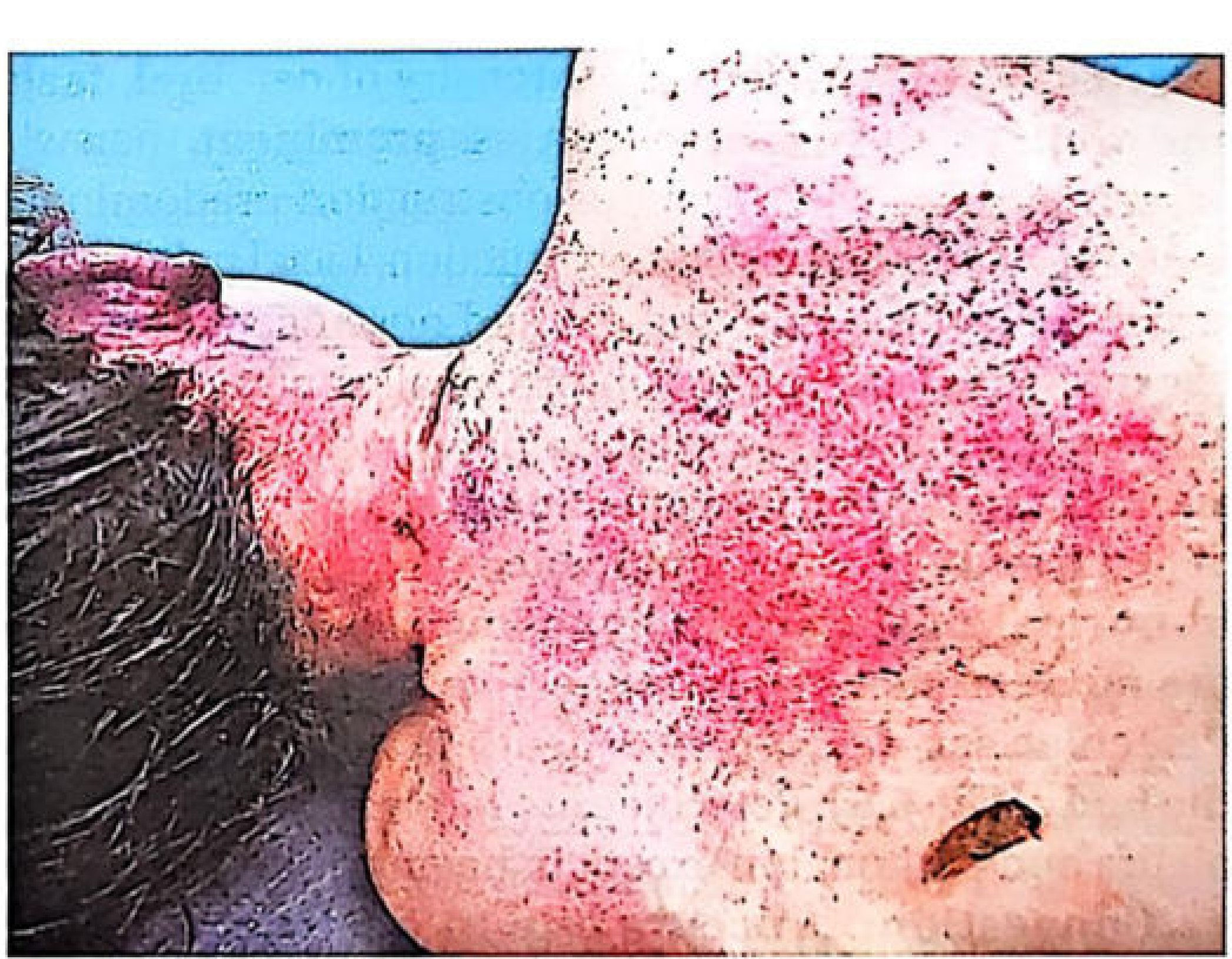
1. Petechial Hemorrhages
- Caused by raised venous pressure → overdistension and rupture of venules (especially in lax tissues)
- NOT due to hypoxia of vessel walls
- Minimum 15-30 seconds required to produce congestion and petechiae
- Size: 0.1-2 mm (larger = ecchymoses)
- Location: skin, sclerae, conjunctivae, outer and inner surfaces of eyelids, mucosal surfaces of mouth, serous membranes (especially visceral pleura and epicardium), brain
2. Congestion and Cyanosis
- About 24 hours after death, oxygen dissociates and cyanosis appears in any dead body
- Cyanosis is marked in: skin, hypostatic livid stains, lips, ears, tip of nose, fingernails, cheeks; internally - lungs, liver, spleen, kidneys, meninges
-
5 g/100 mL of reduced hemoglobin is needed to produce cyanosis
- If head is lower than body: marked congestion, cyanosis, and petechiae are common
3. Fluidity of Blood
- Blood stagnation/slowing → tends to clot → fibrinolysin released from endothelium → blood remains fluid (dark blue) after death
- Postmortem clotting takes a minimum of 4-6 hours
4. Visceral Congestion
- Generalized congestion of all viscera
Asphyxial Stigmata (Important Exam Point)
The classic triad:
- Cyanosis
- Facial, palpebral, bulbar, subpleural, and subepicardial petechiae
- Visceral congestion
These are all due to raised venous pressure. They are merely consistent with, but NOT diagnostic of asphyxia. Reliable local indications of fatal obstructing trauma must be demonstrated to establish mechanical asphyxia.
Histological Signs of Asphyxia in Tissues
- Partial disruption of alveolar septa with distinctive alveolar hemorrhage and intra-alveolar oedema
- Brick-red discoloration of nerve cells in the cerebral cortex (stained neurological sections); pallor and vacuolar degeneration of Purkinje cells in cerebellum
- Vacuolar degeneration of liver cells (in prolonged suffocation)
Stages/Symptoms of Mechanical Asphyxia
| Stage | Features |
|---|---|
| Stage of Dyspnoea | Excess CO2 stimulates respiratory center; increased rate/amplitude of respiration; BP rises; pulse rate rises; slight cyanosis |
| Stage of Convulsions | Mostly expiratory efforts; deeply congested face; BP elevated; rapid pulse; swollen neck veins; convulsions |
| Stage of Exhaustion | Respiratory center paralysed; flaccid muscles; complete insensibility; reflexes lost; widely dilated pupils; gasping (mostly inspiratory); BP falls; respiration ceases; heart may continue beating for minutes |
The three stages last 3-5 minutes before death.
Postmortem Appearances in Asphyxial Deaths
External Findings
- Face: Pale (slow asphyxia) OR distorted, congested, cyanosed, purple, swollen/oedematous (rapid asphyxia)
- Eyes prominent; conjunctivae congested; pupils dilated
- Tongue protruded; ears and fingernails bluish
- Petechiae on skin, conjunctivae, sclerae, eyelids
- Food aspiration may be found in 20-25% of all asphyxial deaths (agonal aspiration)
Internal Findings
- Heavy lungs (450-500g or more) → indicate respiratory arrest with continued heartbeat
- Subpleural and subepicardial petechiae (Tardieu's spots)
- Visceral congestion throughout
- Blood is dark, fluid (fibrinolysin-mediated)
- Obsolete cardiac findings (right heart dilatation and fluid blood) should be disregarded as non-specific
Delayed Deaths Following Asphyxia
Higher cortical centers suffer first → basal ganglia → vital centers, causing:
- Delayed deaths after periods of unconsciousness from anoxic cerebral damage
- Hypostatic pneumonia
- Massive subcutaneous and mediastinal emphysema (from tracheal/laryngeal lacerations)
- Pharyngeal/aryepiglottic fold swelling obstructing airway
- When death occurs hours, days, or weeks later - the traumatic incident remains the proximate cause
POSTMORTEM CHANGES
Postmortem changes are changes occurring in the body after death. They are medico-legally important for estimating time since death (TSD).
Classification
| Category | Changes |
|---|---|
| Immediate | Somatic death signs - cessation of circulation, respiration, CNS |
| Early | Cooling (algor mortis), Hypostasis (livor mortis), Rigor mortis |
| Late | Putrefaction, Adipocere, Mummification |
1. Algor Mortis (Postmortem Cooling)
- After death, the body loses heat to the environment until it reaches ambient temperature
- Rate of cooling: approximately 1.5°F (0.83°C) per hour under standard conditions
- Factors affecting cooling: ambient temperature, clothing, body build (obese cool slower), age (infants cool faster), cause of death
2. Hypostasis (Livor Mortis / Postmortem Lividity)
- Settling of blood to dependent parts of the body due to gravity after circulation stops
- Color: bluish-purple (or cherry-red in CO poisoning; pink in cold)
- Appears in 1-2 hours, well established by 6-12 hours, fixed/permanent by 12-24 hours (usually)
- Medicolegal significance:
- Helps establish time since death
- Distribution indicates position of body at time of death
- If lividity does not correspond to position of body when found - the body was moved after death
- If lividity is in unexpected distribution - suspect foul play
3. Rigor Mortis
Definition: Stiffening and shortening of muscles following the period of primary relaxation after death. It is due to chemical changes in the structural proteins of muscle fibers and indicates molecular death of muscle cells.
Mechanism:
- In life: ATP constantly synthesised and utilised; actin and myosin filaments remain dissociated in relaxed state
- After death: ATP resynthesis continues briefly (using remaining glycogen)
- When glycogen depletes → ATP cannot be resynthesised → actin and myosin filaments fuse into a dehydrated stiff gel = rigor mortis
- Muscle pH changes from slightly alkaline to distinctly acid (local lactic acid accumulation)
- Rigor persists until autolysis of actin and myosin occurs during putrefaction → secondary relaxation
Order of development (Nysten's Law):
- First in involuntary muscles (heart: within 1 hour)
- Then in voluntary muscles: jaw and neck → trunk → upper limbs → lower limbs
- Passes off in the same order (head to foot)
Timing (In India):
- Commences: 2-3 hours after death
- Fully developed (head to foot): ~12 hours
- Persists for: ~12 hours
- Passes off over: ~12 hours
- Total duration: ~36 hours
| TSD Estimate | Rigor Status |
|---|---|
| Within 2 hours | Rigor not yet set in |
| 12-24 hours | Whole body affected |
| After 36 hours | Rigor beginning to pass off |
Special features:
- Affects all muscles - voluntary AND involuntary
- Not dependent on nerve supply (also develops in paralysed limbs)
- Can be broken by mechanical force (flexing a stiff limb → remains flaccid thereafter)
- Cadaveric spasm (instantaneous rigor) - rare, occurs at moment of death in muscles actively contracting at time of death; no period of primary relaxation; important as it may preserve grip position
Cutis Anserina (Goose Skin):
- When erector pilae muscles are affected by rigor → skin shows granular puckered appearance ("goose skin")
- Also seen in bodies exposed to cold water (drowning)
4. Putrefaction
- Decomposition of the body by bacterial action (anaerobic bacteria from intestines) and autolysis (by tissue enzymes)
- Begins internally (GI tract) and progresses outward
- Greenish discoloration first appears in the right iliac fossa (caecum) and spreads over the abdomen, then the whole body
- Bloating from gas formation
- Marbling - discoloration of vessels visible through skin (due to haemolysis and gas production)
- Liquefaction of tissues ultimately
Rate affected by:
- Temperature (fastest at 21-38°C), humidity, type of burial, body composition
5. Adipocere (Saponification)
- Post-mortem change where body fat is converted to a white, soap-like substance (calcium and magnesium soaps of fatty acids)
- Occurs in warm, moist conditions (buried in wet soil, or immersed in water)
- Preserves body contours; may persist for centuries
- Medically: confirms identity even after long periods
6. Mummification
- Drying and shrivelling of body in hot, dry conditions
- Skin becomes dry, hard, brown/black, leathery
- Body weight markedly reduced
- May preserve body for very long periods
Medicolegal Importance of Postmortem Changes
- Estimating time since death (using algor mortis + rigor mortis + livor mortis together)
- Establishing body position at time of death (livor mortis pattern)
- Detecting movement of body after death
- Manner of death clues (e.g., cherry-red lividity in CO poisoning)
- Preservation of evidence (adipocere, mummification)
Sources: The Essentials of Forensic Medicine and Toxicology, 36th Edition (2026); Parikh's Textbook of Medical Jurisprudence, Forensic Medicine and Toxicology; DiMaio's Forensic Pathology, 3rd Edition
This is a shared conversation. Sign in to Orris to start your own chat.