
Differential diagnosis of dysphagia
differential diagnosis of dysphagia oropharyngeal esophageal causes
dysphagia differential diagnosis classification diagram
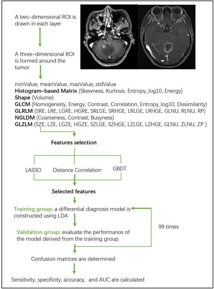
A medical flowchart outlining a radiomics-based MRI classification workflow for differentiating brain tumors, such as glioblastoma and anaplastic astrocytoma. The process begins with axial T1-weighted contrast-enhanced MRI images, followed by 2D and 3D Region of Interest (ROI) segmentation around the tumor. The diagram details the extraction of high-dimensional texture features, including histogram-based metrics (Skewness, Kurtosis), shape parameters (Volume), and various gray-level matrices: GLCM, GLRLM, NGLDM, and GLZLM. The pipeline proceeds to feature selection using LASSO, Distance Correlation, or GBDT methods. A differential diagnosis model is then constructed using Linear Discriminant Analysis (LDA) on a training dataset and evaluated with a validation group. This machine-learning cycle is iteratively performed 99 times to ensure robustness. The final stage involves diagnostic performance evaluation through confusion matrices and the calculation of sensitivity, specificity, accuracy, and Area Under the ROC Curve (AUC). This visual resource illustrates the application of texture analysis and artificial intelligence in neuroradiology and oncology.
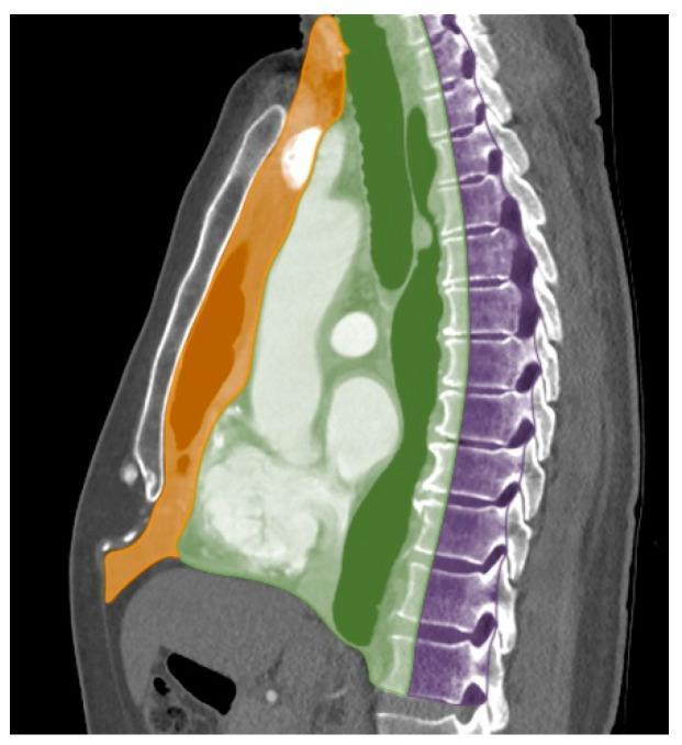
This diagnostic anatomical diagram features a sagittal reformatted computed tomography (CT) scan of the thorax with a color-coded overlay illustrating the International Thymic Malignancy Interest Group (ITMIG) classification system for mediastinal compartments. The image delineates three distinct zones: 1) The Prevascular (Anterior) Compartment, highlighted in orange, which is bounded anteriorly by the sternum and posteriorly by the pericardium and great vessels; it includes the thymic bed. 2) The Visceral (Middle) Compartment, highlighted in green, which encompasses the heart, trachea, and esophagus, extending from the posterior border of the prevascular space to the anterior aspect of the vertebral bodies. 3) The Paravertebral (Posterior) Compartment, highlighted in purple, which covers the paraspinal regions and the spine. This visual aid is designed for radiology and thoracic surgery education to facilitate the accurate localization of mediastinal masses, such as thymomas, lymphomas, or neurogenic tumors, which is the primary step in developing a clinical differential diagnosis.
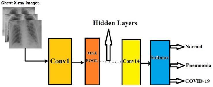
A flowchart diagram illustrating a Deep Learning Model (DLM) architecture for chest X-ray (CXR) image classification. The process begins on the left with the input of CXR images, which transition into a yellow block labeled 'Conv1' (Convolutional Layer 1). This is followed by an orange 'MAX POOL' layer. A series of dots and an upward-pointing arrow indicate intermediate 'Hidden Layers' within the neural network. The sequence continues to a final yellow convolutional layer labeled 'Conv14'. The data then enters a blue 'Softmax' layer, representing the final activation function for multi-class classification. The output culminates on the far right, where the model categorizes images into three clinical diagnostic groups: 'Normal', 'Pneumonia', or 'COVID-19'. This diagram summarizes a 46-layer ResNet-based model used in medical informatics to assist in the differential diagnosis of respiratory conditions via radiological imaging.
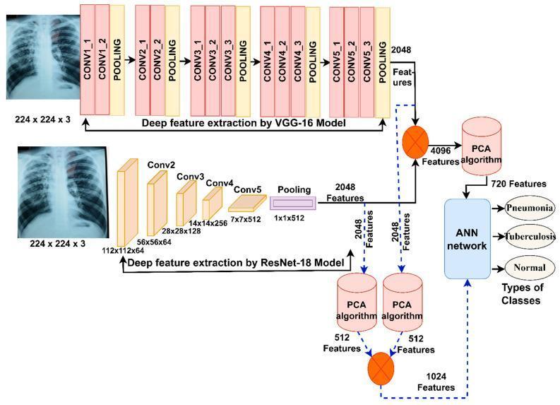
This medical flow diagram illustrates a deep learning architecture for the differential diagnosis of pneumonia and tuberculosis using chest X-rays. The technical algorithm utilizes two parallel feature extraction pathways: a VGG-16 model and a ResNet-18 model. Each model processes an input X-ray image (224x224x3) into 2048 deep features. The VGG-16 pathway details sequential convolutional layers (CONV1_1 through CONV5_3) interspersed with pooling layers, while the ResNet-18 pathway shows its specific block architecture (Conv2 to Conv5). Following feature extraction, two data processing routes are depicted. The first route merges features into a 4096-feature vector, which is then reduced to 720 features via Principal Component Analysis (PCA). The second route applies independent PCA to each model's features (reducing to 512 each) before combining them into a 1024-feature vector. Both routes converge at an Artificial Neural Network (ANN) for final classification. The clinical output categorizes findings into three distinct classes: Pneumonia, Tuberculosis, or Normal. This flowchart highlights advanced diagnostic informatics for respiratory infectious diseases.
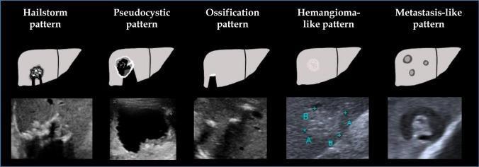
This educational comparison chart illustrates the Echinococcus multilocularis Ulm classification—ultrasound (EMUC-US) for hepatic alveolar echinococcosis (AE). The diagram presents five distinct ultrasonographic patterns using side-by-side schematic liver illustrations and corresponding B-mode clinical ultrasound frames. 1) Hailstorm pattern: Characterized by a heterogeneous cluster of bright echoes with significant posterior acoustic shadowing. 2) Pseudocystic pattern: Depicts a large, well-defined anechoic (liquid-filled) area with irregular borders. 3) Ossification pattern: Shows focal, highly echogenic calcifications with sharp, dense posterior shadowing. 4) Hemangioma-like pattern: Displays a circumscribed, homogeneously hyperechoic lesion with a smooth texture, mimicking a benign hemangioma. 5) Metastasis-like pattern: Presents multiple, discrete, hypoechoic nodules with a target-like or 'bulls-eye' appearance. This tool is used in radiology and hepatology for the differential diagnosis of parasitic liver lesions and to categorize disease morphology according to established clinical criteria.
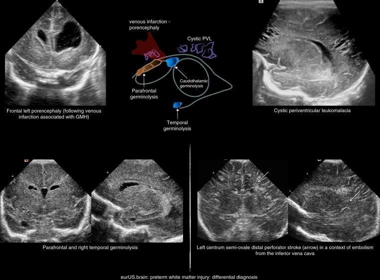
This composite educational graphic presents cranial ultrasound (CUS) images demonstrating a differential diagnosis of preterm white matter injury (WMI). The top-left panel shows frontal left porencephaly following venous infarction associated with germinal matrix hemorrhage (GMH). A central schematic diagram maps the locations of venous infarction, cystic periventricular leukomalacia (PVL), and various sites of germinolysis (parafrontal, caudothalamic, and temporal). The top-right CUS image displays cystic PVL, characterized by multiple hypoechoic, fluid-filled cystic spaces in the periventricular white matter, indicating macroscopic focal necrosis. The bottom-left panels illustrate parafrontal and right temporal germinolysis. The bottom-right panels demonstrate a distal perforator stroke in the left centrum semi-ovale, indicated by arrows pointing to focal hypoechoic lesions amidst relatively hyperechoic brain parenchyma. This collection serves to distinguish between different patterns of neonatal brain injury, including localized vascular events and diffuse leukomalacia, emphasizing sonographic morphology and anatomical distribution for clinical classification.
Differential Diagnosis of Dysphagia
Step 1: Classify the Type
| Feature | Oropharyngeal (Transfer) | Esophageal |
|---|---|---|
| Site of perceived hold-up | Neck / suprasternal notch | Lower sternum / epigastrium |
| Timing after swallow | Immediately (within 1 second) | Seconds later |
| Associated symptoms | Coughing, choking, nasal regurgitation, drooling, dysarthria | Food "sticking" in chest, regurgitation, heartburn |
| Main causes | Neurological, muscular | Structural, motility |
Key rule: solids and liquids equally affected = motility disorder; solids first, then liquids = progressive mechanical obstruction.
Part A: Oropharyngeal Dysphagia
1. Neuromuscular Causes
| Condition | Notes |
|---|---|
| Stroke | Most common cause of oropharyngeal dysphagia; often with aspiration |
| Parkinson disease | Bradyphrenia of swallowing; silent aspiration common |
| Amyotrophic lateral sclerosis (ALS) | UMN + LMN; bulbar involvement; early sign |
| Multiple sclerosis | Brainstem plaques |
| Myasthenia gravis | Fatigable weakness; can be the initial presentation |
| Polymyositis / Dermatomyositis | Proximal muscle inflammation |
| Muscular dystrophy (oculopharyngeal, myotonic) | Genetic; oculopharyngeal dystrophy affects pharyngeal muscles |
| Bulbar / pseudobulbar palsy | LMN vs. UMN - distinguish by jaw jerk, palatal reflex |
| Post-polio syndrome | Late degeneration |
| CNS tumors | Brainstem or posterior fossa |
| Idiopathic UES dysfunction / cricopharyngeal incoordination | Failure of UES to relax; can lead to Zenker diverticulum |
| Thyroid dysfunction | Can cause myopathy affecting pharyngeal muscles |
2. Structural Causes
| Condition | Notes |
|---|---|
| Zenker diverticulum | Posterior pharyngeal pouch; gurgling noise on swallowing, halitosis, regurgitation of old food |
| Carcinoma of pharynx / hypopharynx | Progressive, with weight loss; smoking/alcohol risk |
| Proximal esophageal web (Plummer-Vinson / Paterson-Kelly) | Iron-deficiency anemia + dysphagia + web; middle-aged women |
| Cervical osteophytes | Anterior spur compression; elderly males |
| Thyromegaly / goiter | External compression; especially retrosternal extension |
| Infections of pharynx/neck | Peritonsillar abscess, Ludwig's angina, retropharyngeal abscess |
| Prior surgery or radiation | Fibrosis, scarring |
Part B: Esophageal Dysphagia
1. Motility (Neuromuscular) Disorders
Primary
| Condition | Key Feature |
|---|---|
| Achalasia | Loss of myenteric neurons; absent peristalsis + failed LES relaxation; affects solids AND liquids from the outset; dilated esophagus on CXR ("bird-beak" on barium) |
| Distal esophageal spasm (DES) | Intermittent, non-peristaltic contractions; dysphagia + chest pain; "corkscrew" on barium |
| Hypercontractile (Jackhammer) esophagus | Very high-amplitude contractions (DCI >8000 mmHg/s/cm); chest pain + dysphagia |
| Esophagogastric junction outflow obstruction (EGJOO) | Insufficient LES relaxation with preserved peristalsis; may be achalasia precursor |
| Ineffective esophageal motility | Low-amplitude contractions; often associated with GERD |
Secondary (Systemic Diseases)
| Condition | Notes |
|---|---|
| Scleroderma | Smooth muscle fibrosis; low/absent LES pressure; reflux-induced stricture common |
| Chagas disease | Trypanosoma cruzi destroys myenteric plexus; achalasia-like picture; endemic in South America |
| Reflux-related dysmotility | Chronic GERD alters motility |
| Diabetes mellitus | Esophageal neuropathy |
| Amyloidosis | Infiltration of smooth muscle |
2. Structural (Mechanical) Disorders
Intrinsic (within the esophagus)
| Condition | Key Feature |
|---|---|
| Esophageal carcinoma | Progressive dysphagia: solids → liquids; weight loss; squamous (mid) vs. adenocarcinoma (lower/GEJ); main alarm symptom |
| Peptic stricture | Long-standing GERD; smooth, tapered narrowing at lower esophagus |
| Eosinophilic esophagitis (EoE) | Young men; food impaction; rings/furrows on endoscopy; >15 eos/hpf on biopsy; may present with poor distensibility rather than visible narrowing |
| Schatzki ring (lower esophageal ring) | Mucosal ring at squamocolumnar junction; intermittent dysphagia for solids ("steakhouse syndrome") |
| Esophageal webs | Thin mucosal shelf; cervical or mid-esophagus; associated with Plummer-Vinson |
| Medication-induced stricture | Doxycycline, tetracycline, bisphosphonates, NSAIDs, potassium chloride; pill esophagitis |
| Benign tumors | Leiomyoma (most common benign); smooth, submucosal |
| Foreign body | Acute, clear history |
| Esophageal diverticula | Mid-esophageal (traction) or epiphrenic; regurgitation of undigested food |
| Radiation esophagitis / stricture | History of thoracic/cervical radiotherapy |
| Infectious esophagitis | Candida (immunocompromised), CMV, HSV; odynophagia often > dysphagia |
Extrinsic (compressing from outside)
| Condition | Notes |
|---|---|
| Mediastinal mass | Lymphoma, thymoma, bronchogenic carcinoma |
| Vascular compression | Dysphagia lusoria (aberrant right subclavian artery - most common vascular cause); aortic aneurysm ("dysphagia aortica") |
| Spinal osteophytes | Anterior cervical or thoracic |
| Enlarged mediastinal lymph nodes | TB, sarcoidosis, metastases |
Part C: Special / Functional Causes
| Condition | Notes |
|---|---|
| Globus sensation | Feeling of lump in throat without true dysphagia; no bolus hold-up; associated with anxiety, GERD, UES hyperpharyngeal tone |
| Functional dysphagia (Rome IV) | No structural or motility cause found; abnormal visceral perception; often waxing and waning |
| Medication-induced | Dry mouth (anticholinergics, antihistamines); direct pill esophagitis (bisphosphonates, doxycycline); esophageal dysmotility (calcium channel blockers) |
Diagnostic Clues - "Solids vs. Liquids" Rule
Dysphagia
   |
   ├─ Only to SOLIDS
   │      |
   │      ├─ Progressive → Carcinoma, peptic stricture, EoE
   │      └─ Intermittent → Schatzki ring, EoE (food bolus impaction)
   │
   └─ BOTH solids & liquids
          |
          ├─ Progressive → Achalasia, scleroderma (motility)
          └─ Intermittent → DES, spastic disorders
Alarm Features Requiring Urgent Investigation
- Progressive dysphagia (especially solids → liquids)
- Unintentional weight loss
- Odynophagia (suggests mucosal disease - malignancy, infection)
- Age >50 with new onset
- Hematemesis or melena
- History of head-and-neck radiation or Barrett's esophagus
Initial Investigation Pathway
- Modified barium swallow - best for oropharyngeal dysphagia (with swallowing therapist)
- Upper GI endoscopy (OGD) - preferred for esophageal dysphagia; allows biopsy
- High-resolution manometry (HRM) - for suspected motility disorders (Chicago Classification v4.0)
- CT thorax/neck - for extrinsic compression, mediastinal mass
- Videofluoroscopy - detailed dynamic oropharyngeal assessment
- Functional lumen imaging probe (FLIP) - for esophageal distensibility and EGJ function