
Full details about fistula in ano
Reading File
Now let me search the medical library for fistula in ano comprehensively.
Finding Sources
Searching Images
fistula in ano types Parks classification diagram
Excellent - multiple great sources. Let me read the most comprehensive ones in parallel.
Reading File
Reading File
Searching Images
fistula in ano anatomy sphincter intersphincteric suprasphincteric
Great content and images found. Let me fetch the key images and read more content in parallel.
Loading Image
Loading Image
Reading File
Reading File
Excellent content and beautiful images. Let me read more of the surgical management details and fetch a few more images.
Reading File
Reading File
Loading Image
Loading Image
I now have very rich content from multiple authoritative textbooks and excellent images. Let me read a bit more on surgical options from Bailey & Love and then compile the comprehensive answer.
Reading File
Reading File
I now have comprehensive content from multiple authoritative textbooks. Let me compile the full detailed answer.
Fistula-in-Ano
A fistula-in-ano (anal fistula) is a chronic abnormal communication lined by granulation tissue extending from the anorectal lumen (the internal opening, at or near the dentate line) to an external opening on the perineal skin or buttock (or rarely the vagina).
Aetiology
The majority are cryptoglandular in origin - arising from infection of the anal glands that open into the crypts of Morgagni at the dentate line. The sequence is:
- Infection of an anal gland in the intersphincteric space
- Perianal abscess formation
- Spontaneous or surgical drainage - leaving a persistent fistulous track
~50% of patients with a perianal abscess will eventually develop a fistula.
Secondary causes
-
Crohn's disease (most common secondary cause; suspect with multiple/complex tracts)
-
Tuberculosis
-
Lymphogranuloma venereum
-
Actinomycosis
-
Rectal duplication
-
Foreign body
-
Malignancy (rarely arising within a longstanding fistula)
-
Trauma / obstetric injury
-
Bailey and Love's Short Practice of Surgery 28th Ed, p. 1456-1457
Pathological Anatomy
The internal opening is almost always at the dentate line (the site of the anal gland duct). The tract passes through or around the sphincter muscles to reach the perianal skin. The course of the tract defines the classification.
Classification
Parks' Classification (most widely used)
Based on the relationship of the primary tract to the external anal sphincter:
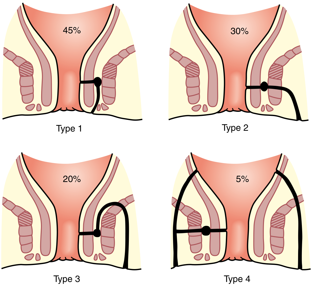
Parks classification - Sabiston Textbook of Surgery
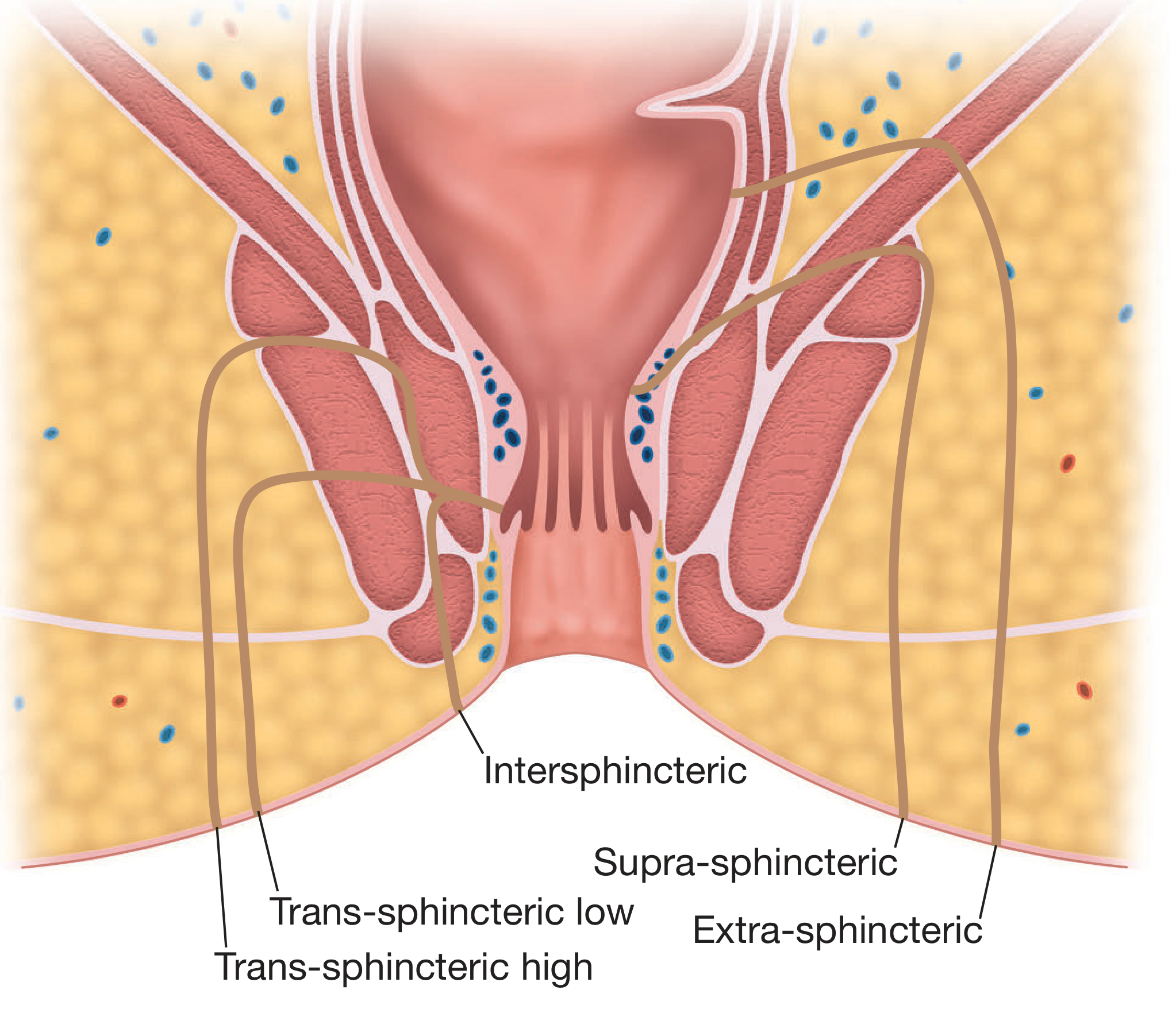
Coronal anatomy of the anal sphincter complex showing all fistula types - Bailey and Love
| Type | Description | Frequency |
|---|---|---|
| Type 1 - Intersphincteric | Track confined to the intersphincteric plane, passes between internal and external sphincters to perianal skin | ~45% |
| Type 2 - Transsphincteric | Track crosses both sphincters, passes through the ischiorectal fossa to the skin | ~30% |
| Type 3 - Suprasphincteric | Track passes up above puborectalis, then curves down through levators and ischioanal fossa to skin | ~20% |
| Type 4 - Extrasphincteric | Track passes without specific relation to sphincters, usually from pelvic disease or trauma | ~5% |
Suprasphincteric fistulae are often caused by excessive probing of an abscess during examination under anaesthesia. Extrasphincteric fistulae usually result from pelvic disease or trauma.
AGA (American Gastroenterological Association) Classification
Clinically useful simplification:
| Category | Features |
|---|---|
| Simple fistula | Low (superficial/low intersphincteric/low transsphincteric), single external opening |
| Complex fistula | High (high inter- or transsphincteric), extrasphincteric/suprasphincteric, associated abscess/collection, anovaginal, or with anal stricture, or Crohn's-related |
- Bailey and Love, Table 80.2
Clinical Features
Symptoms
- Intermittent purulent discharge from the external opening (may be blood-stained)
- Cyclical pain - builds up until drainage occurs, followed by relief
- History of prior anorectal abscess
- Passage of flatus or faeces through the external opening suggests a rectal (rather than anal canal) internal opening
Signs
- One or more chronic wounds or discharging openings on the perianal skin
- Palpable cord-like subcutaneous induration between the external opening and the anal margin (suggests superficial track)
- Supralevator induration - suggests high primary track or secondary extension
- Bilateral external openings - think deep postanal space involvement
- Multiple external openings ("watering-can perineum") - strongly suspect Crohn's disease
Goodsall's Rule
Used to predict the location of the internal opening from the external opening:
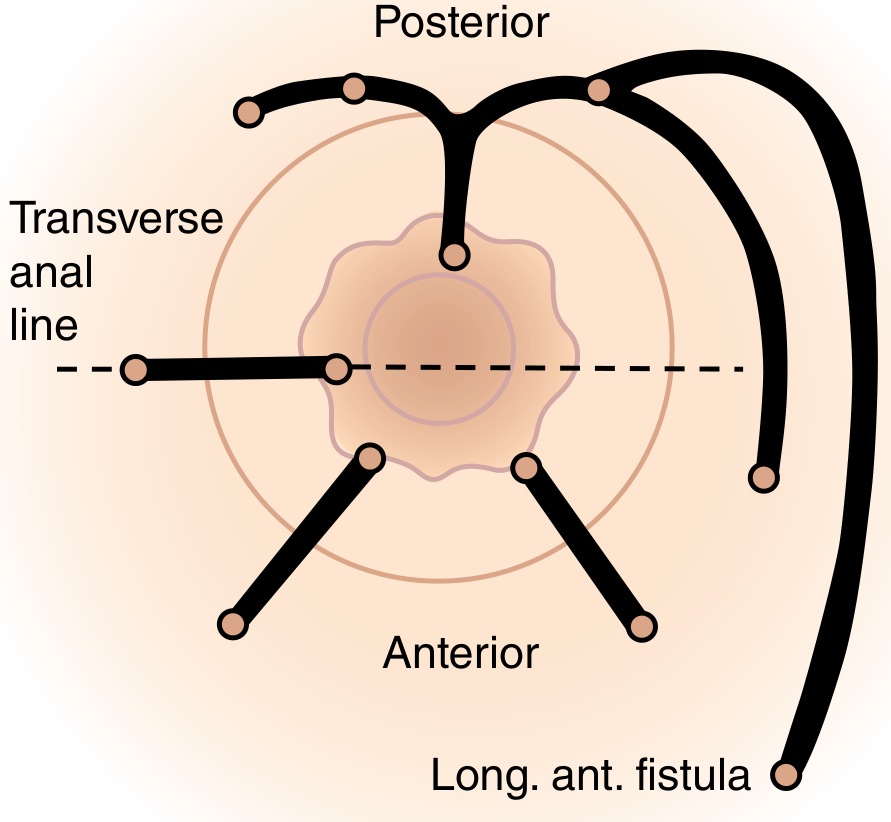
Goodsall's rule - Sabiston Textbook of Surgery
- External opening ANTERIOR to transverse anal line → track runs radially/directly into the anal canal (except when >3 cm from anal verge, which may be an anterior extension of a posterior horseshoe fistula)
- External opening POSTERIOR to transverse anal line → track curves in a curvilinear fashion to a posterior midline internal opening
The majority of internal openings are in the midline (anterior or posterior).
Investigations
Clinical Examination
- Full history including obstetric, GI, and continence history
- Proctosigmoidoscopy
- Gentle palpation for subcutaneous cord; digital rectal examination for supralevator induration
- Identification of internal opening: palpable induration, enlarged papilla at the dentate line
Intraoperative Tools
- Anal fistula probe - gentle probing from external opening; care to avoid creating false passages
- Dilute hydrogen peroxide / methylene blue / milk - injected via the external opening to identify the internal opening
- Anoscopy - direct inspection of the dentate line for erythematous crypt or visible internal opening
Imaging
| Modality | Use |
|---|---|
| Endoanal ultrasound (EAUS) | First-line imaging; assesses sphincter integrity, identifies primary and secondary tracts; enhanced with hydrogen peroxide injection |
| MRI (pelvic, fistula protocol) | Gold standard for complex fistulae; delineates anatomy, identifies secondary extensions missed clinically; T2-weighted images show fistula as high signal |
| Fistulography / CT | Useful when extrasphincteric fistula is suspected |
- MRI is especially valuable for complex or recurrent fistulae and high-level tracts
Principles of Surgery
The five goals of fistula treatment are:
- Treat any undrained infection
- Define fistula anatomy
- Remove or ablate epithelialized tracts
- Avoid or minimize the risk of fecal incontinence
- Prevent recurrence
The most important determinant of continence after fistulotomy is the amount of sphincter muscle preserved rather than the amount divided. At least 2 cm of functional external sphincter should be retained.
Surgical Options
1. Fistulotomy (Lay-Open)
- Probe is passed through the tract; overlying tissue is divided; wound heals by secondary intention
- Best for: Simple, low-lying fistulae (intersphincteric or low transsphincteric) involving less than one-third of the external sphincter
- Recurrence rate: 2-8%; clinically significant incontinence: <5% in patients with normal preoperative sphincter function
- Historically described by John of Arderne in De Arte Phisicali et de Cirurgia
2. Seton
Two modes of use:
- Draining seton (silastic vessel loop or rubber band): Controls the fistula, prevents cyclical symptoms, allows fibrosis and narrowing of the tract, may enable partial fistulotomy through soft tissue to the level of the sphincter
- Cutting/tightening seton: Progressively tightened to slowly divide the tract, allowing fibrosis before division - reduces incontinence risk; however, seton tightening has largely fallen out of favour due to unpredictable incontinence
3. Endorectal Advancement Flap
- Fistula tract is excised; the rectal/internal opening is closed with a vascularized mucosal flap advanced from proximal rectum
- Used for transsphincteric fistulae and complex fistulae
- Healing rates: 66-87%
- Risk: mild-moderate incontinence in up to 35% if sphincter fibers are included in the flap
4. LIFT (Ligation of Intersphincteric Fistula Tract)
- The intersphincteric plane is accessed, the fistula tract is identified, divided, and suture-ligated within the intersphincteric space; the sphincter is approximated
- Sphincter-preserving technique
- If it fails, the result is often an intersphincteric fistula amenable to simple fistulotomy
5. Fibrin Glue / Anal Fistula Plug
- The tract is obliterated with fibrin glue or plugged with a bioresorbable device
- Sphincter-preserving; however, long-term success rates are poor; not recommended as definitive treatment
6. VAAFT (Video-Assisted Anal Fistula Treatment)
- Fistulascope inserted into the external opening; tract and all branches are visualized under direct endoscopic vision
- Internal opening closed with suture/stapling; granulation tissue debrided; tract ablated with an electrode
- No sphincter muscle divided
- Reported healing rates: 71-85% at 1-2 years
7. FiLaC (Fistula Laser Closure)
- Radially emitting laser probe ablates the fistula tract from within under direct vision
- Similar principle to VAAFT but uses laser energy
- Recent meta-analysis: healing in ~65% at median 24 months
8. Stem Cell Therapy (Emerging)
- Mesenchymal stem cells transplanted into peri-fistular tissue
- Promising results in small studies and RCTs, particularly in Crohn's perianal fistulae (darvadstrocel/Alofisel has regulatory approval in some countries)
Special Situations
Crohn's-Related Fistula
- Often complex, multiple, and recurrent
- Medical management is first-line: anti-TNF agents (infliximab/adalimumab), setons for drainage
- Surgery is reserved for palliation; sphincter-sacrificing procedures are generally avoided
Horseshoe Fistula
- Deep postanal space abscess spreads circumferentially through the ischiorectal fossae
- Both ischiorectal fossae are involved with a common posterior communication
- Treatment: drain the deep postanal space (posterior fistulotomy) plus counter-incisions over each ischiorectal component (Hanley's procedure)
Fistula with Abscess
- The acute phase should be drained first; primary fistulotomy at the time of abscess drainage remains controversial (anatomy is distorted and sphincter involvement may be underestimated)
Continence Risk Factors
- Amount of sphincter divided (the dominant factor)
- Preoperative sphincter function
- Prior fistula surgery
- Female sex (shorter anterior sphincter)
- Anterior fistulae in women (highest risk)
- Obstetric injury history
Prognosis
- Simple fistulae treated by fistulotomy: low recurrence (2-8%), excellent continence outcomes
- Complex fistulae: higher recurrence rates, ongoing risk of continence impairment; often require staged or repeat procedures
- Crohn's fistulae have the highest rates of recurrence and complexity
Sources:
- Sabiston Textbook of Surgery: The Biological Basis of Modern Surgical Practice, Ch. 97 (Fistula-in-Ano)
- Bailey and Love's Short Practice of Surgery 28th Edition, Ch. 80
- Sleisenger and Fordtran's Gastrointestinal and Liver Disease
- Harrison's Principles of Internal Medicine 22E (2025)
This is a shared conversation. Sign in to Orris to start your own chat.