
Complementary pathway microbiology
complement pathway activation classical alternative lectin microbiology diagram
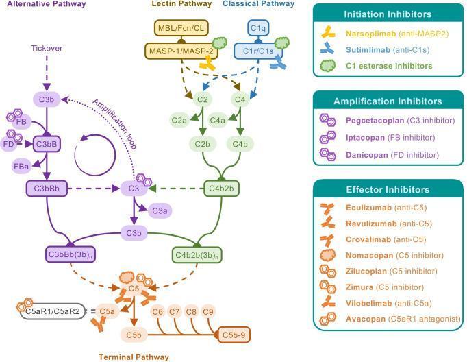
A comprehensive pathophysiology diagram illustrating the three activation pathways of the human complement system—Alternative, Lectin, and Classical—and their convergence into the Terminal Pathway. The Classical pathway is initiated by C1q and C1r/s complexes, the Lectin pathway by MBL/Fcn/CL and MASP-1/2 complexes, and the Alternative pathway via spontaneous 'tickover' starting with C3b. The diagram details the formation of C3 convertases (C3bBb and C4b2b) and C5 convertases, leading to the terminal assembly of the Membrane Attack Complex (MAC, C5b-9) and the release of anaphylatoxins C3a and C5a. Superimposed on the biochemical cascade are clinical therapeutic targets and their respective inhibitors. 'Initiation Inhibitors' include Narsoplimab and Sutimlimab; 'Amplification Inhibitors' include Pegcetacoplan, Iptacopan, and Danicopan; and 'Effector Inhibitors' include Eculizumab, Ravulizumab, and Avacopan. This educational infographic serves as a guide for understanding complement-driven diseases and the pharmacological mechanisms of current and emerging biological treatments.
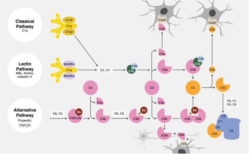
A comprehensive pathophysiology diagram illustrating the three activation pathways of the human complement system: Classical, Lectin, and Alternative. The Classical pathway is initiated by C1q (complexed with C1r2 and C1s2); the Lectin pathway utilizes MBL, ficolins, and collectin-11 with MASPs; and the Alternative pathway involves Properdin and C3(H2O) with Factors B and D. All three pathways converge at the enzymatic cleavage of C3 into C3a and C3b. The C4b2b complex (C3 convertase) is shown mediating this in the classical/lectin routes, while C3bBb performs this in the alternative route. The downstream cascade leads to C5 cleavage, producing the anaphylatoxin C5a and fragment C5b. Educational highlights include the assembly of the Membrane Attack Complex (MAC, C5b-9) depicted as a blue pore-forming structure, and the production of opsonins (iC3b, C3d). The diagram specifically contextualizes the immune response within the nervous system, showing receptors like C3aR, C5aR, and CR3 on glial-like cells, illustrating the role of complement in neuroinflammation or synaptic pruning.
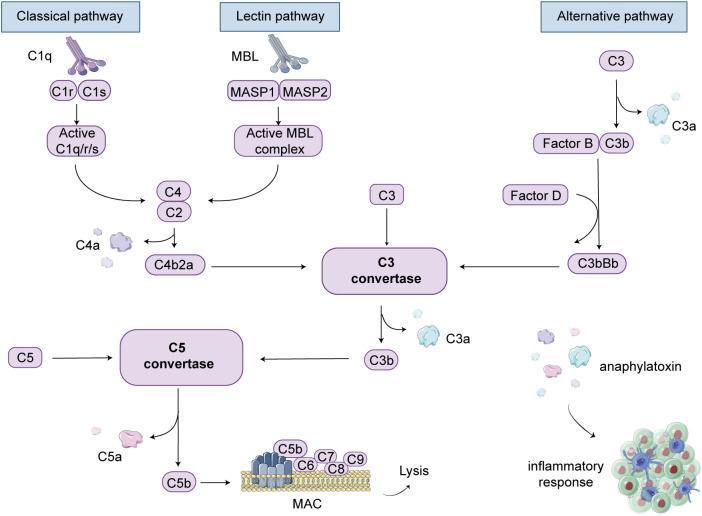
A pathophysiology diagram illustrating the human complement system cascade, organized into three primary activation pathways: Classical, Lectin, and Alternative. The Classical pathway is shown initiating via C1q, C1r, and C1s to form an active C1q/r/s complex. The Lectin pathway begins with MBL and MASP1/2, forming an active MBL complex. Both pathways converge to cleave C4 and C2 into the C4b2a complex. The Alternative pathway depicts the spontaneous cleavage of C3 into C3a and C3b, the latter interacting with Factor B and Factor D to form C3bBb. These processes converge at the central C3 convertase node, which further cleaves C3 into C3a (anaphylatoxin) and C3b. The cascade proceeds to C5 convertase, which cleaves C5 into C5a (anaphylatoxin) and C5b. Terminal events include the assembly of C5b with C6, C7, C8, and C9 to form the Membrane Attack Complex (MAC) embedded in a lipid bilayer, leading to cell lysis. The diagram also highlights the role of C3a and C5a as anaphylatoxins that trigger an inflammatory response involving immune cell recruitment.
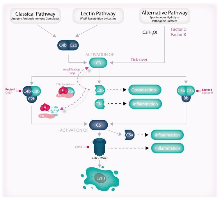
This pathophysiology diagram illustrates the three activation pathways of the human complement system: Classical, Lectin, and Alternative. The Classical pathway is triggered by antigen-antibody complexes, the Lectin pathway by PAMP recognition, and the Alternative pathway via spontaneous hydrolysis ('tick-over') involving Factors B and D. All three pathways converge at the activation of C3. Key downstream events include the cleavage of C3 into C3a (driving inflammation) and C3b (mediating opsonization). The diagram highlights an 'amplification loop' where C3b reinforces C3 activation, alongside regulatory mechanisms such as C3b breakdown by Factor I (FI) and Factor H (FH). The cascade progresses to C5 activation, resulting in C5a-mediated inflammation and the formation of the C5b-9 Membrane Attack Complex (MAC), which is regulated by CD59 and ultimately leads to cell lysis. The illustration serves as an educational summary of innate immune system proteolytic signaling, highlighting the balance between effector functions (inflammation, opsonization, lysis) and inhibitory regulation.
The Complement System - Microbiology
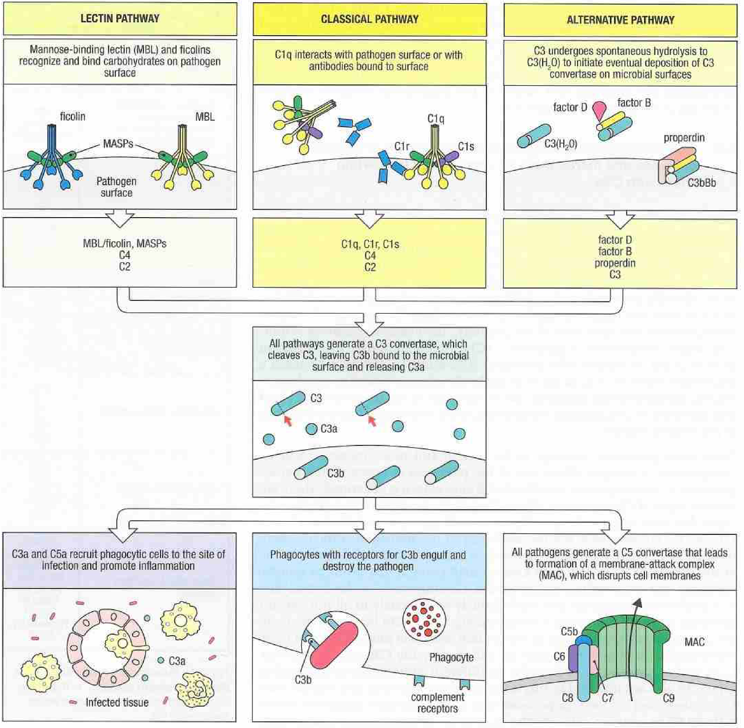
Overview of the Three Pathways
1. Classical Pathway
- C1q binds to Fc regions of IgM or IgG already attached to a pathogen
- IgM (pentameric) is a particularly potent activator - binding multiple identical epitopes on the pathogen surface "bends" it into a conformation that exposes binding sites for C1q
- IgG requires at least two adjacent IgG molecules bound to the surface (a single IgG in solution cannot activate C1)
- C1q binding activates the serine proteases C1r and C1s within the C1 complex
- C1s cleaves C4 → C4a + C4b (C4b binds covalently to pathogen surface)
- C1s cleaves C2 → C2a + C2b
- C4b + C2a assemble → C3 convertase (C4b2a) on the pathogen surface
- C4b2a cleaves C3 → C3a (released) + C3b (covalently deposited on surface)
2. Lectin Pathway
- Microbes display repeating carbohydrate arrays (mannose, fucose, N-acetylglucosamine) that MBL recognizes
- Vertebrate cells terminate their glycans in sialic acid, so MBL does not attack host cells
- MBL (a collectin, synthesized in liver) circulates at low concentrations but rises as an acute-phase protein during infection
- MBL is complexed with serine proteases MASP-1 and MASP-2 (functionally analogous to C1r and C1s)
- MASP-2 cleaves C4 and C2 → forms C3 convertase (C4b2a) - same as classical pathway from this point forward
- Ficolins (ficolin-1, -2, -3) act similarly to MBL and bind N-acetylglucosamine-containing carbohydrates
This pathway is antibody-independent - it acts from the very first exposure to a pathogen.
3. Alternative Pathway
- C3 undergoes spontaneous, low-level hydrolysis in plasma to generate C3(H₂O)
- C3(H₂O) + Factor B → Factor D cleaves Factor B → forms fluid-phase C3 convertase C3(H₂O)Bb
- This cleaves C3 → C3b, which can bind any nearby surface (host or microbial)
- On host cells, regulatory proteins (DAF/CD55, Factor H, Factor I) rapidly degrade C3b - preventing attack on self
- On microbial surfaces (lacking these regulators), Properdin stabilizes the C3bBb complex
- Stabilized C3bBb (solid-phase C3 convertase) generates massive amounts of C3b in an amplification loop
- C3b + C3bBb → C5 convertase (C3bBbC3b) for the alternative pathway
Convergence: The Terminal Pathway (MAC)
| Step | Event |
|---|---|
| C5 convertase cleaves C5 | → C5a (anaphylatoxin, released) + C5b (stays bound) |
| C5b binds C6, C7 | → C5b67 complex inserts into lipid bilayer |
| C8 joins | → initiates pore formation |
| Multiple C9 molecules polymerize | → Membrane Attack Complex (MAC = C5b-9) |
| MAC | → creates ~10 nm transmembrane pore → osmotic lysis |
Effector Functions of Complement (Summary)
| Function | Mediator | Effect |
|---|---|---|
| Opsonization | C3b (+ iC3b) | Binds CR1/CR3 on phagocytes; promotes phagocytosis |
| Inflammation | C3a, C5a (anaphylatoxins) | Mast cell degranulation, neutrophil chemotaxis, increased vascular permeability |
| Cell lysis | MAC (C5b-9) | Direct killing of Gram-negative bacteria, some viruses |
| Immune complex clearance | C3b | Binds CR1 on RBCs; shuttles complexes to liver/spleen for disposal |
| B cell activation | C3d | Binds CR2 (CD21) on B cells; lowers threshold for activation |
C3 and C5 Convertases Compared
| Classical / Lectin | Alternative | |
|---|---|---|
| C3 convertase | C4b2a | C3bBb |
| C5 convertase | C4b2a3b | C3bBbC3b |
| Stabilized by | - | Properdin |
| Inhibited by | C1-INH (classical) | Factor H, Factor I, DAF, MCP |
Complement Regulatory Proteins
- C1-inhibitor (C1-INH): Blocks C1r and C1s. Deficiency → Hereditary Angioedema (HAE)
- C4b-binding protein (C4BP): Accelerates decay of C3 convertase in classical pathway
- Factor H / Factor I: Degrade C3b on host surfaces (alternative pathway)
- DAF (CD55): "Decay accelerating factor" - dissociates C3 convertases on host cells
- CD59 (protectin): Inhibits C9 polymerization; blocks MAC formation on host cells. Deficiency → Paroxysmal Nocturnal Hemoglobinuria (PNH)
- Properdin: Unique positive regulator - stabilizes C3bBb on microbial surfaces
Complement Deficiencies and Clinical Significance
| Deficiency | Consequence |
|---|---|
| C1q, C2, C4 (classical) | Increased susceptibility to immune complex disease (SLE-like syndrome) |
| MBL | Recurrent bacterial infections in infancy |
| C3 | Severe, recurrent pyogenic infections (most serious deficiency) |
| C5-C9 (terminal) | Recurrent Neisseria infections (meningococcemia, disseminated gonococcal infection) |
| C1-INH | Hereditary Angioedema |
| CD59 (or PIG-A mutation) | Paroxysmal Nocturnal Hemoglobinuria |
| Factor H or I | Uncontrolled alternative pathway activation, C3 glomerulopathy, aHUS |
Key Microbiological Points
- Gram-negative bacteria are more susceptible to MAC than Gram-positive (thick peptidoglycan shields Gram-positive bacteria)
- Encapsulated bacteria (S. pneumoniae, H. influenzae, N. meningitidis) resist complement by hiding their surfaces from C3b deposition - this is why capsule is a major virulence factor
- Streptococcus pyogenes M protein inhibits Factor H binding and blocks alternative pathway regulation
- Staphylococcus aureus produces SCIN (Staphylococcal Complement Inhibitor), which freezes C3bBb and C4b2a convertases
- Complement C3b on IgG-antigen complexes also promotes dendritic cell and B cell priming, bridging innate and adaptive immunity