

These things came out of my skin Why are these forming in my skin so much and how can I prevent them it's like pimples and Comedones Help me with these
milia keratin plugs skin comedones extracted
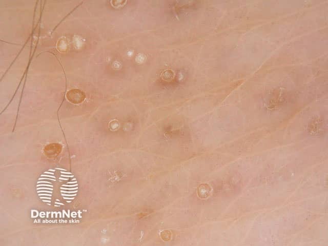
This dermatoscopic image captures a close-up view of multiple small, round papules within a sun-exposed cutaneous surface. The epidermal lesions display keratin-filled centers with pale to light-brown cores and surrounding translucent halos. Several openings resemble Milia-like cysts (small, bright-white to skin-colored droplets) and comedo-like openings (keratin plugs intrafollicular). The background skin is pink to beige with fine surface scaling and occasional fine hairs evident. The overall pattern is scattered, with heterogeneous sizes ranging from 0.5 to 3 mm. Pigmentary features are mild or absent, aligning with benign keratinous lesions rather than vascular or pigmented neoplasms. The morphology suggests seborrheic keratosis or sebaceous hyperplasia spectrum; however, absence of marked vascularization or crusting reduces suspicion for melanoma, basal cell carcinoma, or actinic keratosis in this field. Dermoscopic clues include keratin-filled structures (comedones/milia), sharply demarcated borders, and a lack of pigment networks. This image is valuable for educational demonstrations of common in vivo keratinous skin lesions and can aid in differential diagnosis among seborrheic keratosis, milium, and keratinous cysts. Potential clinical applications: lesion characterization in clinics, tele-dermatology triage, and dermoscopy training modules. Ideal for automated keyword indexing and comparative datasets; compatible with AI-based lesion classifier studies and medical education repositories.
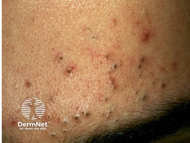
This is a high-resolution clinical photograph of the facial skin showing features characteristic of comedonal acne. Modality: Clinical photography (in vivo, dermoscopy not applied). The image presents a dense pattern of open comedones (blackheads) and occasional closed comedones (whiteheads) scattered across the cheek and perioral region, with minimal surrounding erythema and subtle post-inflammatory changes. The follicles are dilated with keratinous plugs protruding at the ostia, producing a reticulate texture and follicular papules. The skin surface is slightly oily, reflecting sebaceous activity. No pustules or nodules are evident, consistent with non-inflammatory acne predominance. The distribution is facial, bilateral or regionally on the lower malar area, typical for adolescent and early adult acne. The appearance may be influenced by measurement distance and lighting, but the core finding remains follicular plugging with keratin debris. This image is useful for educational illustration of comedone morphology, differential diagnosis (e.g., milia, keratosis pilaris), and guiding therapy decisions. Clinically, management would emphasize keratolytic and comedolytic strategies—topical retinoids, benzoyl peroxide, salicylic acid, and avoidance of comedone extraction to reduce inflammation risk. The photo provides a visual reference for patient counseling and dermatologic training in recognizing mild-to-moderate comedonal acne phenotypes. Useful for teaching, clinical case discussions, and reference libraries.
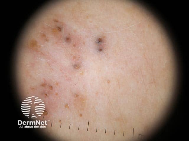
High-resolution dermoscopy of sun-exposed facial skin reveals multiple open, keratin-filled follicular openings consistent with solar comedones. The individual plugs appear as rounded to oval, yellow to brown structures embedded within comedone-like follicles, often surrounded by a lightly erythematous or pale epidermis. The background demonstrates photodamage markers such as fine telangiectasia and subtle actinic elastosis, with reduced skin elasticity. Notably, there is an absence of prominent vascular structures within the plugs, and inflammatory signs are minimal or absent, helping distinguish these lesions from acne vulgaris. The keratin plugs may coalesce into clusters or align along follicular openings, producing a nonuniform topography across the scanned field. Under dermoscopy, these features correlate with chronic ultraviolet exposure and degenerative changes in pilosebaceous units. The image is diagnostic for solar comedones and supports a broader Favre-Racouchot spectrum when accompanied by extensive actinic damage. Clinically, this finding underscores photodamage rather than infectious or neoplastic processes. Diagnostic significance lies in differentiating from actinic keratosis or pigmented lesions in sun-damaged skin. Potential differential diagnoses include open comedones of acne, keratin-filled follicles, and rare follicular tumors. Management emphasizes sun protection, regular surveillance, and keratolytic or exfoliating therapies as appropriate. Educational use: clinicopathologic correlation and photodamage grading for teaching purposes.
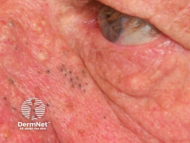
Clinical photography of left periorbital-cheek skin showing solar comedones in actinically damaged facial skin. High-resolution color digital close-up reveals numerous small, dilated follicular openings filled with keratin plugs (open comedones) appearing as black‑brown puncta. Surrounding skin is coarse and parchment-like with flat to slightly raised yellowish areas of elastosis; there are fine telangiectasias and minimal inflammatory erythema. The distribution is characteristic of sun‑exposed facial regions, particularly the malar and periorbital zones; severity suggests Favre-Racouchot syndrome when combined with deep solar elastosis. Visual features include multiple discrete comedones, some coalescing, with minimal surrounding pustulation. The image conveys actinic damage and premature photoaging. Diagnostic significance lies in distinguishing solar comedones from acne vulgaris and other open comedones; clinicians should correlate with patient age, outdoor exposure, smoking history, and ocular surface signs. Differential considerations include acneiform open comedones and Favre-Racouchot syndrome; keratoacanthoma is unlikely in this sun-damaged distribution. Management implications involve sun protection, topical retinoids to promote follicular turnover, comedone extraction, and, in extensive disease, systemic therapy such as isotretinoin. This image is valuable for dermatology education, photodamage assessment, and teaching the appearance of actinic damage-related follicular plugging. Note features include preserved eyelid margin integrity with underlying sun damage; image supports teaching patient counseling on sun avoidance, sunscreen efficacy, and early lesion recognition to prevent progression.
keratin plugs milia sebum plugs coming out of skin causes prevention treatment
What You're Seeing: Keratin/Sebum Plugs (and possibly Milia)
What Each One Is
| Type | What it looks like | Where it forms |
|---|---|---|
| Keratin plug | Firm, white/yellowish cylindrical plug | Hair follicles anywhere on the body |
| Sebum plug | Soft to semi-firm, creamy white | Oily skin areas (face, back, chest, some areas of arms) |
| Milium (milia) | Hard, round, white bead - pops out as a perfect sphere | Face, around eyes, cheeks, but can occur elsewhere |
| Closed comedone (whitehead) | White bump under skin | Anywhere with pores/follicles |
Why They Keep Forming
-
Excess keratin production - Your skin sheds dead cells constantly. Sometimes those cells don't shed properly and instead compact inside the follicle/pore opening.
-
Excess sebum (oil) - Oil produced by sebaceous glands can mix with dead cells and harden inside the pore. This is especially pronounced in people with naturally oily skin.
-
Poor skin cell turnover - When the skin's natural exfoliation cycle slows down (due to age, dehydration, or genetics), dead cells pile up and plug pores.
-
Thick/occlusive products - Heavy creams, sunscreens, or oils that block pores (comedogenic products) trap dead cells inside.
-
Genetics - Some people's follicles are simply shaped in a way that traps material more easily (e.g. keratosis pilaris is a genetic condition causing widespread keratin plugs on the arms).
-
Hormones - Hormonal fluctuations (puberty, menstrual cycle, androgens) increase sebum production dramatically.
-
Sun damage - Chronic UV exposure can cause solar comedones (Favre-Racouchot syndrome), where damaged follicles accumulate keratin plugs, particularly in sun-exposed skin.
How to Prevent and Treat Them
Daily Skincare Routine
- Gentle cleanser twice a day - Use a mild, non-comedogenic face/body wash. Avoid harsh bar soaps that strip the skin barrier, as this triggers even more oil production.
- Do not over-wash - Stripping the skin stimulates compensatory oil production.
Chemical Exfoliants (most effective long-term)
- Salicylic acid (BHA, 0.5-2%) - Oil-soluble, so it penetrates inside the pore and dissolves the plug from within. Use in a cleanser or toner daily or every other day. Best for oily, combination skin.
- Glycolic or lactic acid (AHA, 5-10%) - Water-soluble, works on the surface to loosen dead skin cells. Good for drier skin types. Use 2-3x per week.
Retinoids (gold standard for prevention)
- Retinol (OTC) or tretinoin (prescription) - These speed up cell turnover so dead cells don't accumulate and block pores. They are the most proven treatment for comedones and keratin plugs. Start slowly (2-3x per week at night) to avoid irritation.
Moisturize properly
- Use a lightweight, non-comedogenic moisturizer. Dehydrated skin paradoxically produces more oil and sheds cells unevenly. Look for "non-comedogenic" on the label.
Physical exfoliation
- A gentle exfoliating scrub or brush 1-2x per week can help, but don't be aggressive - micro-tears in skin worsen plugging and cause scarring.
Avoid squeezing or picking
Lifestyle factors
- Stay hydrated - proper hydration helps skin cell turnover
- Change pillowcases frequently - oil and dead cells from your skin transfer back
- Avoid heavy, greasy products on affected areas
- Sun protection - daily SPF prevents UV-induced follicular damage that worsens plugging
When to See a Dermatologist
- The bumps become red, painful, or inflamed (suggests bacterial infection/acne)
- They appear on unusual locations like the palms or soles
- They are hard, white, chalky and very numerous (could be calcinosis cutis, a calcium deposit condition linked to certain medical conditions)
- Home treatments haven't helped after 8-12 weeks
- You're concerned about scarring
Disclaimer: This is general educational information. For a confirmed diagnosis and personalized treatment, consulting a dermatologist is always the best step - especially if these are widespread or persistent.