
Signs of swelling reduction in post traumatic cases. Need extensive research
post-traumatic edema assessment clinical signs reduction
limb edema measurement circumference volumetry trauma
soft tissue swelling fracture surgery timing clinical assessment
post traumatic swelling monitoring lymphedema injury assessment
clinical signs of swelling reduction post traumatic edema assessment wrinkle test skin 2024
wrinkle test soft tissue swelling reduction fracture ankle foot
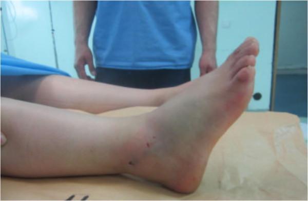
This clinical photograph shows a lateral view of a patient's right lower leg and foot following the reduction of a medial subtalar dislocation. The image demonstrates significant soft tissue swelling (edema) localized around the ankle joint and the distal third of the lower leg, resulting in a loss of normal bony landmarks such as the lateral malleolus. The foot is positioned in a state of moderate plantar flexion. On the anterolateral aspect of the ankle and lower leg, several small, punctate dark red marks are visible, consistent with minor skin abrasions or puncture wounds sustained during the traumatic event. Faint ecchymosis (bruising) is also noted near the posterior aspect of the heel. The overall alignment appears improved compared to a pre-reduction state, though the diffuse swelling masks deep anatomical definition. This image serves as a clinical example of post-reduction monitoring for high-energy ankle trauma and associated soft tissue injury in an emergency or orthopedic setting.
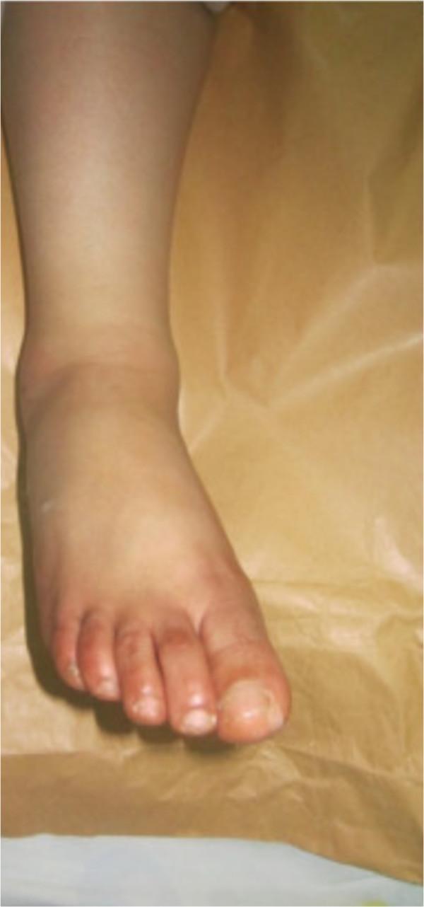
This clinical photograph shows an anterior view of the right foot and lower leg following the closed reduction of a medial subtalar dislocation. The image demonstrates restored anatomical alignment of the foot relative to the leg, resolving the previous clubfoot-like deformity. Significant soft tissue swelling (edema) is visible around the ankle joint and dorsum of the foot, which is a common finding post-trauma and post-reduction. The skin remains intact with no evidence of open wounds or ulcerations. The toes are neutrally aligned in a forward-facing position, and the hallux appears anatomically positioned. This image serves as a visual record of successful conservative management for a high-energy foot injury, highlighting the importance of immediate reduction to prevent skin necrosis and long-term neurovascular complications in subtalar dislocations.
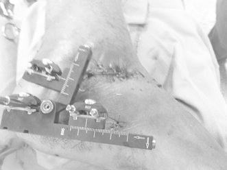
A clinical photograph of an external fixator applied to the hindfoot and ankle region, likely for the management of a calcaneal fracture. The metal device features a primary horizontal rail and a secondary adjustable arm, both containing graduated measurement scales for controlled distraction and fragment reduction. Multiple percutaneous pins are seen entering the soft tissue, secured to the fixator via adjustable locking clamps. Adjacent to the hardware, a linear surgical incision closed with sutures is visible over the lateral aspect of the foot (sinus tarsi area). The surrounding skin shows mild postoperative edema and bruising, consistent with acute surgical trauma. This visual demonstrates the mechanical setup for ligamentotaxis and anatomical alignment in orthopedic trauma surgery, emphasizing pin placement and skin clearance for postoperative swelling.
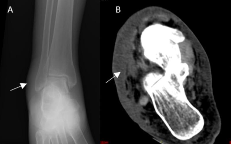
This composite diagnostic image displays a comparison between a conventional radiograph (A) and a computed tomography (CT) scan (B) of a right foot, focusing on the ankle and hindfoot region. Panel A is an anteroposterior X-ray showing marked soft tissue swelling adjacent to the lateral malleolus (indicated by a white arrow), with no evidence of acute fracture, periosteal reaction, or bony erosions. Panel B is an axial CT slice demonstrating high-resolution detail of the same region; it reveals significant subcutaneous edema, skin thickening, and inflammatory fluid infiltration within the soft tissues (arrow). The CT scan confirms the absence of intra-articular collections, gas (emphysema), or cortical bone involvement. These imaging findings are clinically significant for the assessment of cellulitis or soft tissue infection, highlighting the utility of CT in differentiating soft tissue pathology from primary bone disease in the distal lower extremity.
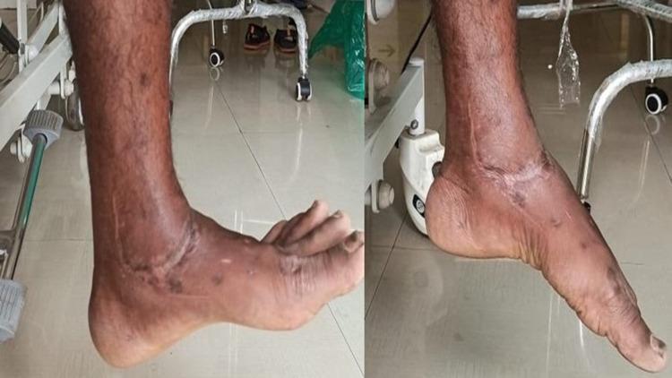
This clinical photograph consists of two side-by-side views of a left ankle and foot, documenting the postoperative range of motion six months following the surgical repair of a complex talar body fracture-dislocation. The images demonstrate the ankle in two distinct positions: a neutral position (left) and active plantarflexion (right). Visible on the anterior and medial aspects of the ankle are well-healed surgical scars, consistent with a direct medial approach used for fracture reduction and internal fixation. There is mild residual soft tissue swelling and hyperpigmentation/discoloration around the ankle joint and dorsum of the foot, indicative of tissue remodeling post-trauma. The photograph serves as clinical evidence of successful functional recovery, showcasing the patient's ability to achieve significant plantarflexion and maintain structural alignment of the foot relative to the lower leg after a severe orthopedic injury. This image is used in medical education to illustrate postoperative outcomes in complex foot and ankle trauma management.
edema reduction signs clinical orthopedic fracture rehabilitation
https://pmc.ncbi.nlm.nih.gov/articles/PMC8310870
post traumatic edema swelling limb clinical photograph
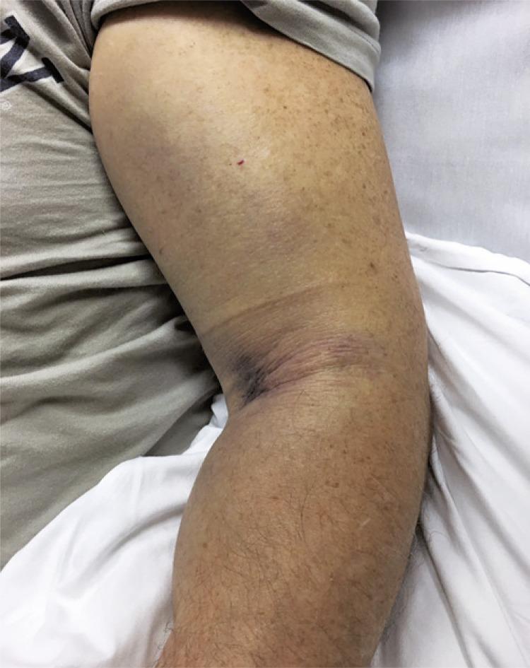
Clinical photograph of a patient's upper limb, demonstrating significant pathology indicative of acute traumatic injury and potential compartment syndrome. The image shows massive edema and soft tissue swelling extending from the mid-humerus to the forearm. A prominent area of ecchymosis and deep purple discoloration is localized to the antecubital fossa and medial elbow region, suggesting a significant underlying hematoma. The skin in the affected area appears taut and shiny due to increased interstitial pressure, with exaggerated skin folds at the elbow crease. Additional features include scattered lentigines and a small puncture or petechial spot on the lateral upper arm. This visual serves as a critical clinical marker for musculoskeletal trauma involving tendon rupture or vascular injury, where intense swelling and pressure sensation necessitate urgent surgical evaluation to prevent neurovascular compromise. The photograph highlights the clinical presentation of pre-operative compartment syndrome risk in the upper extremity.
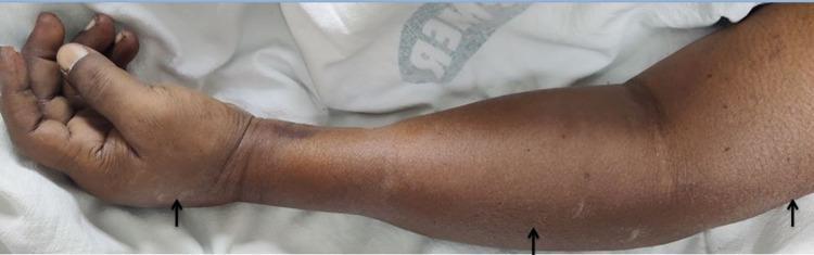
Clinical photograph of a left upper limb demonstrating generalized edema and localized areas of subcutaneous emphysema. The image captures the limb from the hand to the upper arm, showing significant diffuse swelling that distends the anatomical contours. Black arrows highlight specific regions of subcutaneous air accumulation, manifesting as visible puffiness and uneven skin texture over the thenar eminence of the hand, the mid-forearm, and the proximal upper arm. A dark, ecchymotic patch is visible on the dorsal aspect of the wrist joint. This visual finding is characteristic of soft tissue air infiltration, which clinically presents with palpable crepitus. The image serves as a teaching tool for recognizing non-traumatic or traumatic subcutaneous emphysema and the resulting alteration in limb morphology and skin surface integrity in an acute clinical setting.
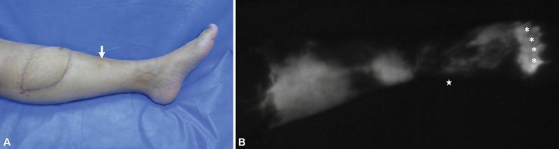
Two-panel image demonstrating post-traumatic lymphedema after reconstructive surgery. Panel A is a clinical photograph of a left lower extremity featuring a previously placed Anterolateral Thigh (ALT) flap on the anterior shin. The leg distal to the flap shows generalized swelling and a white arrow pointing to an indentation, indicating pitting edema. Panel B shows indocyanine green lymphography (ICG-L) of the same limb in a dark field. The image displays a pathological 'stardust' or diffuse fluorescent pattern, signifying lymphatic dysfunction and dermal backflow. Asterisks (*) denote the ICG injection sites in the foot web spaces, and a star indicates the anatomical landmark of the medial malleolus. This composite illustrates how non-lymphatic surgical reconstruction can lead to obstructive lymphedema by disrupting the axiality of lymphatic drainage pathways. Target audience: medical students and surgeons specializing in lymphatic and reconstructive surgery.
Signs of Swelling Reduction in Post-Traumatic Cases
1. The Wrinkle Sign (Skin Wrinkling Test)
- Tibial pilon (plafond) fractures
- Ankle fractures (especially bimalleolar and trimalleolar)
- Calcaneal fractures
- Lisfranc injuries
- Distal tibial fractures
2. Skin Surface and Texture Changes
| Feature at Peak Swelling | Feature During Reduction |
|---|---|
| Taut, shiny, stretched skin | Skin becomes dull, slack, and pliable |
| Loss of normal skin creases | Return of skin folds, creases, and wrinkles |
| "Orange peel" texture (peau d'orange) in severe cases | Smooth, normal skin texture restores |
| Skin blanches with stretching | Blanching disappears; normal capillary refill returns |
| Skin feels warm and tense | Skin temperature normalizes toward the contralateral side |
3. Pitting Edema - Transition and Grading
- The pit depth decreases progressively (from Grade 3-4 to Grade 1 to non-pitting)
- The duration of pitting shortens (the pit rebounds faster)
- Eventually the swelling becomes non-pitting, indicating protein deposition and early fibrosis if untreated, OR complete resolution if managed well
- Stage 0: Asymptomatic, detectable only on lymphoscintigraphy or impedance spectroscopy
- Stage I: Clinically visible, pitting, fully reversible with elevation and compression - this is the typical acute post-traumatic state
- Stage II: Non-pitting; fibrosis begins; no longer fully reversible
- Stage III (Lymphostatic elephantiasis): Severe, irreversible tissue changes
4. Limb Circumference and Volume
- Measured circumference progressively decreases toward the contralateral (uninjured) limb's measurement
- A reduction of >2 cm in circumference compared to peak measurement is a clinically significant sign of improvement
- The "Figure of 8" measurement method is commonly used for ankle and foot edema, measuring a continuous tape around bony landmarks
5. Return of Bony Landmarks
- Malleoli become palpable again in ankle injuries
- Metacarpal heads and knuckles reappear in hand injuries
- Tibial crest and subcutaneous border become visible in leg injuries
- Extensor tendons of the foot or hand become visible under the skin
- The normal concavity around the ankle joint, Achilles tendon, and medial arch of the foot returns
6. Fracture Blister Resolution
- Blood-filled (hemorrhagic) blisters are more serious than clear (serous) blisters and indicate deeper dermal injury
- Re-epithelialization of blister surfaces - confirmed when the blister roof dries, contracts, and the base shows a healthy epithelial surface - signals adequate swelling resolution
- Surgery is deferred until blisters re-epithelialize
7. Pain and Tenderness on Compression
- Resting pain decreases before positional pain
- Pain on gentle palpation (distinct from bony tenderness) reduces as tissue pressure falls
- Pain on passive stretch (an important sign of compartment syndrome) disappears as intracompartmental pressure normalizes
- The limb transitions from a "tight, bursting" quality of pain to a dull ache localized to the fracture/injury site
8. Temperature and Color Normalization
- Skin erythema fades - the skin transitions from red/pink to normal skin tone, then occasionally to a yellow-brown (residual hemosiderin/bruising) before normalizing
- Skin temperature decreases toward baseline - can be tracked with a handheld infrared thermometer (>2°C asymmetry between limbs suggests ongoing inflammation)
- Cyanosis/mottling disappears - in severe swelling, venous outflow compromise can produce a dusky hue; normalization of color signifies improved venous and lymphatic drainage
9. Improved Range of Motion
- Passive range of motion improves before active ROM, as muscle guarding decreases
- Joint line becomes palpable (previously masked by swelling)
- The joint no longer feels "woody" or "boggy" on examination - softening of tissue turgor is a key palpatory sign
- This is particularly well-documented in knee, ankle, and wrist injuries
10. Objective Measurement Tools
| Tool | What It Measures | Clinical Use |
|---|---|---|
| Tape circumference | Limb girth at defined landmarks | Daily/weekly bedside monitoring |
| Water volumetry | Total limb volume displacement | Gold standard for hand/forearm |
| 3D volumetric scanning | Digital surface mapping | Research; increasingly clinical |
| Bioimpedance spectroscopy | Extracellular fluid ratio | Early lymphedema detection (Stage 0) |
| Infrared thermometry | Skin temperature asymmetry | Monitoring inflammatory activity |
| Doppler ultrasound | Venous flow and fluid collections | Rules out DVT complicating edema |
11. Context-Specific Signs by Injury Region
Ankle/Foot
- Return of the wrinkle sign on dorsum and medial/lateral aspects
- Malleoli become visible and palpable
- Tendon outlines of extensors and Achilles return
- Figure-of-8 tape measurement decreases
Hand/Wrist
- Metacarpal head knuckle contours reappear
- Skin over the dorsum softens and wrinkles
- Finger pulp softens; digital ROM improves
- Carpal tunnel pressure-related paresthesias resolve (if not from direct nerve injury)
Knee
- Suprapatellar pouch softens and reduces in volume
- Medial and lateral joint lines become palpable
- Ballottement test reduces (fluid volume decreases)
- Patellar tap sign diminishes as effusion resolves
Thigh/Hip
- Femoral triangle contour returns
- Proximal thigh circumference decreases toward the contralateral limb
- Hip range of motion improves as capsular pressure decreases
12. Pathophysiology Underpinning the Signs
- Trauma triggers the release of histamine, bradykinin, and prostaglandins
- These cause capillary vasodilation and increased permeability
- Protein-rich fluid leaks into the interstitium - dynamic insufficiency of the lymphatic system follows when the fluid load exceeds its transport capacity
- The lymphatics become overwhelmed, and if swelling is not treated promptly, secondary post-traumatic lymphedema may develop, which is irreversible in advanced stages
Clinical Images
Summary Table: Signs of Swelling Reduction
| Sign | Mechanism of Appearance | Clinical Significance |
|---|---|---|
| Positive wrinkle test | Skin regains elasticity as interstitial fluid resorbs | Surgical readiness for ORIF |
| Decreased pitting depth | Reduced interstitial fluid volume | Graded edema regression |
| Return of bony landmarks | Overlying fluid volume reduced | Landmark-based surgical planning |
| Blister re-epithelialization | Dermal-epidermal junction restores as pressure drops | Surgery safe to proceed |
| Decreased circumference | Objective volume reduction | Quantifiable, reproducible progress |
| Skin temperature normalization | Resolution of inflammatory hyperemia | Reduced active inflammation |
| Erythema fading | Decreased prostaglandin/histamine activity | Moving from acute to reparative phase |
| Improved passive ROM | Decreased capsular/periarticular pressure | Rehabilitation readiness |
| Pain reduction on compression | Lower intracompartmental/interstitial pressure | Risk of compartment syndrome reducing |
| Skin softens from taut to pliable | Interstitial pressure normalization | Key pre-operative check |
- Rockwood and Green's Fractures in Adults, 10th Ed. (2025) - soft tissue considerations in tibial pilon and compartment syndrome chapters
- AOFoundation Surgical Timing Guidelines - wrinkle sign for midfoot trauma
- ISL Staging for post-traumatic lymphedema (OAE Plastic and Aesthetic Research, 2021)
- PMC8310870 - perioperative anti-edematous treatment and swelling quantification