
Autosomal polycystic kidney disease
"Polycystic Kidney, Autosomal Dominant"[MeSH Terms]
autosomal dominant polycystic kidney disease
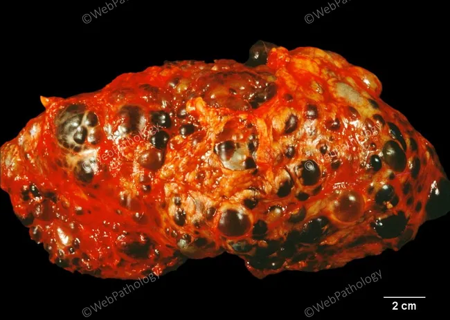
Macroscopic gross pathology photograph of a kidney with autosomal dominant polycystic kidney disease (ADPKD). The external surface is markedly lobulated and studded with variably sized cysts, ranging from small microcysts to several-centimeter cavities. Cyst contents are described as turbid to hemorrhagic fluid, producing a mottled red–orange, sponge-like appearance with darker intracystic fluid in several compartments. The renal cortex and medulla are distorted by an open-ended cystic network, with thinning of intervening parenchyma and areas of compressed, translucent parenchyma between larger cysts. The overall mass is enlarged relative to a normal kidney, reflecting polycystic transformation. Scale bar indicates 2 cm, underscoring that many cysts are several millimeters to a few centimeters in diameter. Such macroscopic morphology is classic for adult-onset, autosomal dominant polycystic kidney disease, often accompanied by progressive loss of functional nephrons and potential rupture of cyst walls with hemorrhage. The surface is irregular, with fibrotic patches and occasional hemostatic clots adherent to the capsule; these features may correspond to chronic hemorrhage and inflammatory changes. Clinically, this gross appearance correlates with a bilateral, enlarged polycystic kidney phenotype and a high risk of renal insufficiency, hypertension, and eventual end-stage renal disease. Radiologic and pathologic correlation with imaging and family history is recommended for definitive diagnosis and guidance of management.
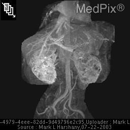
**Imaging Modality:** Magnetic Resonance Angiography (MRA) / Magnetic Resonance Imaging (MRI), Maximum Intensity Projection (MIP) reconstruction.
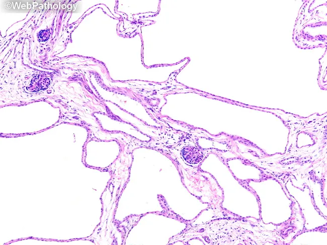
Light microscopic kidney histology depicting a polycystic-appearing parenchyma. The specimen is renal cortex/medulla tissue prepared for hematoxylin and eosin staining and viewed under brightfield illumination. The architecture is distorted by multiple irregularly shaped, dilated tubules that form microcysts, some approaching cystic dilation in diameter. These tubules are separated by very thin, delicate septa containing entrapped glomeruli and residual nephrons, a pattern compatible with collecting duct‑derived cysts. The tubular epithelium lines are cuboidal to flattened, often focusing apically with minimal cytoplasmic detail, and the lumina vary from slit-like to round. Intraluminal contents are scant; the interstitium shows little overt fibrosis or inflammatory infiltrate at this magnification. Overall, the image demonstrates early-to-mid spectrum polycystic kidney changes with preserved, but haphazard cortical-medullary organization. The appearance is consistent with diseases that feature dilated collecting ducts, such as autosomal recessive polycystic kidney disease (ARPKD) or related ciliopathies; differential diagnosis should consider early autosomal dominant polycystic kidney disease, severe hydronephrosis, multicystic dysplastic kidney, glomerulocystic disease, bilateral mesoblastic nephroma, Wilms tumor, lymphoma, tuberous sclerosis, and renal vein thrombosis. Clinically, this finding may reflect neonatal or pediatric renal cystic disease with potential implications for renal function and hypertension management. Correlation with imaging and family history is strongly recommended.
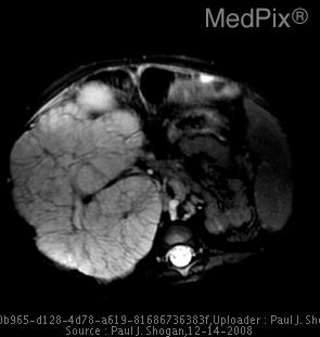
**Imaging Modality:** Magnetic Resonance Imaging (MRI), Axial T2-weighted fat-saturated sequence.
Autosomal Dominant Polycystic Kidney Disease (ADPKD)
Overview
Genetics
- Inheritance is autosomal dominant with complete penetrance for cyst formation. Each child of an affected parent has a 50% risk. At least 10% of families trace to an apparent de novo mutation.
- PKD1 (16p13.3) accounts for about 78% of clinic-based pedigrees. PKD2 (4q21) accounts for about 15%, and no mutation is found in about 7%.
- PKD1 disease is more severe than PKD2, and fully inactivating mutations are worse than incompletely penetrant ones. Patients with both a PKD1 and a PKD2 mutation have worse disease than those with one.
- Rarer genes with an ADPKD-like phenotype are GANAB and DNAJB11.
- About 1% or fewer present in utero or in infancy with enlarged echogenic kidneys that mimic ARPKD. This is often due to hypomorphic alleles in trans. A PKD1/TSC2 contiguous gene deletion gives childhood PKD plus tuberous sclerosis features.
- Intrafamilial variability is marked, which points to modifier genes, environment, and mosaicism. - Brenner and Rector's The Kidney, p. 1983
Pathogenesis
- Cysts arise by clonal proliferation of single tubular epithelial cells, in fewer than 1% of tubules. A "two-hit" model explains this. The patient inherits one mutated allele, and a somatic mutation later inactivates the normal allele. Haploinsufficiency and dosage effects probably contribute too.
- The cysts arise from the collecting duct and other tubular segments. Vasopressin V2 receptor signalling raises cAMP, which drives epithelial proliferation and fluid secretion into the cyst. This is the rationale for tolvaptan and for high water intake. - Brenner and Rector's The Kidney, p. 1998; National Kidney Foundation Primer on Kidney Diseases, 8e
Clinical features
- Renal:
- flank or abdominal pain
- hematuria from cyst hemorrhage
- cyst or urinary tract infection
- nephrolithiasis
- early urinary concentrating defect
- hypertension, which is early and common because of intrarenal RAAS activation
- progressive CKD
- Extrarenal:
- liver cysts (the most common)
- intracranial aneurysms with the risk of subarachnoid hemorrhage
- mitral valve prolapse
- colonic diverticulosis
- hernias
- cysts in the pancreas and spleen
- Children: Few have symptoms before age 15. Hypertension is the earliest and most common systemic feature, in up to 44%. Concentrating defects occur in about 60%. Inguinal hernias are common. Total kidney volume (TKV) grows exponentially at about 7.4% per year. - National Kidney Foundation Primer on Kidney Diseases, 8e
Diagnosis
- Diagnosis is usually by ultrasound plus family history. The Pei-Ravine criteria for a positive family history are:
- age 15 to 39: at least 3 cysts in total
- age 40 to 59: at least 2 cysts in each kidney
- age 60 or over: at least 4 cysts in each kidney
- For at-risk people under 40 with a negative scan, MRI or genetic testing is more sensitive.
- Genetic testing helps when the family history is absent, the presentation is atypical, or a living related kidney donor is being assessed.
- Height-adjusted TKV (HtTKV) on MRI, together with the Mayo imaging class (1A to 1E), predicts the rate of progression.
Management
- Blood pressure:
- ACE inhibitors or ARBs are first line.
- The target is below 130/80 mmHg overall. For adults under 50 with preserved function, the target is below 110/75 mmHg.
- HALT-PKD Study A showed that strict control slowed TKV growth, cut albuminuria, and reduced left ventricular mass. The effect was greatest in those with the most cysts.
- Neither ACE inhibitors nor protein restriction protected kidney function when started late. - National Kidney Foundation Primer on Kidney Diseases, 8e
- Diet and fluids:
- Restrict salt to under 2 g/day. Urinary sodium correlated with TKV growth in the CRISP study.
- Drink more than 3 L/day of water to suppress vasopressin if kidney function is preserved. Avoid extreme intake in advanced CKD because of the risk of hyponatremia.
- Limiting caffeine is theoretical only.
- Tolvaptan (V2 receptor antagonist):
- TEMPO 3:4 (n=1445) showed TKV growth of 2.8% per year versus 5.5% with placebo. The eGFR decline over 3 years was 6.8 versus 10.1 mL/min/1.73 m². Kidney pain and albuminuria also improved.
- REPRISE (n=1370, eGFR 25 to 65) showed a slower eGFR decline: -2.34 versus -3.61 mL/min/1.73 m² per year.
- Aquaresis (thirst, polyuria, nocturia) is the main side effect.
- Transaminases rose above 3× the upper limit of normal in 4.4% of tolvaptan patients versus 1% on placebo. A few patients met Hy's law criteria, so monthly liver tests are needed at the start.
- The FDA approved it in April 2018 for adults at risk of rapid progression. - Brenner and Rector's The Kidney, p. 1998-1999; National Kidney Foundation Primer on Kidney Diseases, 8e
- Complications:
- Treat cyst infection with lipophilic antibiotics that penetrate cysts, such as fluoroquinolones.
- Manage pain stepwise. Cyst decompression is reserved for refractory cases.
- Screen for intracranial aneurysm only in high-risk patients (family history of aneurysm or SAH, a prior rupture, or a high-risk occupation).
- Kidney replacement:
- Peritoneal dialysis is feasible in ADPKD. Outcomes are similar to or better than in other patients, with no excess enteric peritonitis. Hernias and leaks are the main mechanical concerns.
- Transplantation is the preferred treatment. A native nephrectomy is sometimes needed for space or recurrent infection. - National Kidney Foundation Primer on Kidney Diseases, 8e
- Family: Offer genetic counselling. Guidelines do not currently support routine screening of asymptomatic at-risk children. Check their blood pressure and urine regularly. - National Kidney Foundation Primer on Kidney Diseases, 8e
Recent evidence check
- A 2026 review of very early-onset ADPKD in children (PMID 42401882).
- A 2026 review of serum and urine prognostic biomarkers (PMID 42237252).
- A 2024 review of renal neoplasia prevalence in ADPKD (PMID 38301614).