
Children suffering from fever and cold
"fever" AND "upper respiratory infection" AND "children"
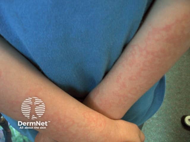
Fever and Cold (Upper Respiratory Tract Infection) in Children
Overview
- Tintinalli's Emergency Medicine, p. 788
- Harrison's Principles of Internal Medicine 22E, p. 297
1. What Is Fever?
- Any temperature above normal is considered a fever.
- In infants, the classic threshold of concern is 38°C (100.4°F) rectally.
- In neonates/infants <2-3 months, hypothermia (<36°C / 96.8°F) is equally alarming.
- Historically, fever >39°C (102.2°F) in children aged 3-36 months raises concern for occult bacteremia.
- Axillary temperatures run ~0.6°C lower than oral, which run ~0.6°C lower than rectal.
Note on moderate fever: Moderate fever is not always harmful - it is associated with decreased microbial reproduction and an enhanced immune response. Temperatures below 39°C in a healthy, comfortable child generally do not require drug treatment.
2. Common Cold (Nonspecific URI) - Causes
- Rhinoviruses (100+ serotypes) - most common
- Coronaviruses
- Parainfluenza virus
- Respiratory syncytial virus (RSV)
- Influenza virus
- Adenovirus
- Metapneumovirus
- Harrison's Principles of Internal Medicine 22E
3. Symptoms
- Nasal fullness, congestion, rhinorrhea (runny nose)
- Sore throat, laryngitis
- Low-grade fever
- Cough
- Lymphadenopathy
- Possible mild myalgias (muscle aches)
4. Management
A. Fever Treatment
| Drug | Dose | Notes |
|---|---|---|
| Paracetamol (Acetaminophen) | 15 mg/kg/dose PO or PR every 4-6 hours (max 80 mg/kg/day, up to 5 doses/day) | First-line; safe from birth |
| Ibuprofen | 10 mg/kg/dose every 6-8 hours (max 40 mg/kg/day) | Use only in children >6 months of age |
-
Aspirin is CONTRAINDICATED in children with viral URIs - it is associated with Reye syndrome (a rare but serious liver and brain condition).
-
Do not alternate acetaminophen and ibuprofen - current evidence shows no benefit and it may be unsafe.
-
Tepid sponge bathing is helpful as a supportive measure when used alongside antipyretic therapy.
-
Tintinalli's Emergency Medicine, p. 788-789
-
Swanson's Family Medicine Review
B. Nasal Congestion (Cold Symptoms)
| Supportive Measure | Details |
|---|---|
| Saline nasal drops | Safe and effective for loosening secretions |
| Bulb syringe suctioning | Especially helpful before feeds and sleep |
| Cool-mist humidifier | Safer than warm-steam humidifiers (avoids burn risk) |
| Warm steam (e.g., shower steam) | Effective for nasal congestion relief |
| Honey (children >1 year) | Can soothe a sore throat |
| Adequate fluids and rest | Prevents dehydration, supports recovery |
Caution: Bulb suctioning done too frequently can cause nasal trauma, mucosal swelling, and worsening congestion.
C. Drugs to AVOID in Young Children
| Drug/Drug Class | Reason to Avoid |
|---|---|
| OTC cough and cold preparations (decongestants, antihistamines) | No proven benefit; significant side effects and toxicity risk in young children |
| Antihistamines | Cold-related rhinorrhea is NOT histamine-mediated; antihistamines do not help and can cause tachycardia, agitation, hyperactivity, and seizures in toxic doses |
| Intranasal/oral decongestants | Not proven effective in young children; can cause significant side effects |
| Codeine/opioids | Not recommended for cough in children |
| Aspirin | Risk of Reye syndrome |
For older children and adolescents, adult-style symptomatic treatment (acetaminophen/NSAIDs for fever, ibuprofen for sore throat, oxymetazoline nasal spray for congestion, dextromethorphan for cough) may be considered.
- Swanson's Family Medicine Review, p. 549-550
- Harrison's Principles of Internal Medicine 22E
D. Antibiotics
5. Red Flags - When to Seek Urgent Care
- Fever >39°C (102.2°F) in a young child, or any fever in a neonate (<1 month)
- Fever lasting >5 days without an obvious source
- Rash with fever (especially petechiae or purpura - possible meningococcemia)
- Stiff neck or bulging fontanelle (possible meningitis)
- Shortness of breath or labored breathing
- Chest pain
- Lethargy, confusion, or the child appears "very ill"
- New ear pain or sinus pain (possible otitis media or sinusitis)
- Symptoms lasting >14 days
- Febrile seizure
6. Age-Specific Considerations
| Age Group | Key Points |
|---|---|
| Neonates (<1 month) | Any fever is an emergency - admit for full sepsis workup (CBC, blood/urine cultures, LP); relative immunodeficiency |
| Infants 1-3 months | High risk for serious bacterial illness; full workup often needed |
| 3-36 months | Source of infection may be unclear; fever >39°C warrants investigation |
| >36 months | Children are easier to evaluate; complaints are usually more localizing; group A Streptococcal pharyngitis becomes more common in school-age children |
7. Complications to Watch For
- Otitis media - ear pain, irritability, tugging at ear
- Bacterial sinusitis - persistent purulent nasal discharge >10-14 days with facial pain
- Pneumonia - cough + fever + respiratory distress
- Meningitis - fever + stiff neck + photophobia + rash
- Kawasaki disease - fever >5 days with strawberry tongue, conjunctivitis, cracked lips, rash, lymphadenopathy (especially in children <5 years; requires aspirin + IV immunoglobulin)
Summary Table: Management at a Glance
| Situation | Action |
|---|---|
| Mild fever (<39°C), child comfortable | Supportive care, reassurance, fluids |
| Fever ≥39°C or child uncomfortable | Acetaminophen 15 mg/kg; Ibuprofen 10 mg/kg if >6 months |
| Nasal congestion (any age) | Saline drops + bulb suction + humidifier |
| Cough/runny nose in <6 yr | NO OTC cough/cold drugs; supportive care only |
| Aspirin for cold/fever | NEVER give to children |
| Antibiotic | Only if bacterial infection confirmed |
| Any fever in neonate <1 month | Emergency - go to hospital immediately |
6 year child hypothermia emergency medicine
"hypothermia" AND "children" AND "emergency"
PMID: 41122889
hypothermia ECG J wave Osborn wave
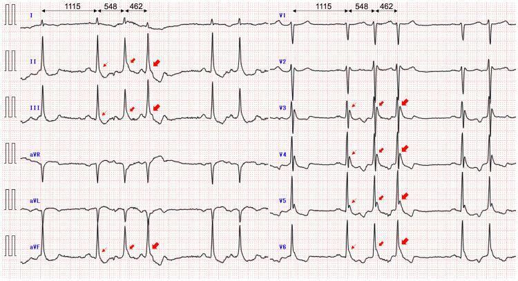
A standard 12-lead electrocardiogram (ECG) demonstrating characteristic findings of hypothermia (recorded at a core body temperature of 28.1°C). The most prominent finding is the presence of Osborn waves (J waves), visualized as a positive deflection or hump at the junction between the QRS complex and the ST segment. These are most marked in the inferior leads (II, III, aVF) and lateral precordial leads (V4-V6), indicated by red arrows. The image shows a dynamic variation in J-wave amplitude, particularly following premature atrial contractions (PACs), where the morphology and height of the deflection change in response to varying RR intervals (labeled as 1115ms, 548ms, and 462ms). Additional features include giant R waves in leads with prominent J waves, a vertical P-wave axis, and a rightward QRS axis shift. The baseline shows minor tremors, common in hypothermic patients. This visual is an educational example of J-point elevation morphology in systemic hypothermia and its relationship with cardiac cycle length.
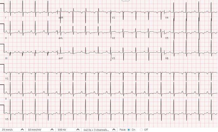
A standard 12-lead electrocardiogram (ECG) displayed on a grid with a paper speed of 25 mm/s and voltage of 10 mm/mV. The tracing demonstrates sinus tachycardia with a heart rate of approximately 105 beats per minute. P waves are present and consistently associated with a narrow QRS complex, suggesting a normal cardiac conduction pathway. The R-R intervals are regular throughout the recording, indicating a stable rhythm without arrhythmias or ectopic beats. Notable morphological features include non-specific T-wave changes in the lateral leads (I, aVL, V5, V6), which are characterized by slight flattening or subtle variations in shape. The precordial leads show normal R-wave progression from V1 to V5. This diagnostic image represents a follow-up ECG following rewarming in a patient with previous hypothermia-related changes, showing the resolution of Osborn (J) waves and first-degree atrioventricular block, returning to a baseline clinical state.
Hypothermia in a 6-Year-Old Child - Emergency Medicine
Definition and Why Children Are at Higher Risk
- Higher body surface-area-to-mass ratio compared to adults = faster heat loss
- Less subcutaneous fat (insulation)
- Limited capacity for sustained shivering thermogenesis
- Children classified as "age extremes" are a mandatory indication for active rewarming
Harrison's Principles of Internal Medicine 22E documents the lowest recorded core temperature with successful resuscitation in a child: 11.8°C in a 2-year-old - demonstrating that profound hypothermia can be survived with aggressive management.
Classification by Severity
| Severity | Core Temperature | CNS | Cardiovascular | Respiratory | Neuromuscular |
|---|---|---|---|---|---|
| Mild | 35°C - 32.2°C (95°F - 90°F) | Amnesia, impaired judgment, dysarthria, apathy | Tachycardia then progressive bradycardia; vasoconstriction | Tachypnea then declining respiratory volume | Shivering (effective) |
| Moderate | <32.2°C - 28°C (90°F - 82.4°F) | EEG abnormalities, progressive LOC, pupil dilation, hallucinations, paradoxical undressing | Pulse and CO falling; atrial/ventricular arrhythmias; J (Osborn) waves on ECG | Hypoventilation; loss of airway reflexes | Hyporeflexia; shivering fails; rigidity |
| Severe | <28°C (<82.4°F) | Coma, loss of ocular reflexes, declining EEG | Severely reduced BP/HR/CO; VF risk maximal; asystole | Apnea; pulmonary edema | No movement; areflexia; no corneal reflexes |
ECG Finding: The Osborn (J) Wave
Emergency Management
Step 1 - Immediate ABC + Stop Heat Loss
- Remove wet/cold clothing immediately
- Dry the child and cover with blankets (including the head - covers ~40% heat loss)
- Move to a warm environment (ambient temp >21°C / 69.8°F)
- Continuous cardiac monitoring (VF risk)
- Core temperature measurement (rectal or esophageal - peripheral thermometers are unreliable)
- IV access; draw labs (CBC, glucose, electrolytes, ABG, coagulation)
Step 2 - Assess Perfusion Status
- Do NOT assume death - in hypothermia, pulses may be undetectable even in perfusing patients; palpate for a full 60 seconds or use bedside echocardiography / ETCO2
- If no signs of life → begin CPR (see cardiac arrest section below)
- Do not withhold CPR based on temperature alone: "No one is dead until they are warm and dead"
Step 3 - Rewarming Strategy
Passive External Rewarming (PER)
- Insulate child in warm environment; cover entire body including head
- Rate: 0.5-2°C per hour
- Appropriate only for: mild hypothermia (>32°C), previously healthy child, able to shiver, no cardiovascular instability
Active External Rewarming (AER)
- Cardiovascular instability
- Core temperature <32°C
- Inadequate rate of passive rewarming
- Age extremes (children are explicitly listed)
- Endocrine insufficiency
- Trauma or secondary hypothermia
| Method | Details |
|---|---|
| Forced-air warming blanket | Most practical and efficient; no afterdrop; safe in ED |
| Warm water circulating pads | Thermostatic systems around trunk |
| Radiant heat source | Useful for infants/young children |
| Hot water bottles | Apply to trunk/axillae/groin only - NOT extremities |
Critical warning: Do NOT apply heat to extremities in chronic/severe hypothermia - this causes peripheral vasodilation, sends cold blood to the core, and causes core temperature afterdrop (continued drop in core temperature after removal from cold). Apply heat to the truncal region only.
Active Core Rewarming (ACR)
| Technique | Details |
|---|---|
| Heated humidified oxygen (40-45°C) | Via mask or ETT; simple, widely available; adds 1-2°C/hr; eliminates respiratory heat loss |
| Warmed IV crystalloids (40-42°C) | Significant heat gain during volume resuscitation |
| Warmed bladder/gastric irrigation | Minimal heat transfer alone; adjunct only |
| ECMO (extracorporeal rewarming) | For cardiac arrest with refractory VF; most effective technique |
Rosen's Emergency Medicine states: "Aggressive treatment of hypothermia is indicated in infants [and children]. Rapid rewarming is advantageous because it minimizes energy expenditure." A neonate with core temp 14.8°C and in CPR made a full neurologic recovery after active external rewarming + warmed IV fluids + heated humidified ventilator gases.
Step 4 - Cardiac Arrest Protocol in Hypothermia
| Core Temp | Defibrillation | Action |
|---|---|---|
| >28°C | Attempt up to 3 shocks at max power | Standard ACLS/PALS |
| <28°C | Defibrillation usually fails | Rewarm to ≥30°C first, then retry |
| Any | CPR | Continue CPR throughout rewarming; do NOT stop |
- The 2025 AHA Guidelines for CPR and Emergency Cardiovascular Care (PMID 41122889) include specific guidance for hypothermic cardiac arrest in children alongside adults.
- ECMO is preferred for refractory hypothermic cardiac arrest when available.
Lab and Monitoring Pitfalls (Critical Pearls)
| Pitfall | Explanation |
|---|---|
| Coagulation tests are falsely normal | Tests run at 37°C; in vivo coagulopathy is present. Treatment is rewarming, NOT plasma/platelets. |
| Hematocrit falsely elevated | Rises 2% for every 1°C drop - anemia may be masked |
| WBC falsely low | White cell sequestration and bone marrow suppression mask infection |
| Electrolytes unpredictable | Hyperkalemia enhanced cardiac toxicity; ECG changes obscured |
| Blood gases: do NOT correct for temperature | An uncorrected pH 7.42 and PCO2 40 mmHg = appropriate at any core temp; over-ventilating causes dangerous alkalosis |
| Hypoglycemia | Common and must be treated immediately; check bedside glucose in every hypothermic child |
| Drug effects are temperature-dependent | Medications work unpredictably when cold; overdosing for "no effect" causes toxicity on rewarming |
Causes to Consider in a 6-Year-Old
- Cold exposure (environmental - most common)
- Near-drowning (cold water submersion)
- Sepsis (infection impairs thermoregulation)
- Hypoglycemia or malnutrition
- CNS injury (head trauma, seizure)
- Toxicological (drug ingestion - alcohol, sedatives, opioids)
- Endocrine (hypothyroidism, adrenal insufficiency - rare at 6 years but possible)
Summary: Emergency Management at a Glance
HYPOTHERMIA IN A 6-YEAR-OLD
         │
         ▼
1. Remove cold/wet clothing. Dry. Cover (including head).
   → IV access, glucose check, cardiac monitor, core temp
         │
         ▼
2. Is there a pulse? (Check 60 seconds or use ECHO)
   ├── YES → Go to rewarming
   └── NO  → Start CPR immediately. Do NOT stop.
         │
         ▼
3. Rewarming (children = age extreme = ACTIVE required)
   Mild (>32°C, stable):  Passive + heated humidified O2
   Moderate (<32°C):      AER (forced air blanket, trunk) + heated O2 + warm IV fluids
   Severe (<28°C, VF):    Active core rewarming + ECMO if available
         │
         ▼
4. Defibrillate only if core temp >28°C
   (or after rewarming to ≥30°C)
         │
         ▼
5. Treat precipitating cause
   Glucose, antibiotics (if sepsis), toxicology screen
Can we give Hydrocortisone in this situation?
Hydrocortisone in Hypothermia - When, Why, and How
Direct Answer
Situation 1 - Secondary Hypothermia from Adrenal Insufficiency
- Thermogenesis is impaired
- The stress response to cold fails
- The patient cannot mount normal cardiovascular and metabolic compensation
Key Clinical Clue: Failure to Rewarm
"Although failure to rewarm spontaneously has been noted in victims with hypothyroidism and other endocrine deficiencies, reserve the use of thyroid hormones and corticosteroids for patients with suspected thyroid and adrenal insufficiency, respectively."
Situation 2 - Myxedema Crisis with Hypothermia
- Severe hypothyroidism is commonly associated with concurrent adrenal insufficiency
- Starting thyroid hormone replacement without covering adrenal function can precipitate acute adrenal crisis (by accelerating cortisol metabolism before the adrenal glands can compensate)
- Hydrocortisone prevents this adrenal crisis and also supports hemodynamic stability
Dosing in Myxedema Crisis (from Tintinalli's Emergency Medicine)
| Drug | Dose | Route | Timing |
|---|---|---|---|
| Hydrocortisone | 100-200 mg | IV | Give first, before starting thyroid hormone |
| Followed by maintenance | 25 mg IV every 6 hours | IV | Until adrenal insufficiency is excluded |
"For myxedema crisis, give a stress dose of hydrocortisone (100 to 200 milligrams IV) at the start of therapy. Obtain serum cortisol levels prior to initiation of therapy, but it is not necessary to wait for results."
- Tintinalli's Emergency Medicine
"Hydrocortisone (50 mg every 6 hours until adrenal insufficiency is excluded) are usually added prior to starting L-thyroxine because of the concern of associated adrenal insufficiency."
Why NOT Routine Hydrocortisone for All Hypothermia?
| Reason | Explanation |
|---|---|
| No benefit in pure accidental hypothermia | Hypothermia from cold exposure alone does not involve adrenal failure; rewarming corrects everything |
| Drug effects are temperature-dependent | At low core temperatures, drugs work unpredictably; dosing for "no effect" when cold causes toxicity on rewarming - a key danger emphasized by Rosen's EM |
| Immunosuppression risk | Corticosteroids impair neutrophil function (already impaired by cold); increases infection risk |
| Hyperglycemia risk | Can worsen metabolic derangements |
| Masks diagnostic features | May obscure signs of underlying infection or inflammation |
Practical Algorithm for Hydrocortisone Decision in a Hypothermic 6-Year-Old
Is the child failing to rewarm despite good technique?
         │
         ├── NO → Continue standard rewarming. No steroids.
         │
         └── YES → Consider secondary hypothermia
                        │
                        ├── Evidence of hypothyroidism?
                        │   (bradycardia, puffy face, dry skin,
                        │    delayed relaxation of reflexes, low T4/TSH)
                        │       └── YES → Myxedema crisis
                        │                 Give Hydrocortisone 100-200 mg IV FIRST
                        │                 Then add thyroid hormone replacement
                        │
                        ├── Evidence of adrenal crisis?
                        │   (refractory hypotension, hyponatremia,
                        │    hyperkalemia, hypoglycemia, skin pigmentation)
                        │       └── YES → Give Hydrocortisone 50-100 mg IV
                        │                 (stress dosing)
                        │
                        └── Neither suspected → Standard rewarming +
                                                treat other causes
                                                (sepsis, toxins, CNS injury)
Important Note for a 6-Year-Old Specifically
- Congenital adrenal hyperplasia (CAH) - inadequately treated
- Autoimmune adrenal insufficiency (Addison's disease in older children)
- Long-term steroid use followed by sudden withdrawal (relative AI)
- Panhypopituitarism (from CNS tumor, craniopharyngioma, or trauma)
- Hypothyroidism (acquired autoimmune or untreated congenital)
Summary
| Scenario | Hydrocortisone? | Dose |
|---|---|---|
| Simple accidental hypothermia (cold exposure) | ❌ No | Not indicated |
| Hypothermia + failure to rewarm + suspected AI | ✅ Yes | 50-100 mg IV |
| Myxedema crisis with hypothermia | ✅ Yes (before thyroid hormone) | 100-200 mg IV |
| Septic shock + hypothermia (refractory) | Consider (stress dose) | 1-2 mg/kg IV |
Bottom line: Hydrocortisone treats the endocrine cause of hypothermia, not the hypothermia itself. The treatment of hypothermia is rewarming.
I have seen doctor prescribing hydrocortisone inj in hyperthermia, bradycardia and low spo2 after surgeries like tkr and thr
Hydrocortisone After TKR/THR - The Clinical Reasoning
The Post-Arthroplasty Triad: What Causes It?
| Condition | Hyperthermia | Bradycardia | Low SpO2 | Timing |
|---|---|---|---|---|
| Fat Embolism Syndrome (FES) | Yes (fever) | Reflex/compensatory | Yes (key feature) | 12-72 hrs post-op |
| Bone Cement Implantation Syndrome (BCIS) | Possible | Yes (cardiac depression) | Yes (V/Q mismatch) | Intraoperative/immediate |
| Pulmonary Embolism (PE) | Yes (low-grade) | Reflex bradycardia in massive PE | Yes | Hours to days post-op |
| SIRS / Post-op Systemic Inflammatory Response | Yes | Can occur | Yes | 24-48 hrs post-op |
| Anaphylaxis (cement, antibiotics, latex) | Yes | Possible | Yes (bronchospasm) | Intraoperative/immediate |
1. Fat Embolism Syndrome (FES) - The Most Likely Culprit
What Happens in TKR/THR?
- Lodge in the pulmonary microvasculature → mechanical obstruction → hypoxia, low SpO2
- Trigger free fatty acid release → toxic damage to capillary-alveolar membrane → ARDS
- Release vasoactive amines and prostaglandins → systemic inflammatory response → fever
- Can cross to the systemic circulation (through a patent foramen ovale) → cerebral involvement
Classic Triad of FES (Gurd's Criteria)
- Respiratory - hypoxemia, low SpO2, ARDS pattern on chest X-ray (bilateral diffuse infiltrates)
- Neurological - confusion, restlessness, deteriorating consciousness
- Petechial rash - conjunctiva, axillae, neck skin folds (pathognomonic)
Role of Hydrocortisone / Corticosteroids in FES
"There is currently no evidence supporting the use of steroids, heparin, or dextran in the management of FES."
- Miller's Anesthesia 10e
"The use of corticosteroid therapy in preventing or treating fat embolism syndrome is controversial."
- Morgan and Mikhail's Clinical Anesthesiology 7e
- FES involves a massive systemic inflammatory response mediated by free fatty acids, prostaglandins, and vasoactive amines
- Corticosteroids are potent anti-inflammatory and membrane-stabilizing agents
- They reduce capillary leak, attenuate the inflammatory cascade, and may reduce pulmonary edema
- Some older studies showed prophylactic methylprednisolone reduced FES incidence after long bone fractures
- In clinical practice, when a patient is deteriorating rapidly with FES, hydrocortisone is often given empirically as a "stabilizing" measure while definitive support (oxygen, ventilation) is established
2. Bone Cement Implantation Syndrome (BCIS) - The Intraoperative/Immediate Post-Op Cause
Mechanism
- Bone marrow debris, fat, and cement monomer are forced into the venous circulation
- Cause pulmonary embolization → right ventricular failure → hypotension, hypoxia, low SpO2
- Methyl methacrylate monomer is directly cardiotoxic → bradycardia, arrhythmias
- Release of cytokines and cyclooxygenase products → pulmonary vasoconstriction → bronchoconstriction
Management of BCIS (from Miller's Anesthesia 10e)
- 100% oxygen
- Fluid resuscitation
- Vasopressors (epinephrine is preferred to off-load the right ventricle)
- Mechanical ventilation if needed
- The cytokine storm and COX-product release mimics an inflammatory/anaphylactoid reaction
- Corticosteroids help blunt this inflammatory cascade
- It is used alongside epinephrine in the management of anaphylactoid reactions to cement
3. Anaphylaxis/Anaphylactoid Reaction
- Bone cement (methyl methacrylate) - anaphylactoid (non-IgE mediated)
- Prophylactic antibiotics (cefazolin, vancomycin) - true anaphylaxis
- Latex (gloves, tourniquet components)
- Blood products (if transfused)
Classic Anaphylaxis Triad After Surgery
- Hypotension (vasodilation)
- Bronchospasm → low SpO2
- Urticaria/rash (may be missed under surgical drapes)
- Fever can occur in anaphylactoid reactions
Hydrocortisone Dosing in Anaphylaxis - Well-Established Indication
| Drug | Role | Dose (Adult) |
|---|---|---|
| Epinephrine (Adrenaline) | First-line - MUST give first | 0.3-0.5 mg IM |
| Hydrocortisone | Second-line - prevents biphasic reaction | 200 mg IV |
| Chlorphenamine | Second-line antihistamine | 10 mg IV |
| IV fluids | For hypotension | Rapid bolus |
Important: In anaphylaxis, hydrocortisone does NOT act fast enough to treat the acute attack - it takes 4-6 hours to work. Its role is to prevent a biphasic (second-wave) reaction 4-12 hours later. Epinephrine always comes first.
4. Post-Operative SIRS (Systemic Inflammatory Response Syndrome)
- TNF-alpha, IL-1, IL-6 surge in the first 24-48 hours
- This causes: fever, tachycardia, mild hypoxia (from microatelectasis)
- Rarely causes significant bradycardia
Summary: Why Doctors Give Hydrocortisone After TKR/THR
| Indication | Evidence Level | Mechanism | Dose |
|---|---|---|---|
| FES - anti-inflammatory | Controversial; no RCT proof but widely used | Reduces inflammatory cascade, capillary leak | 100-200 mg IV |
| BCIS - anti-inflammatory | Empirical; part of supportive care | Blunts cytokine/COX storm | 100-200 mg IV |
| Anaphylaxis | Strong evidence (second-line) | Prevents biphasic reaction | 200 mg IV |
| Perioperative steroid cover (patient on chronic steroids) | Strong evidence | Replaces suppressed HPA axis | 50-100 mg IV |
| Adrenal insufficiency | Strong evidence | Hormone replacement | 50-200 mg IV |
The Honest Bottom Line
- Empirical anti-inflammatory management in a deteriorating patient
- Rational based on the known mechanism (inflammatory cascade)
- Low-risk relative to the severity of the situation
- Practiced widely despite limited hard evidence
Gentamicin used for??
Gentamicin - Complete Clinical Guide
What Is It?
Mechanism of Action
- Enters the bacterial cell - transported across the bacterial cell membrane via an oxygen-dependent active transport system (this is why it is ineffective against anaerobes - no oxygen, no entry)
- Binds irreversibly to the 30S ribosomal subunit - causes misreading of mRNA codon → production of faulty/nonsense proteins → insertion of wrong amino acids → abnormal proteins disrupt the cell membrane → increased permeability → more drug enters → cell death
Antibacterial Spectrum
| Category | Coverage |
|---|---|
| Gram-negative aerobes (primary use) | Pseudomonas aeruginosa, Klebsiella, E. coli, Proteus, Enterobacter, Serratia marcescens, Acinetobacter |
| Gram-positive (synergy only) | Staphylococcus aureus, Streptococcus, Enterococcus (only when combined with cell-wall agents) |
| No activity | Anaerobes (Bacteroides, Clostridium), Streptococci alone, most intracellular organisms |
Clinical Uses - Where Is Gentamicin Used?
1. Serious Gram-Negative Infections (IV/IM - Main Use)
- Sepsis / bacteremia from gram-negative organisms
- Hospital-acquired (nosocomial) pneumonia - Pseudomonas, Enterobacter, Klebsiella (but NOT as single agent - poor lung penetration due to low pH and oxygen tension in infected lung)
- Complicated urinary tract infections (pyelonephritis, urosepsis) - excreted in urine at high concentrations
- Peritonitis / intraabdominal sepsis - combined with metronidazole for anaerobic cover
- Meningitis from gram-negative organisms (when beta-lactams fail)
- Osteomyelitis and septic arthritis caused by gram-negative organisms
- Neonatal sepsis - a key empiric drug in combination (ampicillin + gentamicin)
- Febrile neutropenia - empiric cover in immunocompromised patients
2. Endocarditis (Synergistic Use) - Gram-Positive Cover
| Organism | Combination |
|---|---|
| Enterococcal endocarditis | Gentamicin + Ampicillin OR Vancomycin |
| Streptococcal endocarditis | Gentamicin + Penicillin (shortens treatment duration) |
| Staphylococcal prosthetic valve endocarditis | Gentamicin + Vancomycin + Rifampicin |
Mechanism: Cell wall agents (penicillin, vancomycin) create pores in the gram-positive cell wall, allowing gentamicin to enter (normally excluded) → synergistic bactericidal killing. This synergy is the reason gentamicin is used even against organisms that seem "resistant" to it alone.
Important: For synergistic dosing in endocarditis, target peak 3-4 mcg/mL, much lower than standard dosing (peak 6-10 mcg/mL).
3. Topical Use
- Infected wounds, burns, ulcers - 0.1% cream or ointment
- Eye infections (conjunctivitis, keratitis, blepharitis) - 0.3% ophthalmic drops or ointment
- Intravitreal injection - for serious bacterial endophthalmitis
4. Intrathecal/Intraventricular (Rare)
- Gram-negative meningitis refractory to beta-lactams or severe beta-lactam allergy
- Largely replaced by 3rd-generation cephalosporins
5. Intratympanic Injection - Special Use in ENT
- Ménière's disease - intratympanic gentamicin selectively destroys vestibular hair cells to ablate the labyrinthine function on the affected side, reducing vertigo attacks
- This is a deliberate ototoxic effect used therapeutically
Dosing (Important Concepts)
Once-Daily (Extended-Interval) vs. Divided Dosing
| Feature | Once-Daily (5-7 mg/kg/24h) | Divided (1.7-2 mg/kg q8h) |
|---|---|---|
| Pharmacology | Exploits concentration-dependent killing | Older method |
| Toxicity | Less nephrotoxic (long drug-free period allows renal recovery) | More cumulative renal exposure |
| Efficacy | Equal or better (higher peak/MIC ratio) | Standard |
| Preferred in | Most adult infections | Endocarditis, neonates, pregnancy |
Pediatric Dosing (Harriet Lane Handbook)
- Children (eGFR >75 mL/min/1.73m²): 7.5 mg/kg/24h divided q8h
- Neonates: dose by postconceptional age (see neonatal table - 4-5 mg/kg q24-48h)
- Cystic fibrosis: 7.5-10.5 mg/kg/24h (higher clearance requires higher dose)
Therapeutic Drug Monitoring (TDM) - Mandatory
| Parameter | Target |
|---|---|
| Peak (standard dosing) | 6-10 mcg/mL (general); 8-10 mcg/mL (severe sepsis, CF, osteomyelitis) |
| Trough | <2 mcg/mL (toxicity threshold) |
| Synergy dosing (endocarditis) | Peak 3-4 mcg/mL; Trough <1 mcg/mL |
| Sampling time | Trough: 30 min before 3rd dose; Peak: 30-60 min after 3rd dose |
Toxicities - The Critical Dangers
1. Nephrotoxicity (Kidney Damage)
- Occurs in 5-25% of patients receiving gentamicin >3-5 days
- Caused by accumulation in renal proximal tubule cells
- Presents as non-oliguric acute tubular necrosis (rising creatinine, falling urine output)
- Usually reversible on stopping the drug
- Risk factors: pre-existing renal disease, volume depletion, concurrent nephrotoxins (NSAIDs, vancomycin, contrast agents, amphotericin)
2. Ototoxicity (Ear Damage)
- Occurs in 1-5% of patients on gentamicin >5 days
- Often irreversible
- Manifests mainly as vestibular toxicity (dizziness, loss of balance, oscillopsia) - more common than cochlear
- Hearing loss (cochlear toxicity) also occurs
- Worsened by concurrent loop diuretics (furosemide) - avoid combination
- Partly genetically determined - linked to mitochondrial DNA mutations
3. Neuromuscular Blockade
- Rare but life-threatening
- Can cause respiratory paralysis - especially dangerous in patients on neuromuscular blocking agents (surgery) or those with myasthenia gravis
- Caution in patients receiving anesthetics or neuromuscular blockers
4. No activity in pregnancy (Category D for IV use) - avoid if possible
Contraindications and Cautions
| Situation | Action |
|---|---|
| Renal impairment | Reduce dose or extend interval; monitor TDM closely |
| Pre-existing hearing loss | Use with extreme caution or avoid |
| Concurrent loop diuretics | Avoid if possible; increases ototoxicity risk |
| Neuromuscular disease (myasthenia) | Avoid or use with extreme caution |
| Pregnancy | Avoid IV use (Category D); topical/ophthalmic = Category C |
| Anaerobic infections alone | Ineffective - add metronidazole |
| Monotherapy for pneumonia | Do not use alone - poor lung penetration |
Quick Summary Reference
| Feature | Details |
|---|---|
| Class | Aminoglycoside antibiotic |
| Source | Micromonospora purpurea |
| Action | Binds 30S ribosome → bactericidal |
| Spectrum | Gram-negative aerobes; synergy with gram-positives |
| No cover | Anaerobes, Streptococci (alone), intracellular organisms |
| Route | IV, IM, topical, ophthalmic, intratympanic, intrathecal |
| Main uses | Gram-negative sepsis, UTI, endocarditis (synergy), neonatal sepsis, eye infections |
| Dose (child) | 7.5 mg/kg/day ÷ q8h (normal renal function) |
| Monitor | Peak and trough levels; renal function; hearing |
| Key toxicities | Nephrotoxicity (reversible), Ototoxicity (irreversible), Neuromuscular blockade |