
patient presents with swelling on the of theaspect anterior experiences tingling in the lateral digits, the thenar sensation over the yeteminence remains intact. To relieve the superficial incision is pressure,nerve is most likely Which wihplanneD- being targeted? Ulnar nerve motSuperficial branch of the median nerve Median nerve Radial nerve
- Swelling on the anterior (volar/palmar) aspect of the wrist
- Tingling in the lateral digits (thumb, index, middle, and radial half of ring finger) - the median nerve's sensory distribution
- Thenar eminence sensation intact - this is the key distinguishing clue. The palmar cutaneous branch of the median nerve arises proximal to the carpal tunnel and travels superficially, so it is spared when the median nerve is compressed within the tunnel. This explains why thenar skin sensation is preserved even though the nerve itself is compressed.
| Nerve | Why excluded |
|---|---|
| Ulnar nerve | Supplies medial 1.5 digits; runs through Guyon's canal, not the carpal tunnel |
| Superficial branch of median nerve | This is the palmar cutaneous branch - it's SPARED here (thenar sensation intact), not the one being compressed |
| Radial nerve | Supplies dorsal (back) of hand; no role in anterior wrist/thenar pathology |
Generate an anatomy diagram showing median nerve branches at the wrist
Detailed anatomy diagram of the median nerve branches at the wrist and hand. Show: the main median nerve trunk entering the carpal tunnel beneath the flexor retinaculum (transverse carpal ligament), the palmar cutaneous branch (superficial branch) arising proximal to the carpal tunnel and traveling superficially over the thenar eminence, the recurrent motor branch (thenar branch) curving back to innervate the thenar muscles (abductor pollicis brevis, opponens pollicis, flexor pollicis brevis), and the digital sensory branches supplying the thumb, index finger, middle finger, and radial half of the ring finger. Label all structures clearly: median nerve, palmar cutaneous branch, carpal tunnel, flexor retinaculum, recurrent motor branch, thenar muscles, common palmar digital nerves, proper palmar digital nerves. Use a clean medical illustration style with anterior (palmar) view of right hand and wrist, color-coded nerve branches.
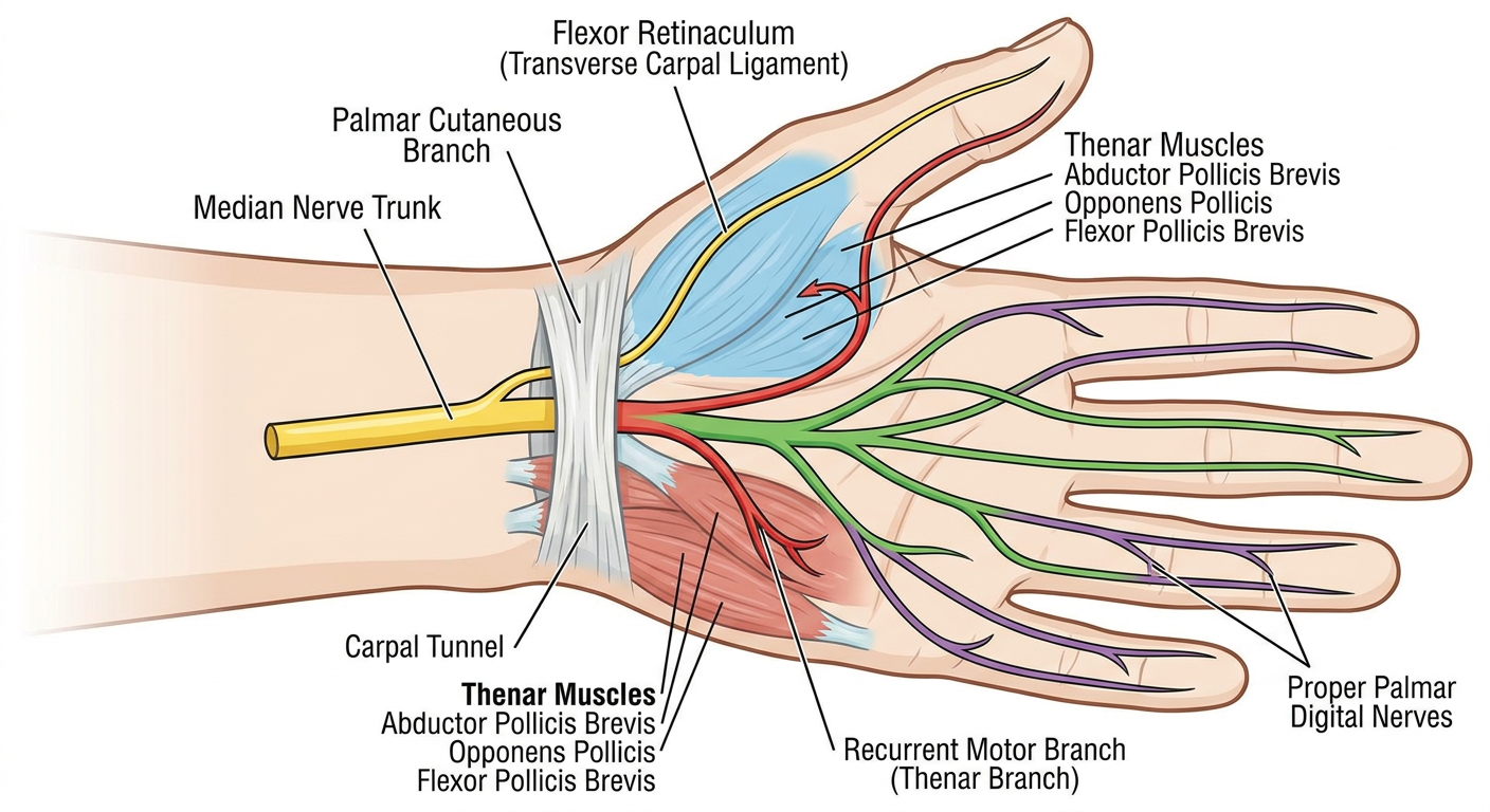
- Palmar cutaneous branch - branches off the median nerve ~5 cm proximal to the wrist crease, travels superficially (outside the tunnel) to supply skin over the thenar eminence. This is why thenar sensation is SPARED in carpal tunnel syndrome.
- Median nerve trunk passes under the flexor retinaculum alongside the 9 flexor tendons. This is the site of compression in CTS.
- Recurrent motor branch (thenar branch) - hooks back radially to innervate the thenar muscles (ABductor pollicis brevis, Opponens pollicis, superficial head of Flexor pollicis brevis). Compression here causes thenar wasting.
- Common palmar digital nerves - divide into proper digital nerves supplying:
- Thumb (both sides)
- Index finger (both sides)
- Middle finger (both sides)
- Ring finger (radial/lateral half only)