
Pathophysiology of myasthenia gravis.. how does pyridostigmine helps
Myasthenia Gravis: Pathophysiology and How Pyridostigmine Helps
1. Normal Neuromuscular Junction (NMJ) - The Baseline
2. Pathophysiology of Myasthenia Gravis
The Core Defect: Postsynaptic Autoimmune Attack
| Antibody | Frequency | Effect |
|---|---|---|
| Anti-AChR (anti-alpha-1 subunit of nicotinic AChR) | 80-90% of generalized MG; 50% of ocular MG | Receptor destruction, blockade, accelerated degradation |
| Anti-MuSK (muscle-specific tyrosine kinase) | Small % of AChR-negative cases | Disrupts AChR clustering at NMJ; more facial/bulbar involvement |
| Anti-LRP4 (lipoprotein receptor-related protein 4) | 1-3% of all patients | Mild-moderate symptoms |
- Bradley and Daroff's Neurology in Clinical Practice; Tintinalli's Emergency Medicine
Mechanisms by Which Anti-AChR Antibodies Cause Damage
- Receptor blockade - Antibodies physically block the ACh binding site, preventing ACh from attaching to its receptor.
- Accelerated degradation - Antibody binding cross-links adjacent AChRs, triggering endocytosis and lysosomal destruction, causing a net reduction in receptor number.
- Complement-mediated destruction - AChR antibodies are complement-fixing; complement activation destroys the postsynaptic membrane and its junctional folds. The normal infolding of junctional folds is reduced, the synaptic cleft is widened, and synaptic architecture is disrupted.
How This Reduces the Safety Factor and Causes Fatigability
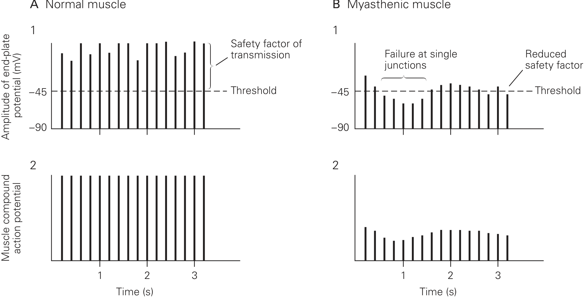
Role of the Thymus
3. Mechanism of Action of Pyridostigmine
- AChE inhibition - Pyridostigmine binds reversibly to AChE at the NMJ, blocking it from hydrolyzing ACh in the synaptic cleft.
- ACh accumulates - Because ACh is not broken down, its concentration in the cleft rises and it persists for longer.
- Increased receptor occupancy - With more ACh present for longer, the probability that ACh molecules find and bind to the reduced number of remaining receptors increases substantially.
- EPP amplitude restored - More receptor activation generates a larger summed EPP that can more reliably exceed the threshold, restoring muscle action potential generation.
- Safety factor partially restored - Fatigability is reduced because even during sustained activity, enough ACh remains in the cleft to continue activating the available receptors.
4. Pyridostigmine Pharmacology
| Property | Detail |
|---|---|
| Class | Reversible AChE inhibitor (carbamate) |
| Route | Oral (primarily for chronic MG management) |
| Duration of action | 3-6 hours (longer than neostigmine's 30 min-2 hrs) |
| CNS penetration | Quaternary nitrogen = does NOT cross the blood-brain barrier; purely peripheral action |
| Preferred over neostigmine | For chronic outpatient MG due to longer duration and oral bioavailability |
- Lippincott Illustrated Reviews: Pharmacology; Barash Clinical Anesthesia, 9th Ed.
- Salivation, lacrimation, urination, defecation (SLUD)
- Abdominal cramping, diarrhea, nausea
- Bradycardia, bronchospasm
- Miosis
5. Why Pyridostigmine is Symptomatic, Not Curative
- Bradley and Daroff's Neurology in Clinical Practice (chapter on NMJ disorders)
- Eric Kandel, Principles of Neural Science, 6th Ed.
- Ganong's Review of Medical Physiology, 26th Ed.
- Costanzo Physiology, 7th Ed.
- Lippincott Illustrated Reviews: Pharmacology
- Tintinalli's Emergency Medicine
Mg vs op poisoning
Myasthenia Gravis vs. Organophosphate Poisoning - A Detailed Comparison
Quick Conceptual Summary
| Myasthenia Gravis | OP Poisoning | |
|---|---|---|
| Core defect | Too few AChRs (postsynaptic) | Too much ACh (AChE irreversibly inhibited) |
| ACh level at NMJ | Normal | Markedly elevated |
| ACh receptor | Blocked/destroyed by antibodies | Normal, but overstimulated |
| AChE | Normal | Irreversibly inhibited |
| Net effect | ACh cannot act effectively | ACh acts excessively and persistently |
1. Pathophysiology
Myasthenia Gravis
- Autoimmune - IgG antibodies target the alpha-1 subunit of nicotinic AChR on the postsynaptic muscle membrane.
- Antibodies cause: receptor blockade, accelerated receptor degradation, and complement-mediated destruction of postsynaptic folds.
- Result: 70-90% reduction in functional AChRs. End-plate potential (EPP) is barely above or below firing threshold. Safety factor of NMJ transmission is severely reduced.
- Presynaptic nerve and ACh release are completely normal.
- Clinically: fatigable weakness that worsens with use and improves with rest. - Bradley and Daroff's Neurology
Organophosphate Poisoning
- OPs (insecticides, nerve agents) irreversibly bind to and inactivate acetylcholinesterase (AChE) at all cholinergic synapses - NMJ, autonomic ganglia, and CNS.
- AChE cannot break down ACh, so ACh accumulates in the cleft and continues stimulating all cholinergic receptors persistently.
- The enzyme undergoes "aging" (irreversible covalent phosphorylation), making the inhibition permanent unless treated early with pralidoxime.
- Carbamates (e.g., neostigmine, pyridostigmine) cause similar but reversible inhibition - they do NOT undergo aging.
- Clinically: features of ACh excess at three receptor sites simultaneously. - Lippincott Illustrated Reviews: Pharmacology; Adams and Victor's Neurology
2. Clinical Features
MUSCARINIC Effects (smooth muscle, glands) - Mnemonic: DUMBELS / SLUDGE
| Feature | OP Poisoning | MG |
|---|---|---|
| Miosis (pinpoint pupils) | ✅ Yes | ❌ No |
| Salivation / hypersecretion | ✅ Yes | ❌ No |
| Lacrimation | ✅ Yes | ❌ No |
| Bronchospasm / bronchorrhea | ✅ Yes | ❌ No (though may get aspiration) |
| Bradycardia | ✅ Yes | ❌ No |
| Diarrhea, urinary incontinence | ✅ Yes | ❌ No |
| Emesis, abdominal cramps | ✅ Yes | ❌ No |
| Sweating | ✅ Yes | ❌ No |
NICOTINIC Effects (NMJ and autonomic ganglia)
| Feature | OP Poisoning | MG |
|---|---|---|
| Muscle weakness | ✅ Yes (depolarizing blockade) | ✅ Yes (insufficient EPP) |
| Fasciculations | ✅ Yes (NMJ over-activation) | ❌ No |
| Tachycardia / hypertension | ✅ Yes (ganglionic) | ❌ No |
| Mydriasis (can occur) | ✅ (ganglionic > muscarinic) | ❌ No |
CNS Effects (OP only - crosses BBB)
| Feature | OP Poisoning | MG |
|---|---|---|
| Anxiety, restlessness | ✅ Yes | ❌ No |
| Seizures | ✅ Yes | ❌ No |
| Coma | ✅ Yes (severe cases) | ❌ No |
| LOC / confusion | ✅ Yes | ❌ No |
MG has no autonomic or CNS features - it is purely a peripheral skeletal muscle weakness problem. OP poisoning causes a pan-cholinergic syndrome affecting everything. - Lippincott Illustrated Reviews: Pharmacology
3. The Critical Overlap - and How to Distinguish Them
| Distinguishing Feature | MG | OP Poisoning |
|---|---|---|
| Pupils | Normal (or slightly large from CN3 palsy) | Miosis (pinpoint) |
| Secretions | Dry | Wet (salivation, bronchorrhea, sweating) |
| Fasciculations | Absent | Present |
| Bradycardia | Absent | Present |
| CNS involvement | Absent | Present (seizures, coma) |
| Response to atropine | No benefit for weakness | Reverses muscarinic features |
| Anti-AChR antibodies | Present (80-90%) | Absent |
| Edrophonium (Tensilon) test | Improves strength | Worsens / no change (already too much ACh) |
| Course | Chronic, fluctuating | Acute, exposure history |
| Smell | Normal | May smell of garlic/petroleum |
- Tintinalli's Emergency Medicine; Goodman & Gilman's Pharmacological Basis of Therapeutics
4. Cholinergic Crisis vs. Myasthenic Crisis (Critical Distinction)
| Myasthenic Crisis | Cholinergic Crisis | |
|---|---|---|
| Cause | Disease exacerbation, infection, under-treatment | Excess AChE inhibitor (same drugs as MG treatment, or OP poisoning) |
| Weakness | Yes | Yes (depolarizing blockade of NMJ) |
| Secretions/bradycardia/miosis | Absent | Present |
| Fasciculations | Absent | Present |
| Edrophonium test | Improves strength | Worsens / causes fasciculations |
| Treatment | More AChE inhibitor, IVIG, plasma exchange | Withhold AChE inhibitor, give atropine |
5. Why Pyridostigmine Helps in MG but is Part of the Problem in OP Poisoning
| MG | OP Poisoning | |
|---|---|---|
| AChE status | Normal - just too few receptors | Already severely inhibited/destroyed |
| Adding AChE inhibitor | Beneficial - raises ACh to compensate for few receptors | Dangerous - piles excess ACh on top of already-maximal ACh, worsening depolarization blockade |
| Treatment goal | Increase ACh at the NMJ | Remove OP from AChE (pralidoxime), block muscarinic excess (atropine) |
6. Treatment Summary
| Treatment | MG | OP Poisoning |
|---|---|---|
| Pyridostigmine/Neostigmine | ✅ First-line symptomatic | ❌ Contraindicated |
| Atropine | Used only for AChI muscarinic side effects | ✅ Primary antidote (blocks muscarinic excess) |
| Pralidoxime (2-PAM) | Not used | ✅ Reactivates AChE if given before "aging" |
| Immunosuppressants | ✅ Steroids, azathioprine | ❌ Not relevant |
| Thymectomy | ✅ If thymoma or refractory disease | ❌ Not relevant |
| Plasma exchange / IVIG | ✅ For myasthenic crisis | ❌ Not relevant |
| Benzodiazepines | Not needed | ✅ For OP-related seizures |
Key Sources
- Bradley and Daroff's Neurology in Clinical Practice
- Goodman & Gilman's Pharmacological Basis of Therapeutics
- Lippincott Illustrated Reviews: Pharmacology
- Adams and Victor's Principles of Neurology, 12th Ed.
- Tintinalli's Emergency Medicine
How do pralidoxime and atropine work together in OP poisoning?
Atropine + Pralidoxime in OP Poisoning: A Complementary Dual Strategy
Step 1: Understand the Problem - Three Sites of Toxic ACh Excess
| Site | Receptor Type | What Goes Wrong |
|---|---|---|
| Parasympathetic end organs (glands, heart, smooth muscle) | Muscarinic | DUMBELS syndrome |
| Neuromuscular junction (skeletal muscle) | Nicotinic (peripheral) | Fasciculations → paralysis |
| Brain | Nicotinic + Muscarinic (central) | Seizures, coma |
Atropine - "Block the Flood"
- Bronchospasm and bronchorrhea (most critical - the primary cause of death)
- Bradycardia and heart block
- Hypersalivation, lacrimation, sweating
- GI hypermotility (diarrhea, urination, vomiting)
- Miosis
- Skeletal muscle weakness, fasciculations, or respiratory muscle paralysis - these are nicotinic effects, and atropine has no activity at nicotinic receptors
- CNS seizures/coma - atropine does cross the BBB (it is a tertiary amine), so it does provide some central muscarinic blockade, but it cannot reverse the nicotinic and other CNS effects of ACh excess
- Start with 1-3 mg IV (0.05 mg/kg in children), doubling the dose every 5 minutes
- Titrate to the endpoint of drying of respiratory secretions and ease of breathing - NOT to heart rate or pupil size (those are unreliable endpoints)
- Severe poisoning may require 200-500 mg in the first hour, and up to 1 gram/day for weeks in extreme cases
- An infusion of 10-20% of the total cumulative atropinization dose per hour maintains effect
"Atropine is not active at nicotinic receptor sites and will not reverse skeletal muscle effects, such as respiratory muscle paralysis." - Rosen's Emergency Medicine
Pralidoxime (2-PAM) - "Restore the Enzyme"
- OPs phosphorylate the serine -OH group at the active site of AChE. This is the same serine that normally performs catalytic hydrolysis of ACh. With serine blocked, AChE is dead.
- Pralidoxime's oxime group (=NOH) competes with the serine -OH for the phosphorus atom.
- The oxime attacks the phosphorylated-enzyme complex, cleaves the phosphorus-serine bond, and regenerates the free serine -OH at the active site.
- AChE is now active again - it can once again hydrolyze ACh in the cleft.
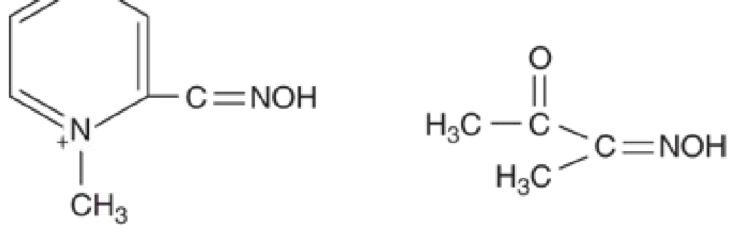
- NMJ effects: fasciculations, muscle weakness, respiratory muscle paralysis
- Reverses the root cause of toxicity (ACh excess) rather than just blocking downstream receptors
- CNS effects - pralidoxime is a quaternary ammonium compound (note the N+ in the structure) and does NOT cross the blood-brain barrier. Central seizures and coma require benzodiazepines.
- It is most effective at the skeletal muscle NMJ, less so at other sites.
The Critical Limitation of Pralidoxime: "Aging"
| Agent | Approximate Aging Time |
|---|---|
| Sarin (GB) | ~5 hours |
| Tabun (GA) | ~14 hours |
| VX | ~40 hours |
| Soman (GD) | 2-6 minutes (extremely rapid - pralidoxime nearly useless) |
| Common insecticides (parathion, malathion) | Hours to days |
The Third Drug: Benzodiazepines
- Atropine has limited central penetration and doesn't address nicotinic CNS excess
- Pralidoxime doesn't cross the BBB
How the Three Work Together: Summary Diagram
OP Poisoning
│
▼
AChE inhibited → ACh accumulates
│
├──► Muscarinic receptors ──────────► ATROPINE (competitive blockade)
│ (glands, heart, smooth muscle) Endpoint: dry secretions
│
├──► Nicotinic NMJ receptors ────────► PRALIDOXIME (enzyme reactivation)
│ (skeletal/respiratory muscle) Give early, before aging
│
└──► CNS receptors ─────────────────► BENZODIAZEPINES (seizure control)
(seizures, coma)
Why You Need Both Atropine AND Pralidoxime
| Scenario | Atropine alone | Pralidoxime alone |
|---|---|---|
| Bronchospasm/secretions | ✅ Controls | ❌ Slow (needs enzyme to act) |
| Respiratory muscle paralysis | ❌ Cannot treat | ✅ Reverses |
| Bradycardia | ✅ Controls | ❌ Not direct |
| Reduces ACh at source | ❌ No (just blocks receptors) | ✅ Yes (restores AChE) |
| Works after aging | ✅ Yes (doesn't depend on enzyme) | ❌ No |
| Time pressure | None | Must give ASAP |
- Katzung's Basic and Clinical Pharmacology, 16th Ed.
- Rosen's Emergency Medicine
- Tintinalli's Emergency Medicine
- Lippincott Illustrated Reviews: Pharmacology
Create a quick reference guide on atropine dosing and endpoints
What format do you want the quick reference guide in? · Which atropine indications to cover?
Create a quick reference guide on atropine dosing and endpoints
What format do you want the guide in? · Which indications to cover?
⚡ ATROPINE QUICK REFERENCE GUIDE
MECHANISM IN ONE LINE
INDICATIONS & DOSING
1. Symptomatic Bradycardia / ACLS
| Patient | Dose | Interval | Max Total |
|---|---|---|---|
| Adult | 0.5-1 mg IV | Every 5-10 min | 3 mg (0.04 mg/kg) |
| Child | 0.02 mg/kg IV/IO/IM | Every 5 min × 2-3 doses | 0.5 mg/dose (child); 1 mg/dose (adolescent); max total: 1 mg (child), 2 mg (adolescent) |
| ET tube (peds) | 0.04-0.06 mg/kg diluted in 1-2 mL NS | Once, may repeat | - |
⚠️ Not recommended for PEA or cardiac arrest. Indicated only for symptomatic bradycardia. ⚠️ Doses < 0.5 mg IV in adults can cause paradoxical bradycardia (central vagal stimulation) - may precipitate VF. - Tintinalli's Emergency Medicine
2. Organophosphate / Nerve Agent Poisoning
| Patient | Initial Dose | Interval | Endpoint |
|---|---|---|---|
| Adult | 2-5 mg IV | Every 3-5 min (keep doubling if no response) | Drying of secretions |
| Adolescent | 1-3 mg | Every 3-5 min | Drying of secretions |
| Child | 0.05-0.1 mg/kg IV/IO/IM | Every 5-10 min | Drying of secretions |
| Weight / Age | AtroPen Dose | Color |
|---|---|---|
| < 7 kg (< 6 mo) | 0.25 mg | Yellow |
| 7-18 kg (6 mo-4 yr) | 0.5 mg | Blue |
| 18-41 kg (4-10 yr) | 1 mg | Dark red |
| > 41 kg (> 10 yr + adult) | 2 mg | Green |
✅ CORRECT ENDPOINT OF ATROPINIZATION
| Correct endpoint | NOT reliable endpoints |
|---|---|
| ✅ Drying of pulmonary/oral secretions | ❌ Heart rate (tachycardia is expected and acceptable) |
| ✅ Ease of breathing / reduced wheeze | ❌ Pupil size (miosis may persist) |
| ✅ Normalized respiratory rate | ❌ Skin dryness alone |
"Tachycardia and mydriasis may occur at these atropine doses but are not an indication to discontinue therapy." - Rosen's Emergency Medicine
"Pupil size and heart rate response are poor indicators of adequate atropinization in organophosphate exposure." - Tintinalli's Emergency Medicine
3. Pre-intubation / Anaesthetic Premedication (antisialogogue)
| Patient | Dose | Route |
|---|---|---|
| Adult | 0.5 mg IV/IM | 30-60 min before induction |
| Child | 0.02 mg/kg IV/IM (max 0.5 mg/dose) | Before premedications |
| Neonate | 0.01-0.02 mg/kg IV over 1 min / IM | Before intubation |
⚠️ Neonatal bradycardia: Routine use for neonatal bradycardia is no longer recommended. ⚠️ Children receiving succinylcholine: Atropine 0.1 mg minimum recommended before succinylcholine to prevent profound vagally-mediated bradycardia (especially for first dose or repeat doses). - Harriet Lane Handbook; Morgan & Mikhail's Clinical Anesthesiology
4. Digoxin Toxicity (Bradycardia)
| Patient | Dose |
|---|---|
| Adult | 1 mg IV |
| Child | 0.02 mg/kg IV (minimum 0.1 mg) |
5. Bronchospasm (Nebulized)
| Patient | Dose | Frequency |
|---|---|---|
| Child | 0.025-0.05 mg/kg in 2.5 mL NS (max 2.5 mg) | Every 6-8 hours via nebulizer |
6. Ophthalmic (Uveitis / Cycloplegia)
| Patient | Solution | Dose |
|---|---|---|
| Adult | 1% | 1-2 drops per eye, once daily to BID |
| Child | 0.5% (dilute 1% 1:1 with artificial tears) | 1-2 drops per eye, once daily to TID |
⚠️ Ophthalmic use not recommended in children < 3 months.
CONTRAINDICATIONS
| Absolute / Caution | Reason |
|---|---|
| Glaucoma (narrow-angle) | Mydriasis raises intraocular pressure |
| Obstructive uropathy / BPH | Urinary retention worsened |
| Tachycardia | Worsens heart rate |
| Thyrotoxicosis | Additive tachycardia risk |
| Myasthenia gravis | Worsens NMJ transmission (reduces secretions but doesn't help weakness) |
ADVERSE EFFECTS (Anticholinergic Toxidrome)
| System | Effect |
|---|---|
| CVS | Tachycardia, palpitations, dysrhythmias |
| Eye | Mydriasis, blurred vision, photophobia |
| Mouth/Skin | Dry mouth, dry flushed skin |
| GI/GU | Constipation, urinary retention |
| CNS | Dizziness, restlessness, hallucinations, delirium, coma (high dose) |
| Temp | Hyperthermia (impaired sweating) |
COMMON PITFALLS AT A GLANCE
| Pitfall | Correct Approach |
|---|---|
| Giving < 0.5 mg IV in adults | Risk of paradoxical bradycardia - always give ≥ 0.5 mg |
| Stopping atropine because of tachycardia in OP poisoning | Tachycardia is acceptable; endpoint is dry secretions |
| Using pupil size to gauge atropinization in OP | Unreliable - use secretion status |
| Using atropine for PEA | Not indicated - no vagal mechanism to block |
| Forgetting that atropine does NOT reverse nicotinic effects | Muscle paralysis in OP needs pralidoxime, not atropine |
| Using minimum 0.1 mg dose rule in neonates | Outdated - can overdose small neonates; dose by weight |
A 38-year-old woman with myasthenia gravis controlled on pyridostigmine presents for thymectomy. During anesthesia, the anesthesiologist notes prolonged neuromuscular blockade after standard-dose rocuronium. Her train-of-four ratio is 0.15 thirty minutes after the last dose. Neostigmine administration provides minimal improvement. Analyze the most likely cause and appropriate management.
Clinical Analysis: Prolonged NMB in a MG Patient After Rocuronium
Step 1: Understanding Why This Happened - Root Cause Analysis
Mechanism A: MG itself causes profound sensitivity to non-depolarizing NMBAs
- Blocks a much larger fraction of the already-diminished receptor pool
- Pushes the EPP far below threshold with a fraction of the normal drug dose
- Produces disproportionately deep and prolonged blockade relative to the drug concentration
"Patients with myasthenia are extremely sensitive to these agents, and the paralytic effects can be expected to persist at least two to three times longer than in normal patients." - Tintinalli's Emergency Medicine
Mechanism B: Why neostigmine fails here - the "ceiling effect"
- Once AChE is fully inhibited, no more ACh can be generated - the available ACh is limited by the amount released per nerve impulse, not by degradation rate
- The maximum attainable TOF ratio neostigmine can achieve from a TOF ratio of ~0.15 is well below 0.9 (the threshold for safe extubation)
- Experimental data demonstrate that even at maximum neostigmine doses from shallow block (TOF count 4, ratio <0.4), neostigmine cannot guarantee TOF ratio ≥0.9 within 10 minutes in 95% of patients
"When acetylcholinesterase is completely inhibited, the concentration of ACh is limited by the amount released - this amount is not increased by further administration of neostigmine. Consequently, neostigmine effectiveness reaches a plateau (ceiling effect)..." - Barash Clinical Anesthesia, 9th Ed.
Step 2: TOF Ratio 0.15 - What Does This Mean?
| Level | Receptor Occupancy | TOF |
|---|---|---|
| Recovered | < 70% | TOF ratio ≥ 0.9 |
| Minimal block | 60-70% | TOF ratio 0.4-0.9 |
| Shallow block | 60-70% | TOF count 4, ratio < 0.4 ← Patient is here |
| Moderate block | 70-90% | TOF count 1-3 |
| Deep block | 90-95% | TOF count 0, PTC ≥ 1 |
- Respiratory failure on extubation
- Aspiration from pharyngeal weakness
- Reintubation
Step 3: Appropriate Management
Immediate Action: Sugammadex
- It is a modified gamma-cyclodextrin - a ring-shaped molecule with a hydrophobic central cavity
- It encapsulates the steroidal nucleus of rocuronium (and vecuronium) in a tight 1:1 complex with no clinically relevant dissociation
- This rapidly removes free rocuronium from plasma, creating a steep concentration gradient that draws rocuronium away from the NMJ back into the bloodstream
- The cycle continues until essentially all rocuronium is sequestered in the plasma-sugammadex complex and excreted renally
- No receptor interaction - completely independent of AChE status, AChR number, or pyridostigmine levels
| Depth of block | Sugammadex dose |
|---|---|
| Moderate (TOF count 1-3) | 4 mg/kg |
| Shallow (TOF count 4, ratio < 0.4) ← this patient | 2-4 mg/kg |
| Minimal (TOF ratio 0.4-0.9) | 2 mg/kg |
| Profound / RSI reversal | 16 mg/kg |
"Sugammadex may be used to reverse rocuronium if necessary [in MG patients]." - Tintinalli's Emergency Medicine
"For a train-of-four count 2 or more, a sugammadex dose of 2 mg/kg will reverse the majority of adult and pediatric patients to a TOF ratio ≥ 0.9 in 2 to 4 minutes." - Barash Clinical Anesthesia, 9th Ed.
While waiting / if sugammadex unavailable:
- Continue mechanical ventilation - do not extubate with TOF < 0.9
- Maintain normothermia (hypothermia prolongs NMB)
- Ensure no residual volatile anesthetic (potentiates NMB)
- Monitor with quantitative TOF (not subjective) - at TOF 0.15, clinical signs are unreliable
Step 4: What Should Have Been Done Differently (Preventive Strategy)
| Principle | Rationale |
|---|---|
| Avoid NMBAs entirely if possible | Use total IV anaesthesia (propofol + remifentanil) + laryngoscopy without paralysis |
| If NMBA needed, use 10-20% of normal dose | Profound sensitivity due to reduced AChRs |
| Avoid long-acting NMBAs; prefer short-acting | Prolonged unpredictable duration in MG |
| Quantitative TOF monitoring throughout | Clinical signs and subjective fade are unreliable |
| Have sugammadex drawn up at induction | Inevitable: if rocuronium is used, sugammadex reversal is the only reliable exit strategy |
| Do NOT rely on neostigmine for reversal | AChE already partially inhibited by pyridostigmine; ceiling effect makes reversal unreliable |
| Succinylcholine: use with caution | Unpredictable response - may show resistance (due to reduced AChRs) or prolonged block |
"Because of the increased sensitivity of myasthenia gravis patients to NMJ inhibitors and an unpredictable reaction to succinylcholine in particular, avoid the administration of depolarizing or nondepolarizing paralytic agents in preparation for intubation." - Tintinalli's Emergency Medicine
Step 5: Post-Operative Considerations
- Myasthenic crisis post-thymectomy is a recognized complication - surgery, pain, and inflammation can destabilize MG
- Pyridostigmine management: some centres hold the morning dose before surgery to reduce cholinergic side effects and allow a "cleaner" NMJ baseline; this is controversial but explains the AChE saturation problem
- ICU admission for post-operative respiratory monitoring is standard
- IVIG or plasma exchange may be given pre-operatively in severe MG to optimize status before thymectomy
- Monitor for cholinergic crisis if pyridostigmine is restarted aggressively post-op
Summary: The Key Chain of Events
MG patient (70-90% AChR loss)
↓
Standard-dose rocuronium administered
↓
Disproportionately deep + prolonged block
(tiny fraction of normal dose occupies all remaining receptors)
↓
TOF ratio 0.15 at 30 minutes
↓
Neostigmine given → FAILS
(AChE already occupied by background pyridostigmine + ceiling effect)
↓
CORRECT RESPONSE: Sugammadex 2-4 mg/kg IV
(encapsulates rocuronium → removes it from NMJ → TOF ratio ≥0.9 in 2-4 min)
↓
Confirm TOF ≥ 0.9 with quantitative monitor → extubate → ICU monitoring
Anterior vs posterior epistaxis
Anterior vs. Posterior Epistaxis
Vascular Anatomy First - The Foundation
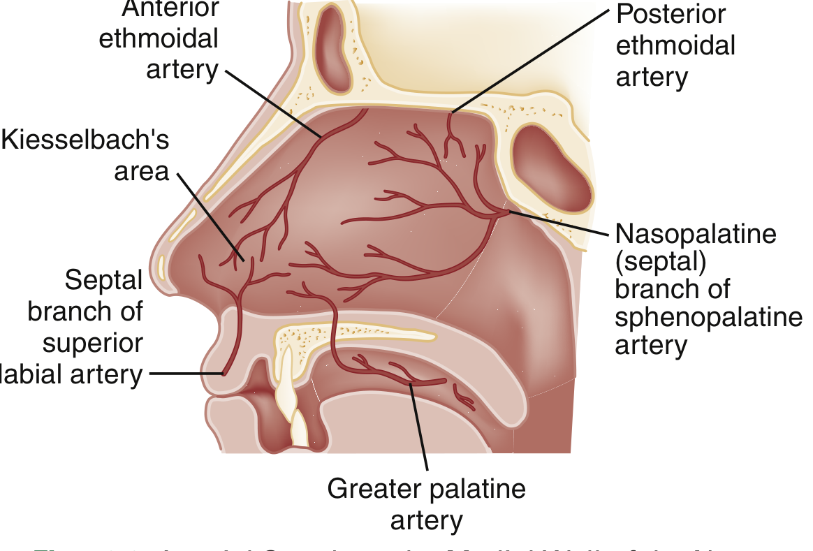
| Artery | Origin | Territory |
|---|---|---|
| Sphenopalatine artery | External carotid (via maxillary a.) | Turbinates, meatus laterally; posterior + inferior septum medially |
| Anterior + posterior ethmoidal arteries | Internal carotid (via ophthalmic a.) | Superior mucosa, medially + laterally |
| Superior labial artery | External carotid (via facial a.) | Anterior mucosal septum + anterior lateral mucosa |
Head-to-Head Comparison
| Feature | Anterior Epistaxis | Posterior Epistaxis |
|---|---|---|
| Frequency | ~90% of all cases | ~10% of cases |
| Location | Anteroinferior septum - Kiesselbach's plexus (Little's area) | Posterior nasal cavity - sphenopalatine artery territory |
| Vessel involved | Small arterioles + venous plexus | Larger branches of sphenopalatine artery |
| Typical patient | Children, young adults | Elderly, hypertensives, anticoagulated patients |
| Common causes | Nose picking, dry air, URI, low humidity, trauma, allergies | Hypertension, anticoagulants (warfarin, rivaroxaban, aspirin), atherosclerosis |
| Laterality | Typically unilateral | Often bilateral or cannot be lateralized |
| Bleeding direction | Out of anterior naris | Down the throat (posterior pharynx) - patient may swallow blood |
| Severity | Usually mild-moderate, self-limiting | More severe, harder to control, higher risk of airway compromise |
| Visibility | Source often visible on anterior rhinoscopy | Source not visible on anterior examination |
| Response to pressure | Usually responds to 10-15 min direct compression | Does NOT respond to anterior compression |
| Risk of airway compromise | Low | Significant - blood pools in pharynx |
| Risk of hemodynamic instability | Rare | Real risk in elderly / anticoagulated |
- Rosen's Emergency Medicine; Bailey and Love's Short Practice of Surgery
Clinical Clue: How to Suspect Posterior Bleeding
"Posterior epistaxis is suggested when bleeding persists in the setting of properly placed anterior nasal packing." - Rosen's Emergency Medicine
- Bilateral, brisk, nontraumatic bleeding not controlled anteriorly
- Blood seen dripping into the oropharynx (use tongue depressor to look)
- Elderly patient with hypertension or on anticoagulants
- No identifiable anterior source on nasal speculum exam
Management - Step-by-Step
Anterior Epistaxis
- Patient leans forward (not back - prevents swallowing/aspiration of blood)
- Pinch the cartilaginous (soft) part of the nose - NOT the bony bridge
- 10-15 min of continuous pressure - nose clip superior to manual pressure alone
- Oxymetazoline 0.05% sprayed into the naris before pressure: vasoconstriction optimizes hemostasis and aids examination
- Topical anesthetic (2% lidocaine via atomization or pledget)
- Open nasal speculum vertically (not laterally) with patient's head straight - floor of nose parallel to floor
- Silver nitrate stick, from periphery inward, superior to inferior
- Contact no longer than 15 seconds (risk of septal damage/necrosis)
- Never cauterize both sides of the septum simultaneously - may compromise septal blood supply and cause perforation
- Absorbable gelatin sponge (Gelfoam), oxidized cellulose (Surgicel)
- Topical tranexamic acid (500 mg IV solution applied to pledget or atomized): meta-analysis shows moderate evidence for reduced bleeding at 10 min and re-bleeding at 7-10 days; superior to packing in antiplatelet patients
- Polyvinyl acetal tampon (Merocel): insert dry, expands with moisture
- Inflatable balloon (Rapid Rhino): coated in procoagulant, inserted along the floor of the nose, inflated with air
- If bleeding persists with one pack → insert second pack in opposite naris
Posterior Epistaxis
- Bleeding persisting despite well-placed anterior pack
- Visualize oropharynx for posterior blood flow
- Double-balloon catheter (e.g., Epistat): insert along floor of nose; inflate posterior balloon in nasopharynx first, then pull anteriorly to seat it; then inflate anterior balloon
- If no commercial device: Foley catheter - insert into nasopharynx, inflate with 5-7 mL water, pull anteriorly, then secure and apply anterior pack around it
- All patients with posterior packing require hospital admission and monitoring (risk of airway compromise, hypoxia, vagal bradycardia from balloon pressure)
- Supplemental oxygen - nasal packing impairs nasal breathing
-
Endoscopic sphenopalatine artery ligation/clipping - effective, preferred first surgical option
-
Endoscopic cautery of sphenopalatine artery under direct vision with rigid nasendoscope
-
Interventional radiology embolization - for life-threatening or surgically inaccessible bleeding
-
Surgical ligation of internal maxillary artery (pterygopalatine fossa, endoscopically) or anterior/posterior ethmoidal arteries
-
External carotid artery ligation above the lingual artery origin - last resort
-
Bailey and Love's Short Practice of Surgery; Rosen's Emergency Medicine
Special Causes to Know
| Cause | Notes |
|---|---|
| Hereditary Hemorrhagic Telangiectasia (HHT / Osler-Weber-Rendu) | Recurrent multifocal bleeding from thin-walled vessels lacking muscle/elastic tissue; refractory to standard treatment |
| Juvenile Angiofibroma | Adolescent boys; massive life-threatening bleeding; do NOT biopsy - risk of uncontrollable hemorrhage; preop embolization before surgical excision |
| Anticoagulants (warfarin, rivaroxaban, aspirin, clopidogrel) | Most common systemic cause in adults; associated with recurrence; check coagulation studies |
| Cocaine use | Chronic vasoconstriction → mucosal ischemia → septal perforation |
| Hypertension | Associated with persistent epistaxis once present; no proven causal relationship for initiating epistaxis |
| von Willebrand disease / haemophilia | Consider in young patients with recurrent epistaxis and family history |
| Leukaemia / thrombocytopenia | Epistaxis may be the presenting feature |
Quick Summary Box
ANTERIOR (90%) POSTERIOR (10%)
───────────────────────────────── ──────────────────────────────────
Site: Kiesselbach's/Little's area Site: Sphenopalatine artery
Vessel: Small arterioles Vessel: Larger branches, higher flow
Patient: Young, healthy Patient: Elderly, HTN, anticoagulated
Bleeds: Out of nose Bleeds: Down throat
Bilateral: No Bilateral: Often yes
Visible: Yes Visible: No
Pressure: Controls it Pressure: Fails
Rx: Pressure → cautery → ant pack Rx: Double balloon → endoscopic → embolization
Risk: Low Risk: Airway, haemodynamic instability
Conn syndrome
Conn Syndrome (Primary Hyperaldosteronism)
1. Pathophysiology - The Core Mechanism
- Upregulate Na+/K+ ATPase and epithelial Na+ channels (ENaC)
- Retain sodium (and with it water) → volume expansion → BP rise
- Excrete potassium and H+ → hypokalemia + metabolic alkalosis
Autonomous aldosterone excess
↓
Na+ and water retention ──────────► Hypertension (volume-dependent)
↓
K+ wasting in distal nephron ──────► Hypokalemia
↓
H+ wasting (K+/H+ exchange) ───────► Metabolic alkalosis
↓
Suppressed renin (negative feedback ► ↓ Plasma renin activity (PRA)
from volume expansion)
2. Causes / Subtypes
| Cause | Frequency (selective screening) | Frequency (non-selective screening) | Management |
|---|---|---|---|
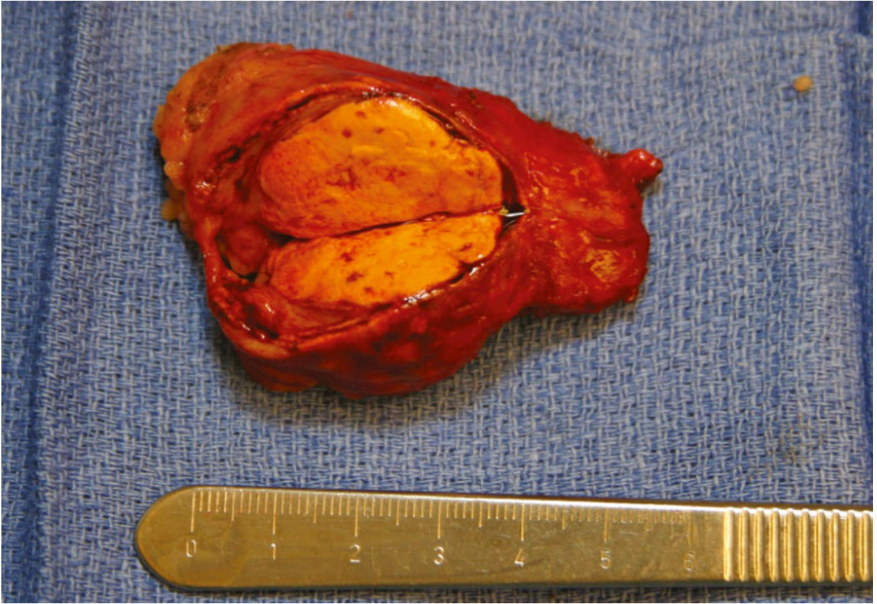
| Aldosterone-producing adenoma (APA) - Conn's syndrome proper | ~60% | ~30% | Surgery (adrenalectomy) |
| Bilateral adrenal hyperplasia (idiopathic hyperaldosteronism) | ~35% | ~65% | Medical (spironolactone/eplerenone) |
| Familial hyperaldosteronism Type I (glucocorticoid-remediable) | Rare | Rare | Glucocorticoids |
| Aldosterone-producing adrenocortical carcinoma | < 1% | < 1% | Surgery |
Important: Non-selective screening of hypertensive populations finds bilateral hyperplasia as the most common cause (65%). Selective screening in specialist centres still finds APA predominating (60%). - Sabiston Textbook of Surgery
3. Clinical Features
- Hypertension - moderate to severe, refractory to 2-4 antihypertensive agents
- Hypokalemia - though note: up to 50% of patients with confirmed primary aldosteronism are normokalemic
- Metabolic alkalosis
| Symptom | Mechanism |
|---|---|
| Muscle weakness, cramps, paresthesias | K+ depletion → hyperpolarized resting potential |
| Polyuria, polydipsia, nocturia | Hypokalemia impairs ADH responsiveness → nephrogenic diabetes insipidus-like state |
| Carbohydrate intolerance | Hypokalemia inhibits insulin secretion from beta cells |
| Tetany (severe cases) | Metabolic alkalosis → reduced ionized Ca2+ |
| Headache | Hypertension |
4. Diagnosis - Step-by-Step Approach
Step 1 - Screening: Aldosterone-to-Renin Ratio (ARR)
- Plasma aldosterone concentration (PAC, ng/dL) ÷ Plasma renin activity (PRA, ng/mL/hr)
- Ratio > 30 is suggestive of primary aldosteronism
- Sensitivity ~85%, widely available
- Hypokalemia should be corrected before testing (K+ suppresses aldosterone)
- Interfering drugs: spironolactone and eplerenone must be stopped 4-6 weeks before; ACE inhibitors and ARBs falsely raise renin (lower ratio); beta-blockers suppress renin (raise ratio)
Step 2 - Confirmatory testing
- Salt loading test (IV saline 2L over 4h): aldosterone fails to suppress in primary hyperaldosteronism (remains > 10 ng/dL)
- Fludrocortisone suppression test
- Captopril challenge test
Step 3 - Localisation: CT/MRI
- APA on CT: Small (mean 1.6-2.2 cm), low attenuation (< 10 HU), minimal enhancement, rarely calcify
- Bilateral adrenal hyperplasia on CT: Adrenal limb width ≥ 5 mm (specificity 100% for BAH if both sides ≥ 5 mm)
- Limitation: CT sensitivity for APA only 88-100% - a normal CT does not exclude APA (microadenomas can be missed). A "mass on CT" in a patient > 40 years may be a non-functioning incidentaloma - aldosterone is coming from the other side.
Step 4 - Adrenal Vein Sampling (AVS) - The Gold Standard for Lateralisation
- All patients ≥ 40 years (incidentalomas are common; CT unreliable)
- Any patient without clear unilateral CT abnormality
- Young patients (< 40 years) with a clear > 1 cm unilateral mass and normal contralateral gland may proceed directly to surgery
- Success rate 40-80% (most common failure: inability to cannulate right adrenal vein)
- Only available at specialised centres
- Performed with/without ACTH stimulation to maximise selectivity
5. Treatment
Unilateral APA → Surgery
- Laparoscopic (minimally invasive) adrenalectomy - preferred
- Outcomes (PASO international consensus):
-
80% of patients: normalization of BP or significant reduction in antihypertensive medications (from 3-4 drugs to 1)
- Hypokalemia resolves within 24 hours post-operatively
- BP improvement may take weeks if there is long-standing sodium overload
-
- Post-operative watch: Hyperkalemia in 5-10% (transient suppression of contralateral adrenal gland, which has been chronically inhibited); check weekly K+ for 1 month
- Stop spironolactone, ACE inhibitors, ARBs immediately post-op; taper beta-blockers gradually
- Male > 45 years
- Family history of hypertension
- Long duration of hypertension
- Requirement for > 2 antihypertensives pre-operatively
- Non-response to spironolactone pre-operatively
Bilateral Hyperplasia → Medical Management
- Spironolactone (first-line): Competitive aldosterone antagonist at the mineralocorticoid receptor; controls both hypertension and hypokalemia. Side effects: gynecomastia, erectile dysfunction, menstrual irregularities (due to anti-androgenic effect)
- Eplerenone: More selective mineralocorticoid antagonist, fewer anti-androgenic side effects; better tolerated but less potent than spironolactone
- Amiloride: K+-sparing diuretic (blocks ENaC directly); alternative if mineralocorticoid antagonists not tolerated
- Additional antihypertensives as needed (calcium channel blockers are well-tolerated)
Primary vs. Secondary Hyperaldosteronism - Key Distinction
| Feature | Primary (Conn's) | Secondary |
|---|---|---|
| Cause | Autonomous adrenal secretion | Elevated renin (renal artery stenosis, heart failure, cirrhosis, diuretics) |
| Renin | Suppressed (low) | Elevated (high) |
| ARR | High | Low |
| Treatment | Treat adrenal cause | Treat underlying renin-driving cause |
Summary Box
CONN SYNDROME AT A GLANCE
────────────────────────────────────────────
Definition: Autonomous aldosterone excess (APA or bilateral hyperplasia)
Triad: HTN + Hypokalemia + Metabolic alkalosis
Key lab: ↑ PAC, ↓ PRA, ARR > 30
Screening: Any hypertensive with hypokalemia, refractory HTN, adrenal incidentaloma
Imaging: CT (APA: small, <10HU) → AVS if ≥40 yrs or no clear unilateral lesion
APA: Laparoscopic adrenalectomy → >80% BP improvement
BAH: Spironolactone / Eplerenone lifelong
Pitfall: Normal K+ does NOT exclude the diagnosis
Pitfall: CT finding does NOT replace AVS for lateralisation in patients ≥40 yrs
Hypertensive encephalopathy
Hypertensive Encephalopathy
1. Definition and Context
"Symptoms such as headache, epistaxis, and dizziness are not, in and of themselves, evidence of acute TOD and do not constitute a hypertensive emergency, nor do they indicate the need for acute BP reduction." - Rosen's Emergency Medicine
2. Pathophysiology - The Core Mechanism
Normal Cerebral Autoregulation
-
Lower limit of autoregulation (LLA): MAP ~50 mmHg - below this, CBF falls passively with BP (ischemia risk)
-
Upper limit of autoregulation (ULA): MAP ~150 mmHg - above this, the arterioles can no longer maintain constriction
-
Miller's Anesthesia
What Happens in Hypertensive Encephalopathy
Severe/acute BP rise → MAP exceeds upper limit of autoregulation
↓
Cerebral arterioles lose vasoconstrictive capacity → "breakthrough" vasodilation
↓
Passive pressure-driven hyperperfusion
↓
Overdistension of arteriolar walls → fibrinoid necrosis of vessel walls
↓
Disruption of blood-brain barrier → endothelial dysfunction
↓
Vasogenic edema (protein-rich plasma leaks into interstitium)
↓
Punctate microhemorrhages + fibrin thrombi in small vessels
↓
Diffuse cerebral dysfunction
3. Causes
| Category | Examples |
|---|---|
| Most common | Rapidly worsening essential hypertension |
| Renal | Acute glomerulonephritis, renal artery stenosis, end-stage renal disease |
| Endocrine | Pheochromocytoma, Cushing syndrome |
| Obstetric | Eclampsia, HELLP syndrome (special form) |
| Drugs/toxins | Cocaine, sympathomimetics, aminophylline, phenylephrine |
| Rare | Chemotherapeutic agents (cyclosporin, cisplatin) - cause PRES |
4. Clinical Features
| Feature | Detail |
|---|---|
| Headache | Severe, progressive, often occipital - from raised ICP and vessel overdistension |
| Nausea/vomiting | From raised ICP |
| Altered mental status | Confusion → agitation → stupor → coma in severe cases |
| Seizures | Focal or generalized; may be more marked on one side |
| Visual disturbances | Blurred vision → visual field defects → cortical blindness (from occipital involvement) |
| Visual hallucinations | Posterior cortex involvement (Balint syndrome in severe cases) |
| Focal neurological signs | Important: do NOT follow a single anatomic distribution - bilateral, patchy; distinguishes from stroke |
| Papilledema | Often present; previously considered mandatory for diagnosis - not always present |
| Hypertensive retinopathy | Flame haemorrhages, exudates, AV nipping |
"Focal neurologic deficits do not follow a singular anatomic pattern and may occur on opposite sides of the body, indicating diffuse cerebral dysfunction rather than an anatomically localized stroke." - Rosen's Emergency Medicine
"The neurologic syndrome is usually dominated by symptoms referable to the occipital and adjacent parietal region." - Adams and Victor's Neurology
5. Investigations
Brain Imaging
- May be normal or show non-specific changes
- Can show diffuse/regional white matter hypodensity, small haemorrhages
- Primary value: exclude haemorrhagic stroke, space-occupying lesion before making presumptive diagnosis
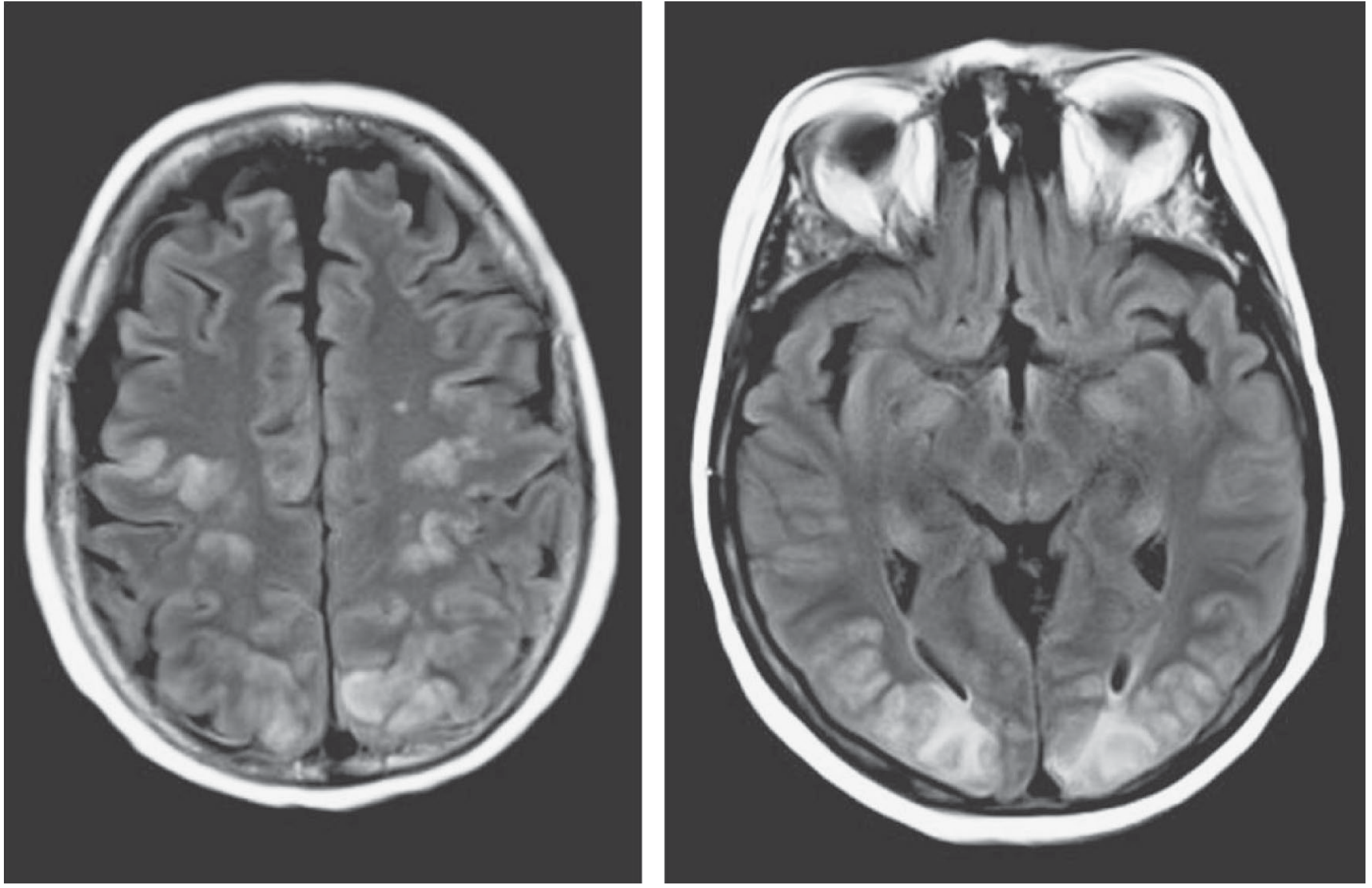
- T2/FLAIR hyperintensity in white matter, predominantly in posterior parietal-occipital regions (bilateral)
- Corresponds to vasogenic oedema
- Little or no mass effect (distinguishes from tumour, stroke)
- Does NOT course along white matter tracts (distinguishes from demyelination)
- Scattered cortical lesions in watershed distributions = small infarctions
- Changes normalise over weeks with BP control
CSF
- Elevated pressure and protein (> 100 mg/dL in some cases)
- No cellular reaction
- Lumbar puncture generally not needed for diagnosis; only to exclude subarachnoid haemorrhage
Other workup
- Urinalysis (proteinuria, casts - renal involvement)
- Renal function, electrolytes
- FBC, coagulation (especially if HELLP suspected)
- Urine/serum catecholamines if pheochromocytoma suspected
6. Diagnosis
- Markedly elevated BP
- Altered mentation or diffuse neurological dysfunction
- Normal or non-specific CT (excluding haemorrhage/mass)
- Objective findings: papilledema, retinal haemorrhages (supportive but not mandatory)
7. PRES - The Radiological Syndrome
- Pre-eclampsia/eclampsia
- Immunosuppressants (cyclosporine, tacrolimus)
- Cytotoxic chemotherapy (cisplatin)
- Vasculitis, TTP, porphyria
| Feature | PRES |
|---|---|
| MRI | Vasogenic oedema, posterior > anterior |
| Distribution | Bilateral occipital-parietal predominant; may involve brainstem/cerebellum |
| Clinical | Headache, confusion, seizures, cortical blindness |
| Reversibility | Fully reversible with BP control / drug withdrawal |
| Mechanism | Endothelial dysfunction + BBB breakdown, similar to HE |
| PRES/HE | Venous thrombosis | Top of basilar | |
|---|---|---|---|
| Onset | Acute, evolves over days | Acute, evolves over days | Sudden, hours |
| Key features | Seizures first, visual aura, cortical blindness | Headache, focal deficits, papilloedema | Cortical blindness, brainstem signs |
| MRI | Posterior white matter oedema, no venous infarct | Haemorrhage + ischaemic infarcts, cord sign | Bilateral paracalcarine/thalamic infarcts |
| Prognosis | Completely reversible with BP control | High mortality in severe cases | Partial recovery at best |
- Plum and Posner's Diagnosis and Treatment of Stupor and Coma
8. Treatment
BP Reduction Goals
| Timeframe | Target |
|---|---|
| First hour | Reduce MAP by 20-25% (Rosen's suggests 30-40% for HE specifically, but with caution) |
| Hours 2-6 | Reach BP ~160/100 mmHg |
| Over 24-48 hours | Gradual normalization |
"A typical target is a pressure of 150/100 mmHg or a 20% reduction in mean pressure." - Adams and Victor's Neurology
Antihypertensive Agents
| Drug | Mechanism | Notes |
|---|---|---|
| Labetalol | Alpha + beta blocker | Smooth, predictable BP reduction; no reflex tachycardia; safe in most contexts |
| Nicardipine | Calcium channel blocker (dihydropyridine) | Titratable infusion; good cerebral vasodilation; preferred in neurological emergencies |
| Sodium nitroprusside | NO donor → arteriolar + venous dilation | Potent, immediately titratable; risk of cyanide toxicity (prolonged use), increases ICP - caution in HE |
| Hydralazine | Direct arteriolar vasodilator | IV bolus; used in eclampsia; less predictable |
| Esmolol | Ultra-short-acting beta-1 blocker | Good for rate control; less hypotensive effect |
| Magnesium sulfate | NMDA antagonism + vasodilation | Specifically for eclampsia - controls both seizures and BP |
"Intravenous sodium nitroprusside; a calcium channel blocker such as nifedipine, or intravenous beta-adrenergic blockers have all been used." - Adams and Victor's Neurology
Seizure Management
- Benzodiazepines for acute seizure control
- Levetiracetam or phenytoin for prevention
- Antiepileptics can often be tapered once BP is controlled (seizures are secondary to the encephalopathy, not primary epilepsy)
Monitoring
- ICU admission
- Continuous arterial line for BP monitoring
- Neurological observations every 30-60 min
- Repeat MRI to confirm reversal of changes
9. Prognosis
- Fully reversible with prompt treatment - MRI changes normalise over days to weeks
- In-hospital mortality < 1% in modern series
- Delayed or inadequate treatment → permanent neurological deficit, haemorrhagic stroke, or death
- Underlying cause must be identified and managed long-term
Summary
HYPERTENSIVE ENCEPHALOPATHY AT A GLANCE
──────────────────────────────────────────────────────
Core mechanism: BP exceeds ULA → breakthrough hyperperfusion
→ endothelial damage → vasogenic oedema
BP threshold: Typically MAP >150 or diastolic >125 mmHg
(lower in acute risers: eclampsia, GN)
Classic triad: Severe HTN + encephalopathy + MRI posterior changes
Key clinical: Diffuse dysfunction (NOT focal), papilledema, visual Δ
MRI: T2/FLAIR posterior parietal-occipital white matter hyperintensity
= PRES; normalises with treatment
Mimics to rule out: Haemorrhagic stroke, venous thrombosis, metabolic coma
Treatment: IV labetalol or nicardipine; reduce MAP by ~20-25% in 1st hour
Target ~150/100 mmHg initially
DO NOT: Reduce BP too fast → ischaemic stroke
Use nitroprusside if raised ICP suspected
Reversibility: FULLY reversible if treated promptly; <1% mortality