
Glide path management in endodontics .give me 25 marks mds theory answer with contents
Reading File
Now let me search the medical library for glide path management content.
Finding Sources
Finding Sources
Finding Sources
Finding Sources
Finding Sources
Searching PubMed
glide path endodontics management
Reading Article
PMID: 39195101
Reading Article
PMID: 35786583
Reading Article
PMID: 40449882
Searching the Web
glide path management endodontics MDS theory instruments techniques 2024
I now have comprehensive information to write a thorough 25-marks MDS theory answer. Let me compose it.
GLIDE PATH MANAGEMENT IN ENDODONTICS
[MDS Theory Answer - 25 Marks]
CONTENTS / OUTLINE
- Introduction and Definition
- Historical Background
- Rationale / Importance of Glide Path
- Anatomy of the Glide Path
- Goals of Glide Path Preparation
- Assessment Before Glide Path Creation
- Instruments Used
- Manual (Stainless Steel) Files
- Nickel-Titanium (NiTi) Rotary Glide Path Files
- Reciprocating NiTi Files
- Techniques of Glide Path Preparation
- Manual Techniques
- Mechanical/Engine-Driven Techniques
- Preflaring and Its Role
- Irrigation During Glide Path Preparation
- Working Length Determination
- Clinical Endpoints / Verification of Glide Path
- Procedural Errors and Their Prevention
- Effect on Subsequent Shaping
- Recent Advances and Future Directions
- Conclusion
1. INTRODUCTION AND DEFINITION
The concept of the glide path is fundamental to contemporary endodontic practice and serves as the cornerstone for safe and predictable root canal shaping. Before any engine-driven nickel-titanium (NiTi) rotary or reciprocating instrument is introduced into the root canal, the creation of a smooth, reproducible, unobstructed radicular tunnel is mandatory.
Definition (Buchanan, 1989): A glide path is defined as "a smooth, reproducible, unobstructed pathway from the canal orifice to the physiological terminus of the root canal." In simpler terms, it is the pre-existing natural space once occupied by the vital pulp, refined to allow instruments to travel smoothly from the coronal access cavity to the apical foramen without binding, transportation, or blockage.
A canal is considered glide-path ready when:
- A size #10 K-file reaches working length without resistance
- The file moves freely in and out of the canal
- This movement is consistently reproducible
- There is no file binding along the canal walls
The 2024 literature review by Lup et al. (PMID: 39195101) synthesized 116 studies confirming that establishing a glide path reduces root canal transportation, postoperative pain, and file separation.
2. HISTORICAL BACKGROUND
| Era | Development |
|---|---|
| Pre-1980s | All root canals shaped exclusively with stainless steel hand files; glide path as a concept was implicit but not formalized |
| 1989 | Buchanan formally introduced and named the "glide path" concept |
| 1990s | Introduction of NiTi rotary instruments (ProFile, Hero 642) made glide path creation a formal prerequisite |
| 2000s | PathFile system (Dentsply Maillefer) - first dedicated NiTi rotary glide path files introduced |
| 2010s | ProGlider (single-file rotary), R-Pilot and WaveOne Gold Glider (reciprocating) - expanded options |
| 2020s | Heat-treated alloys (M-Wire, Gold, Blue), controlled memory (CM) wire - improved fatigue resistance |
The term "glide path" gained prominence with the widespread adoption of NiTi rotary systems, as instrument fracture emerged as a significant clinical concern when entering inadequately prepared canals.
3. RATIONALE / IMPORTANCE OF GLIDE PATH
The rationale for glide path creation is multi-faceted and stems from the biomechanical demands placed on NiTi instruments:
a) Stress Reduction on NiTi Instruments:
NiTi rotary files fail primarily by two mechanisms:
- Cyclic fatigue - repeated bending cycles at the point of maximum curvature
- Torsional failure - when the tip locks in the canal while the shank continues to rotate
A pre-existing glide path reduces both forms of stress by ensuring the file engages dentine with its flutes rather than its tip.
b) Prevention of Procedural Errors:
- Ledge formation
- Canal transportation (loss of working length or apical deviation)
- Canal zipping and stripping
- File separation/fracture
- Apical foramen perforation
c) Maintains Original Canal Anatomy:
The glide path follows the natural canal curvature, ensuring subsequent shaping instruments do not deviate from the true canal path.
d) Facilitates Irrigant Penetration:
A patent, smooth glide path allows irrigants such as NaOCl and EDTA to reach the apical third effectively.
e) Reduces Postoperative Pain:
A 2022 review by Ajina et al. (PMID: 35786583) found that engine-driven glide path preparation causes significantly less postoperative pain compared to manual techniques, attributed to reduced apical extrusion of debris.
4. ANATOMY OF THE GLIDE PATH
Understanding the natural anatomy is essential before glide path creation:
- The glide path is the natural space previously occupied by the vital or necrotic pulp tissue
- It is NOT a manufactured tunnel - it is a pre-existing pathway refined by the clinician
- The glide path must follow all natural canal curvatures, dilacerations, and apical deltas
- It extends from the canal orifice at the level of the pulp chamber floor to the apical constriction (minor apical foramen), approximately 0.5-1 mm short of the radiographic apex
- The apical constriction (0.2-0.35 mm diameter) represents the endpoint of the glide path
Key anatomical considerations:
- Single vs. multiple curvatures (S-shaped canals require incremental scouting)
- Canal calcification (increases difficulty; requires EDTA chelation)
- Apical delta and lateral canals (path must not be forced through accessory foramina)
- Root length and root curvature (Schneider angle used to classify severity)
Schneider's Classification of Canal Curvature:
- Straight: 0-5°
- Moderately curved: 10-20°
- Severely curved: 25-70°
5. GOALS OF GLIDE PATH PREPARATION
- Create a smooth, unobstructed, reproducible tunnel from orifice to apex
- Preserve the natural canal anatomy (no iatrogenic modification of curvature)
- Reduce mechanical stress on subsequent NiTi shaping files
- Facilitate accurate working length determination
- Allow safe and efficient passage of all rotary/reciprocating instruments
- Minimize apical extrusion of debris and bacteria
- Reduce the risk of procedural errors (ledging, transportation, perforation)
- Reduce postoperative pain and complications
6. ASSESSMENT BEFORE GLIDE PATH CREATION
Preoperative Radiographic Assessment:
- Number of roots and canals
- Canal curvature (Schneider angle, Jou's method)
- Canal length estimate
- Presence of calcifications or pulp stones
- Proximity to vital structures
- Previous root canal treatment (separated instruments, ledges)
CBCT (Cone Beam CT):
- Indicated in complex cases: calcified canals, unusual morphology, S-shaped curvatures, C-shaped canals
- Provides 3D anatomical map unavailable on 2D periapical films
Clinical Assessment:
- Tooth isolation with rubber dam (mandatory)
- Adequate access cavity preparation (critical prerequisite)
- Coronal preflaring status
7. INSTRUMENTS USED IN GLIDE PATH PREPARATION
A. Manual (Stainless Steel) Hand Files
Manual negotiation with stainless steel files remains the gold standard for initial canal scouting, particularly in calcified, narrow, or severely curved canals.
Files used (in sequence):
| File | ISO Size | Tip Diameter | Use |
|---|---|---|---|
| C-pilot file / C+ file | 06, 08 | 0.06-0.08 mm | Ultra-fine scouting in calcified canals |
| K-file | #10 | 0.10 mm | Standard initial scouting |
| K-file | #15 | 0.15 mm | Minimal glide path endpoint |
| K-file | #20 | 0.20 mm | Optimal glide path endpoint (recommended) |
| K-file | #25 | 0.25 mm | Large glide path (some authors) |
Properties of Stainless Steel (SS) Files:
- High stiffness - limits use in curved canals beyond size #15
- Good tactile feedback
- Inexpensive and widely available
- Can be pre-curved to follow canal anatomy
B. NiTi Rotary Glide Path Files
Dedicated NiTi rotary instruments developed to overcome limitations of SS hand files in curved canals:
| System | Manufacturer | Number of Files | Taper | Motion | Special Feature |
|---|---|---|---|---|---|
| PathFile | Dentsply Maillefer | 3 files (#13, #16, #19) | 0.02 | Continuous rotation | First dedicated NiTi GP system |
| ProGlider | Dentsply Sirona | Single file | Progressive (2-8%) | Continuous rotation | Controlled Memory (CM) wire |
| G-Files (One G) | Micro-Mega | Single file | 0.03 | Continuous rotation | D-RaCe alloy, single use |
| HyFlex EDM | Coltene | Single file | 0.03 | Continuous rotation | Electro Discharge Machining - superelastic |
| Scout RaCe | FKG Dentaire | 3 files | 0.02 | Continuous rotation | Safe tip design |
| R-Pilot | VDW | Single file | Progressive | Reciprocation | Gold alloy |
| WaveOne Gold Glider | Dentsply Sirona | Single file | Progressive | Reciprocation | Gold alloy - heat treated |
| ReciprocBlue Glider | Dentsply | Single file | 0.03 | Reciprocation | Blue NiTi alloy |
| EdgeGlider | EdgeEndo | Single file | 0.03 | Continuous rotation | FireWire alloy |
C. Properties of NiTi Glide Path Files:
Alloy Generations:
- Conventional NiTi - original superelastic alloy (e.g., standard ProTaper, K3)
- M-Wire - thermally treated; improved fatigue resistance (e.g., ProGlider)
- CM Wire (Controlled Memory) - pre-bent files maintain shape; superior flexibility
- R-Phase / Gold Wire - heat treated; higher fatigue resistance and flexibility (e.g., WaveOne Gold Glider, R-Pilot)
- EDM-processed (Electro Discharge Machining) - e.g., HyFlex EDM; can self-anneal after deformation
Design Features of NiTi GP Files:
- Variable taper (progressive taper files reduce torsional stress)
- Off-centered mass of rotation - reduces screw-in effect
- Safe (non-cutting) tip - reduces risk of ledge formation
- S-shaped or parabolic cross-sections - enhance flexibility
- Short pitch length - increases torsional resistance
8. TECHNIQUES OF GLIDE PATH PREPARATION
A. Manual Technique (Classic Approach)
Step-by-step protocol:
-
Rubber Dam Isolation - non-negotiable; prevents aspiration and contamination
-
Adequate Access Cavity - straight-line access to the canal orifice is essential; remove all coronal obstructions
-
Coronal Preflaring - use GG drills (sizes 1, 2, 3) or orifice shapers to remove coronal dentinal triangles; reduces file deflection and improves tactile feel
-
Initial Scouting:
- Use a size #08 or #10 K-file with a gentle, light watch-winding motion
- Pre-curve the file apically to navigate curved canals
- Irrigate copiously with NaOCl + EDTA gel (e.g., RC-Prep, Glyde) throughout
- Do NOT force - if resistance is felt, remove, re-irrigate, re-curve
-
Establish Provisional Working Length (PWL) - using apex locator + radiograph
-
Sequential Enlargement:
- Progress from #10 → #15 → #20 K-files using watch-winding or balanced force motion
- Each file should be loose and reproducibly reaches working length before advancing
-
Recapitulation - after each file, return to smaller file (#10) to prevent blockage and confirm patency
-
Patency Filing - a small #10 file gently moved 0.5-1 mm past the apical constriction to keep the foramen open (controversial - some advocate, some do not)
Manual Motions Used:
- Watch-winding (balanced force): Clockwise 90° engagement + counterclockwise 90° release - recommended for curved canals
- Reaming: Clockwise rotation with apical pressure - use in straight canals only
- Filing/rasping: Pull-out stroke after engagement - least preferred for curved canals
B. Mechanical / Engine-Driven NiTi Technique
General Principles:
- Use dedicated motor with controlled torque and speed settings
- Always have a manual glide path (at least loose #10 K-file to WL) before introducing NiTi GP files
- Use generous irrigation between each file
- Single-file systems preferred to reduce instrumentation steps
PathFile Technique (3-file system):
- Establish manual glide path with SS #10 K-file (loose at WL)
- PathFile #1 (ISO 13, 0.02 taper) - continuous rotation 300-500 rpm
- PathFile #2 (ISO 16, 0.02 taper) - continuous rotation
- PathFile #3 (ISO 19, 0.02 taper) - continuous rotation
- Proceed to shaping NiTi system (ProTaper Next, WaveOne Gold, etc.)
ProGlider Technique (single-file system):
- Establish manual glide path with SS #10 K-file (loose at WL)
- ProGlider: continuous rotation at 300 rpm, 2 N·cm torque, gentle in-out pecking motion
- Single pass confirms adequate glide path
- Proceed to shaping
WaveOne Gold Glider Technique (reciprocating single-file):
- Establish manual glide path with SS #10 K-file (at least negotiated to WL)
- WaveOne Gold Glider: reciprocating motion, dedicated WaveOne motor program
- Gentle in-out pecking motion (3-4 mm amplitude)
- Proceed to WaveOne Gold shaping
Key Principles for Mechanical NiTi GP Files:
- Never force instruments apically
- Use brushing motion, not plunging
- Clean flutes every 3-4 mm advancement
- Confirm WL after glide path completion before shaping
- Discard single-use files after each patient
9. PREFLARING AND ITS ROLE
Preflaring refers to the enlargement of the coronal and middle thirds of the root canal before apical instrumentation. It is a prerequisite to glide path creation.
Instruments for Preflaring:
- Gates-Glidden (GG) drills: sizes 1 (0.5 mm), 2 (0.7 mm), 3 (0.9 mm)
- Orifice shapers (ProTaper SX file)
- Coronal flaring instruments (e.g., Tri-Auto ZX coronal files)
Benefits of Preflaring:
- Removes the coronal dentinal triangle (restrictive dentin)
- Provides straight-line access to apical third
- Reduces taper lock on NiTi files
- Improves tactile sensation during apical scouting
- Allows better irrigation with wider coronal opening
- Reduces apical extrusion of debris (debris displaced coronally)
- Reduces overall preparation time
GG Drill Sequence (Crown-Down):
GG #3 → GG #2 → GG #1 (progressively deeper, never forced to full working length)
10. IRRIGATION DURING GLIDE PATH PREPARATION
Irrigation is integral to glide path management and cannot be separated from instrumentation.
Irrigants Used:
| Irrigant | Concentration | Role |
|---|---|---|
| Sodium Hypochlorite (NaOCl) | 1-5.25% | Tissue dissolution, antimicrobial, lubricant |
| EDTA (17%) | 17% | Chelation of calcified material, smear layer removal |
| RC-Prep / Glyde / File-EZE (EDTA + urea peroxide) | As supplied | Lubrication + chelation + effervescent debris removal |
| Chlorhexidine (2%) | 2% | Antimicrobial (do not mix with NaOCl) |
| Normal Saline | Isotonic | Flush between irrigants |
Protocol:
- Irrigate before introducing each instrument
- Use EDTA gel on the file tip as a lubricant during scouting
- Alternate NaOCl with EDTA
- Recapitulate with #10 file and irrigate after every instrument
11. WORKING LENGTH DETERMINATION
Working length is defined as the distance from a coronal reference point to the point where canal preparation and obturation should terminate (apical constriction, 0.5-1 mm short of the radiographic apex).
Methods:
- Electronic Apex Locator (EAL) - most accurate; monitors impedance/frequency shift; 4th generation devices (e.g., Raypex 6, Propex Pixi) are most reliable
- Radiographic method - using an ISO #10 K-file at estimated WL on periapical film; less accurate
- Combination - EAL + radiographic confirmation = gold standard
Working length is confirmed BEFORE and AFTER glide path completion, as patency filing can slightly alter the apex locator reading.
12. CLINICAL ENDPOINTS / VERIFICATION OF GLIDE PATH
The glide path is considered complete and satisfactory when:
- A size #15 K-file (minimum) or ideally #20 K-file moves freely to working length without resistance
- The file recapitulates reproducibly - same feel each time it is withdrawn and reinserted
- No binding is felt along the canal walls
- The ISO #10 file is loose at working length (tactile "looseness")
- The apex locator reading is stable at the WL
- A confirmatory radiograph shows the instrument at the correct working length with proper canal curvature maintained
Importance of #20 endpoint:
Most NiTi rotary GP file systems (PathFile, ProGlider) have a tip diameter of approximately 0.13-0.20 mm. A size #20 glide path (0.20 mm) ensures these instruments are not making first contact with the canal wall apically, preventing tip binding and torsional failure.
13. PROCEDURAL ERRORS AND THEIR PREVENTION
Common Errors:
a) Ledge Formation:
- Cause: Forcing a stiff file in a curved canal; inadequate pre-curving
- Prevention: Pre-curve #10 K-file; use watch-winding motion; irrigate copiously
- Management: Negotiate past ledge with a small pre-curved #08 or #10 file
b) Canal Blockage / Apical Plug:
- Cause: Packing of dentinal debris or organic tissue into the apical third
- Prevention: Frequent recapitulation with #10 file; continuous irrigation
- Management: Attempt to pass blockage with pre-curved #08 K-file; use EDTA chelator
c) Canal Transportation / Stripping:
- Cause: Failure to maintain the original canal curvature; using overly stiff files
- Prevention: Adequate preflaring; use of NiTi over SS in curved canals; watch-winding motion
- Significance: Increases risk of perforation, weakens root structure, compromises seal
d) Instrument Separation (File Fracture):
- Cause: Torsional lock in canal; cyclic fatigue; using damaged files; forcing instruments
- Prevention: Inspect files for unwinding before use; single-use protocol for NiTi; establish glide path before using rotary files; do not exceed torque limits
- Management: Attempt bypass; ultrasonic retrieval; surgical endodontics
e) Perforation:
- Cause: Transportation in apical third; misguided preparation in calcified canals
- Prevention: Pre-curved scouting files; CBCT in complex cases; adequate access cavity
- Management: MTA plug placement; surgical repair
f) Zipping and Elbowing:
- Cause: Inadequate glide path; using stiff rotary files in curved canals without prior manual negotiation
- Prevention: Complete glide path preparation before introducing shaping NiTi systems
14. EFFECT ON SUBSEQUENT SHAPING
A well-established glide path has been shown to directly influence the outcomes of subsequent NiTi rotary shaping:
-
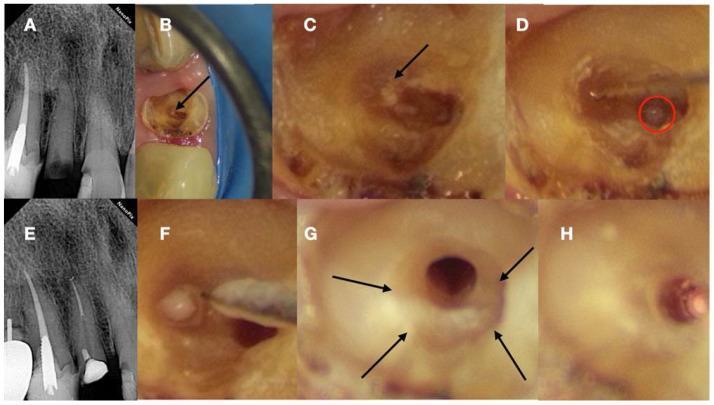
Reduced Canal Transportation: The 2024 review by Lup et al. (PMID: 39195101) confirmed that a glide path reduces root canal transportation, especially when rotary methods are used for GP preparation.
-
Reduced File Fracture: Engine-driven NiTi shaping files (ProTaper, WaveOne, Reciproc) are far less likely to separate when introduced into a pre-existing glide path.
-
Shorter Overall Preparation Time: Single-file NiTi GP systems (ProGlider, R-Pilot) significantly reduce preparation time compared to sequential hand filing.
-
Preserved Taper and Canal Shape: The tapered shape produced by NiTi shaping instruments more faithfully reflects the original canal anatomy when a glide path is present.
-
Less Postoperative Pain: The 2025 meta-analysis by Talekar et al. (PMID: 40449882) demonstrated that rotary glide path preparation produces significantly less apically extruded debris than manual techniques (SMD: 1.35; 95% CI: 0.82-1.87), reducing postoperative flare-ups.
-
No Effect on Dentine Microcracks: Glide path preparation per se does not increase the prevalence of dentine microcracks or wall defects (Lup et al., 2024).
15. COMPARISON: MANUAL vs. ENGINE-DRIVEN GLIDE PATH
| Parameter | Manual (SS K-Files) | Rotary NiTi GP Files | Reciprocating NiTi GP Files |
|---|---|---|---|
| Flexibility in curved canals | Low | High | Very high |
| Tactile feedback | Excellent | Moderate | Moderate |
| Speed of preparation | Slow | Moderate-fast | Fast |
| Apical debris extrusion | Higher | Lower | Lowest |
| Fatigue resistance | N/A | Moderate-high | High |
| Postoperative pain | Higher | Lower | Lowest |
| Canal transportation | Moderate | Low | Very low |
| Cost | Low | Moderate | Moderate-high |
| Learning curve | Low | Moderate | Moderate |
| Best indication | Calcified/complex canals for initial scouting | Most canals | Severely curved/difficult canals |
(Based on Ajina et al. 2022, Talekar et al. 2025)
16. SPECIAL CONSIDERATIONS
Calcified Canals:
- Use ultrasonic tips (e.g., Start-X tips) to locate orifices
- CBCT for pre-treatment mapping
- C-Pilot files (#06, #08) with EDTA gel for initial penetration
- Magnification (dental microscope) essential
Severely Curved Canals (>25°):
- Rely on pre-curved SS #08 or #10 K-file for initial negotiation
- Use CM-wire or M-Wire NiTi GP files (maximum flexibility)
- Reciprocating motion preferred over continuous rotation
- Multiple gentle short strokes rather than single aggressive passes
Previously Treated Canals (Retreatment):
- Remove existing obturation material before glide path can be assessed
- Use solvent (chloroform, eucalyptol) to soften gutta percha
- Rotary retreatment files first, then re-establish glide path
- High risk of ledge or blockage - confirm with apex locator at each step
Immature Apices / Open Apices:
- Standard glide path concepts apply but working length termination is at the apical constriction (may be irregular)
- Avoid apical widening; focus on disinfection
- Use apical plug (MTA/Biodentine) if apex is wide open
C-Shaped Canals:
- Most common in mandibular 2nd molars
- Glide path preparation must be adapted to the irregular C-shaped cross-section
- Use small files with watch-winding; risk of stripping at isthmus
- CBCT mandatory for accurate anatomy
17. RECENT ADVANCES
-
Self-adjusting File (SAF): A hollow, cylindrical NiTi lattice file that adapts to the natural canal shape without a pre-defined glide path; still requires a minimal #20 size before insertion.
-
Trunatomy (Dentsply Sirona): Ultra-slim NiTi system with 0.03 taper; requires a robust glide path (size #20) given its minimal taper design.
-
Heat-Treated Files (Blue/Gold/FireWire Alloys): Latest generation alloys show dramatically improved flexibility and cyclic fatigue resistance, making them safer in severely curved canals.
-
Guided Endodontics (Dynamic Navigation): 3D-printed surgical guides or dynamic navigation systems (Navident, X-Guide) allow precise access cavity preparation in calcified canals, complementing glide path management.
-
Artificial Intelligence in Endodontics: AI-based analysis of periapical radiographs and CBCT to predict canal curvature and guide instrument selection before glide path preparation.
18. CONCLUSION
Glide path management is not an optional or preparatory step - it is the structural foundation of safe, predictable endodontic treatment. The creation of a smooth, reproducible radicular tunnel from the canal orifice to the physiological foramen is an absolute prerequisite before the introduction of any NiTi shaping instrument.
The evolution from manual stainless steel K-files to dedicated NiTi rotary and reciprocating glide path systems has significantly improved:
- Canal centering ability
- Reduction in procedural errors
- Reduction of postoperative pain
- Overall treatment predictability
Current evidence (Lup et al. 2024; Talekar et al. 2025; Ajina et al. 2022) unequivocally supports the superiority of engine-driven NiTi glide path files over manual techniques in terms of debris extrusion, canal transportation, and preparation time. Manual scouting with SS K-files, however, remains indispensable for initial canal negotiation, particularly in calcified or severely curved canals.
The clinician must always remember: "Safe shaping begins with a reliable glide path."
REFERENCES (Key Citations)
- Buchanan LS. The standardized-taper root canal preparation - Part 1. Concepts for variably tapered shaping instruments. Int Endod J. 1989.
- Lup VM et al. Glide Path in Endodontics: A Literature Review of Current Knowledge. Dentistry J. 2024. [PMID: 39195101]
- Ajina M et al. The Effect of Glide Path Preparation on Root Canal Shaping Procedures and Outcomes. Eur Endod J. 2022. [PMID: 35786583]
- Talekar AW et al. Rotary versus Manual Glide Path Preparation: A Systematic Review and Meta-Analysis on Apically Extruded Debris. J Endod. 2025. [PMID: 40449882]
- Patel S et al. European Society of Endodontology Position Statement: Use of Cone Beam CT in Endodontics. Int Endod J. 2019.
- Schilder H. Cleaning and shaping the root canal. Dent Clin North Am. 1974.
Examiner's Note for Answer Structure: This answer covers the full 25-mark scope by addressing definition, historical evolution, rationale, anatomy, instruments (manual and NiTi), step-by-step techniques, irrigation, endpoints, complications with prevention, comparison table, special considerations (calcified/curved/retreatment/open apex/C-shaped canals), recent advances, and conclusion with references. Diagrams of canal anatomy, file cross-sections, and PathFile/ProGlider systems can be added to enhance the answer in written exams.
This is a shared conversation. Sign in to Orris to start your own chat.