
Management of prostate enlargement
"benign prostatic hyperplasia"[MeSH Terms] AND "management"[MeSH Terms]
benign prostatic hyperplasia BPH management treatment diagram
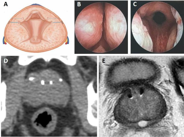
Multi-panel educational figure demonstrating the Prostatic Urethral Lift (PUL) procedure using the UroLift system for Benign Prostatic Hyperplasia (BPH) treatment. Panel A is a cross-sectional anatomical diagram illustrating four implants placed transversely to compress lateral prostatic lobes and widen the urethral lumen. Panels B and C are comparative cystoscopy photographs: B shows the baseline prostatic urethra obstructed by lateral lobes, while C displays the post-procedure 'continuous channel' and reduced obstruction. Panel D is a computed tomography (CT) scan in the axial plane showing the radiopaque UroLift implants as small, hyperdense (bright) points within the prostate. Panel E is a corresponding T2-weighted MRI scan showing the implants as signal voids with small surrounding artifacts, demonstrating their impact on prostatic urethral morphology. This figure provides a multimodal comparison of surgical intervention, illustrating how mechanical compression of glandular tissue relieves urinary obstruction.
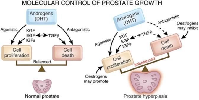
This medical schematic illustrates the pathophysiology of benign prostatic hyperplasia (BPH) compared to a normal prostate, focusing on the molecular signaling pathways that regulate tissue growth. The diagram is divided into two comparative sections: 'Normal prostate' and 'Prostate hyperplasia.' In a normal prostate, a balanced scale shows homeostatic equilibrium between cell proliferation and cell death. Androgens, primarily dihydrotestosterone (DHT), exert agonistic effects via growth factors (KGF, EGF, IGFs) to promote proliferation, and antagonistic effects via TGF-beta to promote apoptosis. In the prostate hyperplasia model, the scale is tilted toward proliferation, indicating an 'imbalanced' state. This pathological state is characterized by increased agonistic signaling and potential hormonal shifts, where estrogens may further promote cell proliferation and inhibit cell death. The illustration uses flow arrows to define the relationships between hormonal inputs (DHT, Oestrogens), intermediary growth factors (KGF, EGF, IGFs, TGF-beta), and the resulting physiological outcomes within the prostatic stroma and epithelium.
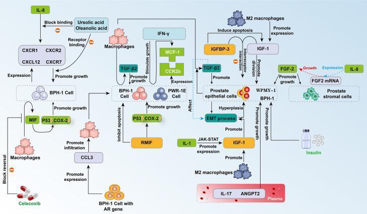
This medical pathophysiology diagram illustrates the complex cytokine signaling network and cellular interactions involved in Benign Prostatic Hyperplasia (BPH). It highlights the interplay between prostate epithelial cells (BPH-1, PWR-1E), stromal cells (WPMY-1), and infiltrating M2 macrophages. Key signaling pathways depicted include the IL-8 axis (involving receptors CXCR1/2 and CXCR7), which promotes BPH-1 growth and is inhibited by Ursolic and Oleanolic acids. The diagram shows M2 macrophages secreting TGF-β2 and IGF-1 to drive epithelial-mesenchymal transition (EMT), hyperplasia, and cell division. Pro-inflammatory signaling through IL-1 activates the JAK-STAT pathway to stimulate IGF-1, while recombinant MIF (rMIF) promotes growth by modulating P53 and COX-2 levels. Additionally, the role of metabolic factors like insulin and plasma cytokines (IL-17, ANGPT2) in stimulating BPH-1 and WPMY-1 cell proliferation is shown. Therapeutic interventions are represented by Celecoxib, which blocks macrophage-induced proliferation via CCL3 reversal. This schematic serves as an educational tool for understanding the immunoinflammatory and endocrine mechanisms driving prostate enlargement.
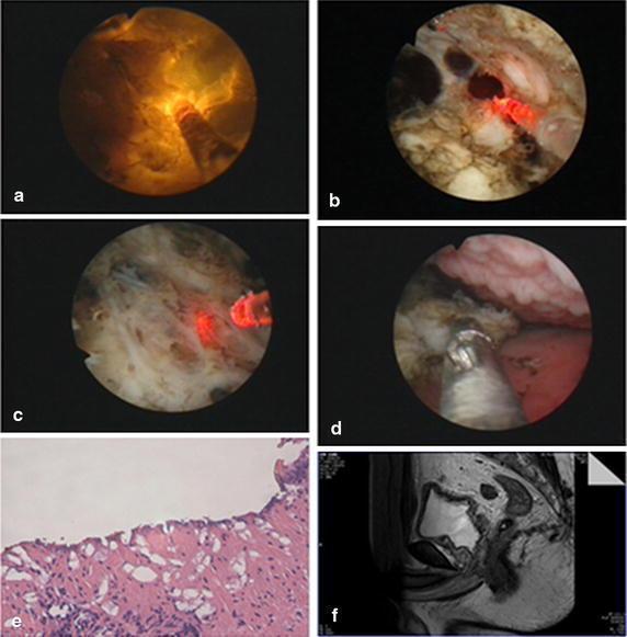
A multi-panel figure illustrating the surgical management and pathology of Benign Prostatic Hyperplasia (BPH) using Plasma-Resection Stripping-Vaporization of the Prostate (PRSVP). Panels (a-d) provide endoscopic views of the procedure: (a) demonstrates a straight beam LBO (green) laser in operation; (b) shows the mechanical stripping of hyperplastic prostate tissue; (c) visualizes the surgical plane at the level of the fibrous prostatic capsule; and (d) depicts the use of endoscopic forceps to retrieve tissue specimens. Panel (e) is a light microscopy image (H&E stain, 10x magnification) of the harvested specimen, confirming prostatic hyperplasia with characteristic glandular and stromal proliferation. Panel (f) is a sagittal T2-weighted MRI of the pelvis, showing the post-operative anatomical results with a widened prostatic urethra and surgical cavity. The collection serves as an educational resource for urological surgery, correlating intraoperative endoscopic findings with histopathology and post-operative radiological assessment.
"benign prostatic hyperplasia"[MeSH Terms] AND treatment
Management of Prostate Enlargement (Benign Prostatic Hyperplasia / BPH)
1. Assessment Before Treatment
- IPSS (International Prostate Symptom Score): Mild (0-7), Moderate (8-19), Severe (20-35)
- Uroflowmetry: Maximum flow rate (Qmax); <10 mL/s for voided volume >200 mL is a strong indication for treatment
- Post-void residual (PVR) ultrasound
- PSA: Offered after counselling; if PSA 4-10 ng/L with free-to-total PSA <15%, malignancy must be excluded with mpMRI +/- biopsy
- Serum creatinine, electrolytes, haemoglobin
- Urinalysis and MSU
- Pressure-flow urodynamics: Reserved for suspected neuropathy, dominant irritative symptoms, young men (<50 yrs), old men (>80 yrs), or recurrent LUTS post-surgery
2. Strong Indications for Surgery (Absolute / Urgent)
| Indication | Details |
|---|---|
| Acute urinary retention | In fit men with no reversible cause (drugs, constipation); accounts for 25% of prostatectomies |
| Chronic retention + renal impairment | Residual urine ≥200 mL, hydronephrosis, uraemia, abnormal renal function; accounts for 15% of prostatectomies |
| Complications of BOO | Bladder stones, recurrent infection, diverticulum formation |
| Recurrent haematuria | Vascular prostate confirmed on cystoscopy with no other cause |
3. Conservative Management (Watchful Waiting)
- Fluid manipulation: Avoid fluid bingeing and late-night fluid intake
- Dietary advice: Reduce caffeine and alcohol consumption
- Bladder training and lifestyle modification
- Regular reassessment with IPSS scoring and uroflowmetry
4. Medical (Drug) Therapy
A. Alpha-1 Adrenoceptor Blockers (alpha-blockers)
- Examples: Tamsulosin, alfuzosin, doxazosin, terazosin
- Mechanism: Relax smooth muscle in the prostate and bladder neck, reducing outflow resistance
- Onset: Fast (days to weeks)
- Best for: Any prostate size; especially mild-to-moderate symptoms
- Side effects: Postural hypotension, retrograde ejaculation (particularly tamsulosin), dizziness
B. 5-Alpha Reductase Inhibitors (5-ARIs)
- Examples: Finasteride (type 2 inhibitor), dutasteride (dual type 1 and 2 inhibitor)
- Mechanism: Block conversion of testosterone to DHT, reducing prostate volume by 20-30%
- Onset: Slow (3-6 months for full effect)
- Best for: Large prostates (>35-40 mL); prevent symptom progression and acute retention
- Side effects: Reduced libido, erectile dysfunction, ejaculatory disorders, gynaecomastia; PSA is halved (must double PSA value when interpreting)
C. Combination Therapy
- Alpha-blocker + 5-ARI together
- Better outcomes than monotherapy for glands >35 g
- Reduces risk of acute retention and need for surgery (evidence from MTOPS and CombAT trials)
D. Antimuscarinics / Beta-3 agonists
- Used when irritative/storage LUTS (urgency, frequency) dominate
- Antimuscarinics (e.g., solifenacin, tolterodine) - caution in elevated PVR
- Mirabegron (beta-3 agonist) - safer alternative in men with elevated PVR
E. Phosphodiesterase-5 Inhibitors
- Tadalafil 5 mg daily: Licensed for LUTS secondary to BPH; also treats co-existing erectile dysfunction
5. Elective Surgical Treatment
- Medical therapy has failed (typically after a trial of alpha-blockers +/- 5-ARI)
- Qmax <10 mL/s
- Elevated residual urine (100-250 mL)
- Severe symptoms (IPSS >20)
- Complications arise (retention, stones, infection, renal impairment, haematuria)
A. Transurethral Resection of the Prostate (TURP)
- Gold standard for surgical management
- Suitable for prostates up to ~80 g (most do <60 g)
- Advantages: Well-established, highly effective
- TURP syndrome (historically): Dilutional hyponatraemia from glycine irrigation - now largely avoided with bipolar/saline systems
- Complications: Haemorrhage, retrograde ejaculation (~80%), urethral stricture, bladder neck stenosis, incontinence (rare ~1%)
B. Laser Surgery
- GreenLight (Photoselective Vaporisation): Haemostatic vaporisation; suitable even in anticoagulated patients; not for very large glands
- HoLEP (Holmium Laser Enucleation of Prostate): Size-independent; laser cuts adenoma from false capsule, morcellated and extracted; equivalent outcomes to open prostatectomy for large glands; requires specialist expertise
C. Open / Robotic Simple Prostatectomy (Millin's / Freyer's)
- Reserved for very large prostates (>80-100 g)
- Millin's (retropubic): Most common open approach
- Increasingly being replaced by robotic-assisted simple prostatectomy
6. Minimally Invasive Surgical Therapies (MISTs)
| Procedure | Mechanism | Key Points |
|---|---|---|
| Prostatic Urethral Lift (UroLift) | Implants retract lateral lobes to widen urethra | Preserves ejaculation; no heat/ablation; for glands <80 g without prominent median lobe |
| Water Vapour Therapy (Rezum) | Convective steam energy causes cell death | Office-based; preserves ejaculation; effective for smaller glands |
| Prostate Artery Embolisation (PAE) | Radiological embolisation of prostatic arteries | Suitable for high surgical risk patients; larger glands; performed by interventional radiologist |
| Aquablation | Robotic waterjet ablation guided by ultrasound | Size-independent; preserves ejaculation better than TURP |
7. Special Situations
Acute Urinary Retention
- Insert urethral catheter urgently to drain bladder
- Commence alpha-blocker
- Trial without catheter (TWOC) after 48-72 hours while on alpha-blocker
- If TWOC fails, or retention recurs, plan prostatectomy
- If renal impairment/hydronephrosis present: Keep catheter for at least 6 weeks before surgery to allow renal recovery
Chronic Retention with Renal Impairment
- Urgent catheterisation
- Monitor for post-obstructive diuresis: High urine output post-catheterisation due to loss of tubular reabsorptive capacity
- Monitor daily weight, fluid balance, creatinine/electrolytes
- IV fluid replacement if patient cannot keep up with losses
- Transfuse if Hb <9 g/L once fluid balance stabilised
- Unfit men or those with dementia: Long-term indwelling catheter (urethral or suprapubic)
8. Summary of Management Pathway
LUTS/BPH Diagnosed
       |
 Absolute indications?  ──Yes──> Urgent prostatectomy / catheterisation
       | No
       ↓
  Mild symptoms (IPSS ≤8)?
       | Yes ──> Watchful waiting + lifestyle modification
       ↓ No (moderate-severe)
  Medical therapy
  - Small prostate: Alpha-blocker alone
  - Large prostate (>35g): Alpha-blocker + 5-ARI (combination)
  - Storage symptoms dominant: Add antimuscarinic or mirabegron
       ↓
  Failed medical therapy / complications?
       | Yes ──> Surgery
       |          ├─ TURP (gold standard)
       |          ├─ HoLEP (large glands)
       |          ├─ Open/Robotic prostatectomy (very large glands)
       |          └─ MISTs (UroLift, Rezum, PAE, Aquablation)
       ↓
  Regular review and reassessment