
How to Read emg report
Reading File
Finding Sources
Finding Sources
Searching Images
EMG needle electrode motor unit action potential waveform interpretation
Searching the Web
how to interpret EMG report step by step fibrillation potentials motor unit action potentials
Reading File
Reading File
How to Read an EMG Report
An EMG report (usually called an "EMG/NCS" report) has two main parts done together: nerve conduction studies (NCS) and needle electromyography (EMG). Reading it means working through each part systematically, then combining them into a pattern.
1. Nerve Conduction Studies (NCS) - the first half of the report
NCS stimulates a nerve electrically and records the response, either from a muscle (motor study) or from the nerve itself (sensory study).
Key numbers to check, each compared against the lab's own reference ranges (age/height/sex-matched):
- Latency - time from stimulus to response. Prolonged latency suggests a focal slowing/demyelination (e.g., at a compression point like the carpal tunnel).
- Amplitude (CMAP for motor, SNAP for sensory) - reflects the number of intact axons. Low amplitude = axon loss.
- Conduction velocity - distance/time between two stimulation points. Normal is roughly 50-70 m/sec in the arms and 40-60 m/sec in the legs. Slowing suggests demyelination; near-normal velocity with low amplitude suggests axonal loss.
- F-waves and H-reflexes - assess proximal nerve segments (useful in radiculopathy, Guillain-Barre).
General pattern reading:
- Low amplitude + near-normal velocity = axonal loss.
- Markedly slowed velocity/prolonged latency, conduction block, or dispersed waveform = demyelinating process.
- Normal sensory but abnormal motor (or vice versa) helps localize to root (sensory often spared in radiculopathy since the lesion is proximal to the dorsal root ganglion) versus peripheral nerve/plexus.
2. Needle EMG - the second half
The needle electrode records muscle electrical activity in three phases:
A. At rest (insertional and spontaneous activity)
- Insertional activity: brief activity from needle movement - normal, unless prolonged.
- Spontaneous activity (abnormal if present at rest):
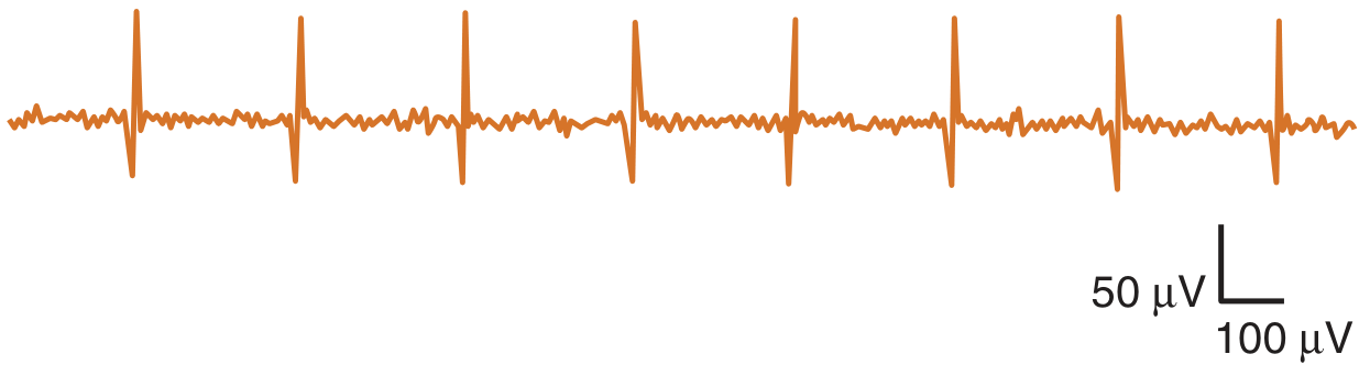
- Fibrillation potentials / positive sharp waves - single muscle fiber discharges indicating denervation or muscle membrane irritability (myositis). Note timing: these typically take 2-3 weeks to appear after acute nerve injury, so an EMG done too early can look falsely normal.
- Fasciculations - whole motor unit discharges, seen in motor neuron disease, radiculopathy, or benign causes.
- Complex repetitive discharges / myotonic discharges - seen in chronic denervation or myotonic disorders respectively.
B. During mild voluntary contraction (motor unit action potential, MUAP, analysis)
- Amplitude, duration, number of phases are assessed:
- Large amplitude, long duration, polyphasic MUAPs = neurogenic/reinnervation pattern (chronic denervation with collateral sprouting).
- Small amplitude, short duration, polyphasic MUAPs = myopathic pattern (fewer functioning fibers per motor unit).
C. During maximal effort (recruitment and interference pattern)
- Reduced recruitment (fewer motor units firing faster than expected for the effort) = neurogenic loss (fewer motor units available).
- Early/rapid recruitment with full interference pattern but low amplitude = myopathic process (more units needed for a given force because each is weak).
- Absent MUAPs despite maximal effort = complete axonal loss or conduction block to that muscle.
3. Putting it together - the "impression"
The electromyographer synthesizes both halves to answer:
- Is the lesion neurogenic, myopathic, or neuromuscular junction (e.g., myasthenia, seen on repetitive stimulation)?
- Where is it located - single nerve (mononeuropathy, e.g., median at wrist), root (radiculopathy - paraspinal muscle involvement is a clue), plexus, or generalized polyneuropathy (distal, symmetric, length-dependent pattern)?
- Axonal vs demyelinating - based on amplitude vs velocity/latency findings.
- Acute vs chronic vs ongoing - fibrillations without large reinnervated MUAPs = acute/subacute; large polyphasic MUAPs with reduced recruitment = chronic with reinnervation; both together = chronic process with ongoing active denervation.
- Bradley and Daroff's Neurology in Clinical Practice: NCS/EMG serve four purposes - localize the lesion, grade severity, determine acuity (acute/subacute/chronic), and determine if it's axonal or demyelinating. It also notes denervation changes may not appear on EMG until up to 4 weeks after axonal injury.
- Pfenninger and Fowler's Procedures for Primary Care: normal motor conduction velocity is roughly 50-70 m/sec in the arms and 40-60 m/sec in the legs; reference ranges are lab-specific and must be demographically matched.
Below is a genuine EMG waveform showing an abnormal spontaneous discharge (fibrillation potential), the kind of finding you'd see flagged in the "spontaneous activity" section of a report:
Practical tip for reading your own or a patient's report
Look for these labeled sections in order: (1) NCS table with latency/amplitude/velocity for each nerve tested, (2) needle EMG table listing muscles tested with columns for insertional/spontaneous activity, MUAP morphology, and recruitment, (3) a written "Impression" or "Conclusion" that states the overall pattern (e.g., "findings consistent with a chronic left C6 radiculopathy" or "moderate axonal sensorimotor polyneuropathy"). The impression is where the electromyographer's clinical correlation lives - always read it alongside the raw numbers, since EMG/NCS results must be interpreted in the context of the clinical exam.
A quick check of recent literature (meta-analyses/systematic reviews, last 2 years) didn't surface anything that changes this standard interpretive framework - it remains the accepted approach in current neuromuscular medicine.
If you have an actual report you'd like help interpreting, share the specific findings (which nerves/muscles, the numeric values, and what abnormalities are listed) and I can walk through what they mean for your specific case.
This is a shared conversation. Sign in to Orris to start your own chat.