
Alar collapse in rhiniplasty
Reading File
Now let me search the medical library and PubMed in parallel.
Finding Sources
Finding Sources
Searching PubMed
"alar collapse" rhinoplasty
Good results. Now let me read the key sections from Cummings:
Reading File
Finding Sources
Reading File
Reading File
Reading File
Reading File
Finding Sources
Reading File
Reading File
Reading File
Reading File
Finding Sources
Now let me fetch the key intraoperative images to enrich the answer:
Loading Image
Loading Image
I now have thorough textbook content from multiple authoritative sources to compose a detailed answer.
Alar Collapse in Rhinoplasty
Definition and Anatomy
The external nasal valve is formed by the lateral ala (skin and soft tissue), the caudal edge of the upper lateral cartilage (ULC), the lower lateral cartilage (LLC / alar cartilage), the nasal sill, and the septum. Alar collapse refers to dynamic inward buckling of this sidewall during inspiration, causing external nasal valve obstruction. The normal internal nasal valve angle is 10-15 degrees in white patients (wider in others); the external valve sits lateral to this and is highly dependent on cartilaginous support.
- Cummings Otolaryngology, p. 568-569
Causes / Aetiology
Primary (Pre-existing)
- Inherent lateral crural weakness
- Recurvature of the lateral crura
- Cephalic malposition of the LLC (acute orientation >35-45 degrees off midline reduces lateral wall support)
- Lax connective tissue (aging)
Iatrogenic (Post-rhinoplasty)
The most common cause is over-resection of the lower lateral cartilages (LLCs) during tip surgery:
- Excessive cephalic strip resection of the lateral crura (minimum 6 mm of residual cartilage must be preserved)
- Inadequate support of the lateral wall after tip modification
- Post-operative scar contracture tightening the sidewall
- Cephalic malposition introduced or worsened surgically
- Caudal septal dislocation
- Previous filler injection to the nasal sidewall (mass effect on the internal nasal valve via inferomedial lateralization of the ULC)
"Alar collapse and retraction is frequently caused by over-resection of the LLCs in an attempt to sculpt the nasal tip." - Cummings Otolaryngology, p. 688
Clinical Features
- Nasal obstruction worsening on inspiration (especially rapid/forceful breathing)
- Exaggerated supra-alar crease visible on frontal view
- Alar-columellar disharmony: excessive columellar show (normal = 2-4 mm on lateral view), notching at the apex of the alar margin
- Aesthetic: pinched tip, parenthesis deformity on frontal view (when LLC is both bulbous and cephalically oriented)
- On examination: dynamic inward collapse of the alar sidewall during sniffing
Diagnosis
Modified Cottle maneuver - an instrument is placed underneath the lateral nasal sidewall to gently support it while the patient inhales. Improvement in nasal airflow confirms a dynamic (i.e., functional) external valve collapse that is amenable to structural support. A positive Cottle suggests alar batten grafts or lateral crural strut grafts (LCSGs) would help.
- Cummings Otolaryngology, p. 685
Peak nasal inspiratory flow (PNIF) testing can underestimate airflow because the inspiratory effort itself can provoke alar collapse, limiting the measurement.
Surgical Correction
Correction is directed at structural support of the lateral sidewall and, when needed, repositioning or augmenting the LLC.
1. Alar Batten Grafts
- Taken from septal or conchal cartilage
- Placed in a subcutaneous pocket at the point of maximal lateral wall collapse, slightly overlapping the lateral crura
- Can be positioned cephalad, caudal, or directly over the lateral crus; may extend over the bony piriform aperture for added stability
- Correct both internal and external nasal valve collapse
- Shown to correct both types of valve collapse even in non-scarred noses
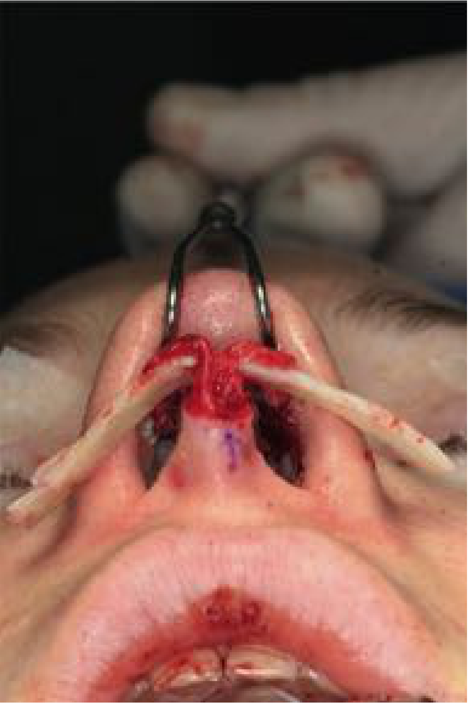
2. Lateral Crural Strut Grafts (LCSGs)
- Typically fashioned from septal cartilage
- Sutured directly to the dissected undersurface of the lateral crura
- Can flatten recurvature of the lateral crus
- Re-orients the caudal margin above the cephalic margin
- Extends to the piriform aperture for structural stability
- Particularly useful when the lateral crus is displaced or malpositioned
A "mini-strut" version can be placed underneath the lateral crus to strengthen it and correct a severely convex or concave configuration.
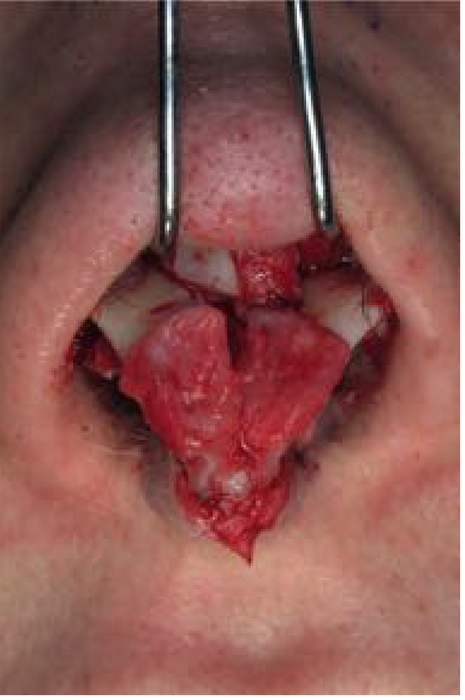
3. LLC Repositioning (for Cephalic Malposition)
- Defined as LLC orientation >35-45 degrees off midline
- The lateral crus is elevated from its bed and reinserted into a more caudal and posterior pocket
- Often combined with a strut graft for added support
- Achieves functional correction of valve collapse AND cosmetic improvement (nasal length, projection, rotation)
- Can be performed via endonasal or external approach (external is technically less demanding)
4. Alar Retraction with Composite Grafts
When retraction (as opposed to simple collapse) is present:
- Mild: Cartilage grafts placed through a marginal incision into a snug subalar pocket
- Severe: Composite graft from the cymba conchae of the opposite ear placed into a pocket dissected between the alar rim and the residual LLC, displacing the alar rim caudally
- An alternative: septal or rib cartilage spacer graft placed in the scroll area (between ULC and LLC) to push the LLC caudally
5. Spanning / Lateral Crural Flip-Over Techniques
- Spanning grafts straddle the dorsal septum attached to the lateral crura, keeping them in the desired orientation
- Flip-over technique: severely convex/concave lateral crus is completely mobilized and re-inserted with the concavity converted to convexity, sutured to the medial segment
6. Turn-In Flap (for alar retraction with soft tissue deficit)
- The strip of cephalic trim can be used as a turn-in flap to splint the remaining lateral crus strip and reduce excessive curvature
Summary of Graft Options
| Graft | Position | Primary Goal |
|---|---|---|
| Alar batten graft | Over point of maximal collapse | Support lateral wall; correct external valve collapse |
| Lateral crural strut graft (LCSG) | Undersurface of lateral crus | Structural support + correction of recurvature/malposition |
| Composite graft (conchal) | Alar rim pocket | Correct severe alar retraction |
| Spacer graft (septal/rib) | Scroll area (ULC-LLC junction) | Caudal displacement of LLC to correct retraction |
| Alar rim graft | Alar margin | Recreate convexity and rounding of nasal tip |
Prevention
- Preserve a minimum 6 mm strip of lateral crural cartilage after cephalic trim
- Preserve vestibular mucosa to reduce contracture risk
- Avoid over-aggressive tip suturing, which can create tension on the lateral crus
- Pre-operatively identify cephalic malposition and plan repositioning rather than simple trimming
- Identify pre-existing alar collapse with the modified Cottle maneuver and address it at the time of primary rhinoplasty
Sources:
- Cummings Otolaryngology Head and Neck Surgery, pp. 568-569, 685, 688-689
- Scott-Brown's Otorhinolaryngology Head & Neck Surgery Vol 1, pp. 1195-1196
- Scott-Brown's Otorhinolaryngology Head & Neck Surgery, p. 1195
- K.J. Lee's Essential Otolaryngology
ENT manifestations in GERD
Searching the medical library and recent literature simultaneously.
Finding Sources
Finding Sources
Searching PubMed
"laryngopharyngeal reflux" ENT manifestations
Good. Now read the key ENT/otolaryngology sections:
Reading File
Reading File
Finding Sources
Reading File
Reading File
Finding Sources
Reading File
Reading File
Finding Sources
Reading File
Reading File
Finding Sources
Reading File
Reading File
Finding Sources
Reading File
Reading File
Loading Image
Loading Image
I now have comprehensive content. Let me compile the full answer.
ENT Manifestations of GERD / Laryngopharyngeal Reflux (LPR)
Terminology and Concept
GERD (gastroesophageal reflux disease) refers to pathological reflux of gastric contents into the esophagus causing typical symptoms (heartburn, regurgitation). When refluxate reaches above the upper esophageal sphincter to contact the larynx, pharynx, and airway, the term laryngopharyngeal reflux (LPR) is used - also called extra-oesophageal reflux (EOR).
A key clinical point: more than 60% of patients with LPR do not have classic GERD symptoms like heartburn. LPR can exist without GERD, though it is more likely when GERD is present. This makes LPR a diagnosis that ENT surgeons must identify independently.
"Laryngopharyngeal reflux (LPR) may not manifest symptoms of gastroesophageal reflux disease (GERD), such as classic heartburn or indigestion." - K.J. Lee's Essential Otolaryngology, p. 919
Pathophysiology
The laryngeal mucosa lacks the protective mechanisms of the esophagus (no peristaltic clearance, no bicarbonate secretion, weaker mucosal defense). Two components of refluxate cause tissue damage:
- Hydrochloric acid - directly corrosive; partially suppressed by PPIs
- Pepsin - activated at pH <4 but remains active almost up to neutrality (pH 7) using human pepsin. Pepsin binds to and is taken up into laryngeal epithelial cells by endocytosis; can be reactivated by subsequent acid exposure. Even acidic drinks (cola, pH <2) can re-activate deposited pepsin. Pancreatic proteases, bile acids, and partially digested food may also contribute.
This is why purely acid-suppressive treatment may not fully resolve LPR - non-acid pepsin-mediated injury continues.
- Scott-Brown's Otorhinolaryngology, p. 1137
ENT Manifestations
1. Laryngeal Manifestations (Most Important)
A. Reflux Laryngitis / Posterior Laryngitis
The posterior larynx (arytenoids, posterior commissure, inter-arytenoid region) is closest to the esophageal inlet and is most vulnerable. First described by Delahunty in 1972.
Endoscopic findings (summarized in the Reflux Finding Score, RFS):
| Finding | Details |
|---|---|
| Infraglottic oedema (pseudosulcus vocalis) | Fold parallel to free edge of vocal cord; high sensitivity for reflux (PPV 67-90%); may extend the full cord length |
| Posterior commissure hypertrophy | Cobblestone/pachydermia of posterior laryngeal wall |
| Arytenoid oedema & erythema | Loss of normal arytenoid contour; pathognomonic of GORD when severe |
| Ventricular obliteration | Effacement of the laryngeal ventricle |
| Vocal fold oedema | Bilateral; may cause Reinke's oedema |
| Diffuse laryngeal oedema | "Active larynx" appearance |
| Thick endolaryngeal mucus | Viscous mucus pooling |
| Granuloma / granulation tissue | Especially posterior glottis/vocal process |
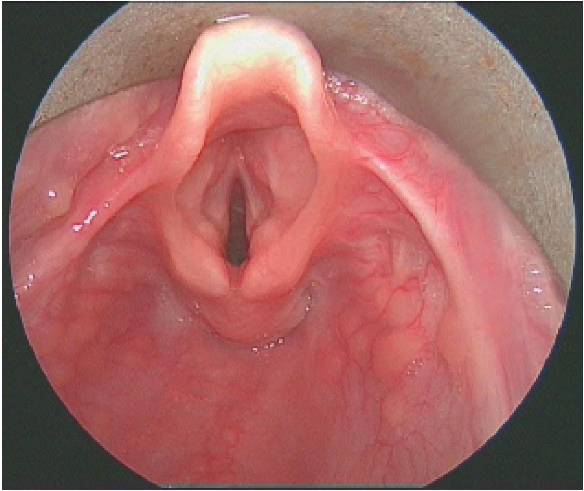
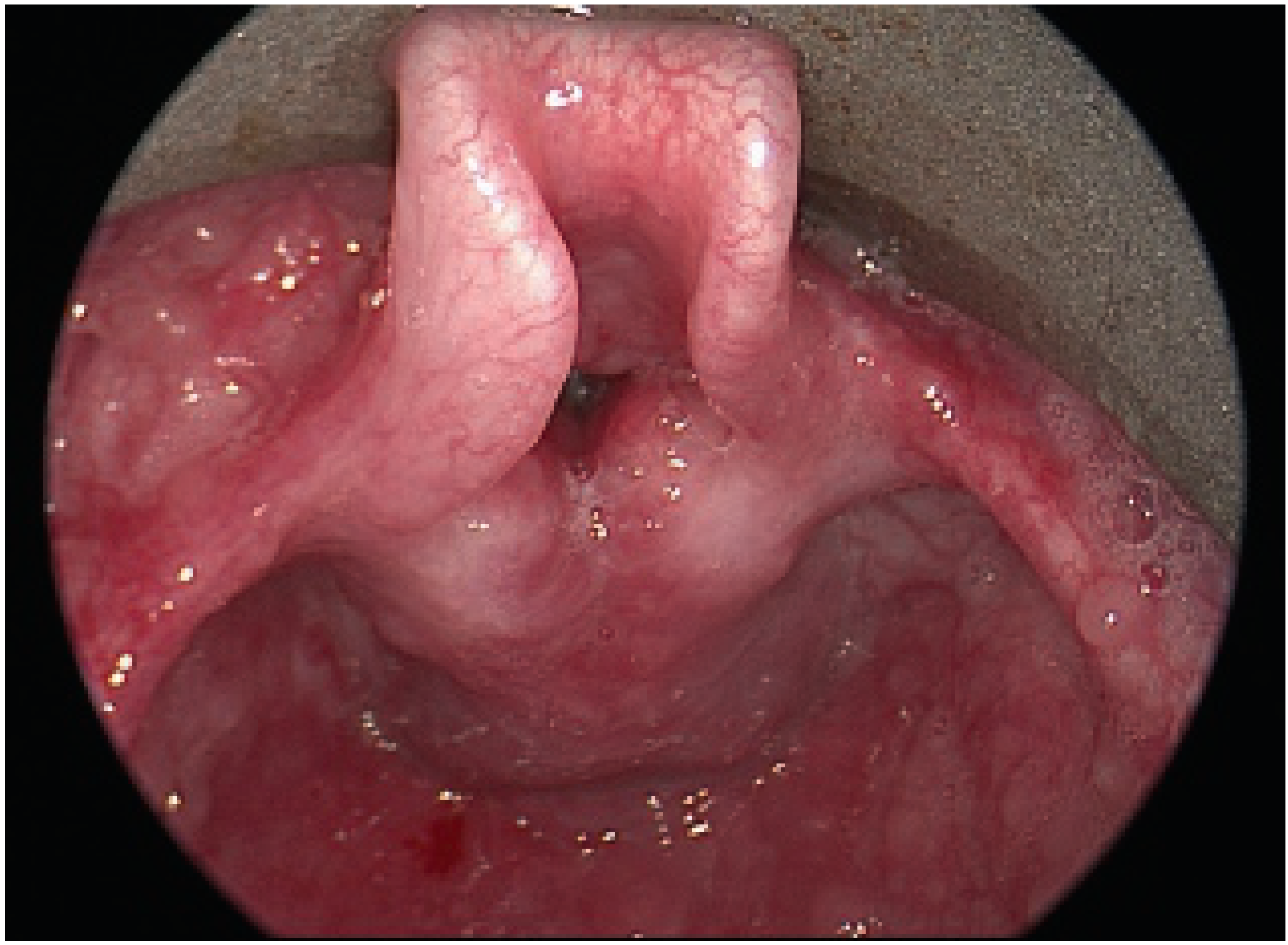
Important caveat: No single finding is pathognomonic. Laryngeal abnormalities are reported in 64-86% of normal controls. Pseudosulcus vocalis has the best PPV (67-90%) but must be interpreted with clinical context.
RFS scoring: Out of a potential maximum of 26 points; score >7 suggests LPR. An RSI (Reflux Symptom Index) score >10 on a 9-item questionnaire also indicates LPR.
B. Contact Ulcer / Vocal Process Granuloma
- Ulceration and granuloma formation at the vocal processes of the arytenoids from repeated acid/pepsin exposure
- Presents with throat pain, foreign body sensation, dysphonia
C. Subglottic Stenosis
- LPR is an established contributing factor to idiopathic subglottic stenosis and to acquired subglottic stenosis post-intubation
- Gastric acid impairs mucosal healing after laryngotracheal injury - Scott-Brown's Vol 2, p. 548
D. Laryngomalacia (Paediatric)
- Up to 90% of infants with laryngomalacia have associated GER
- Increased intrathoracic pressure from airway obstruction promotes reflux; the same vagal dysfunction that affects laryngeal tone also affects the lower esophageal sphincter
- Pepsin has been identified in tracheal aspirates and arytenoid mucosa of infants with severe laryngomalacia
E. Laryngospasm
- Refluxate triggering the larynx can provoke reflex laryngospasm
- May present as episodic choking, throat tightening, or cough syncope
F. Vocal Cord Dysfunction (VCD) / Paradoxical Vocal Fold Movement
- LPR is a recognized trigger for VCD - reflux-induced irritation causes aberrant vocal fold adduction during inspiration
- Presents as inspiratory stridor, often misdiagnosed as asthma
2. Pharyngeal Manifestations
| Symptom/Sign | Description |
|---|---|
| Globus pharyngeus | Constant sensation of a lump/something sticking in the throat; one of the most common LPR symptoms |
| Throat clearing | Chronic, often the dominant complaint; caused by posterior pharyngeal irritation and mucus |
| Postnasal drip sensation | Excess thick mucus, often mistaken for sinusitis |
| Dysphagia | Pharyngeal discomfort/difficulty swallowing; may be from hypopharyngeal irritation or reduced pharyngeal sensation |
| Halitosis | Sour/foul breath from refluxate |
| Recurrent sore throat | Chronic posterior pharyngeal irritation |
| Brackish taste / foul taste on waking | From nocturnal reflux reaching the oral cavity |
3. Nasal and Sinus Manifestations
- Chronic rhinosinusitis: LPR is implicated as a co-factor in refractory chronic rhinosinusitis; refluxate reaching the nasopharynx may trigger mucosal inflammation
- Adenoid hypertrophy: Extra-oesophageal reflux is possibly associated with adenotonsillar hypertrophy; pepsin has been identified in adenoid tissue
4. Ear Manifestations
-
Eustachian tube dysfunction / Otitis media with effusion (OME): LPR is listed among concomitant disorders causing chronic obstructive Eustachian tube dysfunction. Refluxate reaching the nasopharynx can inflame the tubal orifice and Gerlach's tonsil, impairing ET function. Pepsin has been detected in middle ear fluid and adenoid tissue, suggesting a possible direct pathogenic role.
-
Evidence linking gastric reflux to OME exists (Tasker et al.) but the causal relationship remains debated.
-
Cummings Otolaryngology, p. (Eustachian tube chapter)
5. Respiratory / Aerodigestive Manifestations
- Chronic cough: GERD is the 3rd most common cause of chronic cough (after postnasal drip and asthma), accounting for ~21% of cases. Mechanism: irritation of upper respiratory tract (with/without microaspiration) + esophageal-bronchial vagal cough reflex. 43-75% of patients with GERD-related cough have no typical reflux symptoms.
- Asthma / reactive airways: Reflux can trigger or worsen asthma via aspiration and vagal reflex bronchospasm
- Recurrent croup / stridor in infants: LPR can present as stridor, croup, apnea, and hoarseness in infants
- Recurrent laryngeal papillomatosis: Laryngopharyngeal acid exposure is increasingly recognized as a contributor to papilloma recurrence
6. Dental Manifestations
- Dental erosion: Prevalence 17-68% in GERD patients. Chronic acid exposure causes loss of enamel - painless at first, then sensitivity. Palatal surfaces of upper teeth affected earliest (from regurgitation).
Diagnostic Tools
| Tool | Notes |
|---|---|
| RSI (Reflux Symptom Index) | 9-item questionnaire, score >10 suggests LPR; sensitive but not specific |
| RFS (Reflux Finding Score) | Laryngoscopic scoring (0-26); score >7 suggests LPR; some interobserver variability |
| 24-hour dual-probe pH monitoring | Gold standard for GERD; pH <4 = significant; reflux index >4% = abnormal. Less reliable for LPR (no gold standard) |
| pH/MII (impedance monitoring) | Detects acid and non-acid reflux; best test for refractory EOR |
| Pepsin assay (saliva/BAL) | Promising biomarker; detects non-acid pepsin-mediated injury; more specific than lipid-laden macrophages for aspiration |
| Empirical PPI trial | First-line diagnostic/therapeutic approach; must be maintained for at least 3 months for cough |
Summary of ENT Manifestations
GERD / LPR Extra-esophageal ENT Manifestations
│
├── Larynx
│   ├── Reflux laryngitis (posterior laryngitis)
│   ├── Contact ulcer / vocal process granuloma
│   ├── Subglottic stenosis
│   ├── Laryngomalacia (paediatric)
│   ├── Laryngospasm
│   └── Vocal cord dysfunction
│
├── Pharynx
│   ├── Globus pharyngeus
│   ├── Chronic throat clearing
│   ├── Postnasal drip sensation
│   ├── Dysphagia
│   └── Halitosis / recurrent sore throat
│
├── Nose/Sinuses
│   ├── Chronic rhinosinusitis (refractory)
│   └── Adenoid hypertrophy
│
├── Ear
│   ├── Eustachian tube dysfunction
│   └── Otitis media with effusion
│
├── Airway/Respiratory
│   ├── Chronic cough (3rd most common cause overall)
│   ├── Asthma/reactive airways
│   └── Recurrent croup/stridor (paediatric)
│
└── Oral/Dental
    └── Dental erosion (enamel loss)
Sources:
- Cummings Otolaryngology Head and Neck Surgery, Chapters 29, 47, 68, 131
- Scott-Brown's Otorhinolaryngology Head & Neck Surgery Vol 2, Chapter 44 (Extra-oesophageal Reflux)
- Scott-Brown's Otorhinolaryngology, Chapter 77 (LPR)
- K.J. Lee's Essential Otolaryngology, pp. 919, 1260
- Textbook of Family Medicine 9e, Reflux Laryngitis section
- Recent guideline: Spanish multidisciplinary ReFaL Consensus on LPR diagnosis and management (2026, PMID 41391506)
This is a shared conversation. Sign in to Orris to start your own chat.