
Bronchial asthma pathology with pictures
Reading File
Now let me search the medical library for bronchial asthma pathology content and images simultaneously.
Finding Sources
Finding Sources
I can see Murray & Nadel's Respiratory Medicine has the best asthma pathology content. Let me now read the key sections.
Finding Sources
Reading File
Let me find the dedicated pathology section in the Murray & Nadel's respiratory medicine book and also read the immunology/T2 section which had images.
Finding Sources
Reading File
Excellent! Found a great image. Now let me search for the main pathology chapter on asthma and look for gross/histological images.
Finding Sources
Finding Sources
Reading File
Finding Sources
Excellent! Robbins Pathology has a dedicated MORPHOLOGY section with Figures 15.10 and 15.11. Let me read that section along with other key content.
Reading File
Reading File
Now let me read the Robbins Pathology morphology section and fetch all the key images in parallel.
Reading File
Loading Image
Loading Image
Loading Image
I have excellent images and content. Let me also get the histological images from Robbins (Fig 15.11) and the mast cell diagram.
Finding Sources
Loading Image
Now let me get the Robbins Basic Pathology section which has the histological morphology (Fig 15.11 with Curschmann spirals, Charcot-Leyden crystals, etc.).
Reading File
I have the histological biopsy image URL. Let me fetch it along with the Robbins full pathology diagram.
Loading Image
I now have all the key content and images needed to deliver a comprehensive, well-illustrated response on bronchial asthma pathology. Let me compile the full answer.
Bronchial Asthma - Pathology
Bronchial asthma is a chronic inflammatory disorder of the airways characterized by episodic, reversible airway obstruction, bronchial hyperresponsiveness, and structural airway remodeling. The pathology involves both acute and chronic changes.
1. Pathogenesis Overview
Asthma is fundamentally an exaggerated Th2-mediated immune response to environmental antigens in genetically susceptible individuals. The central cytokines driving pathology are:
| Cytokine | Effect |
|---|---|
| IL-4 | IgE production by B cells, vessel wall priming (VCAM-1/ICAM-1) |
| IL-5 | Eosinophil development and activation |
| IL-13 | Goblet cell metaplasia, mucus hypersecretion, bronchial hyperreactivity |
| TSLP/IL-33 | Activation of ILC2s and mast cells |
The diagram below (from Robbins Pathology) shows the full pathogenic sequence:
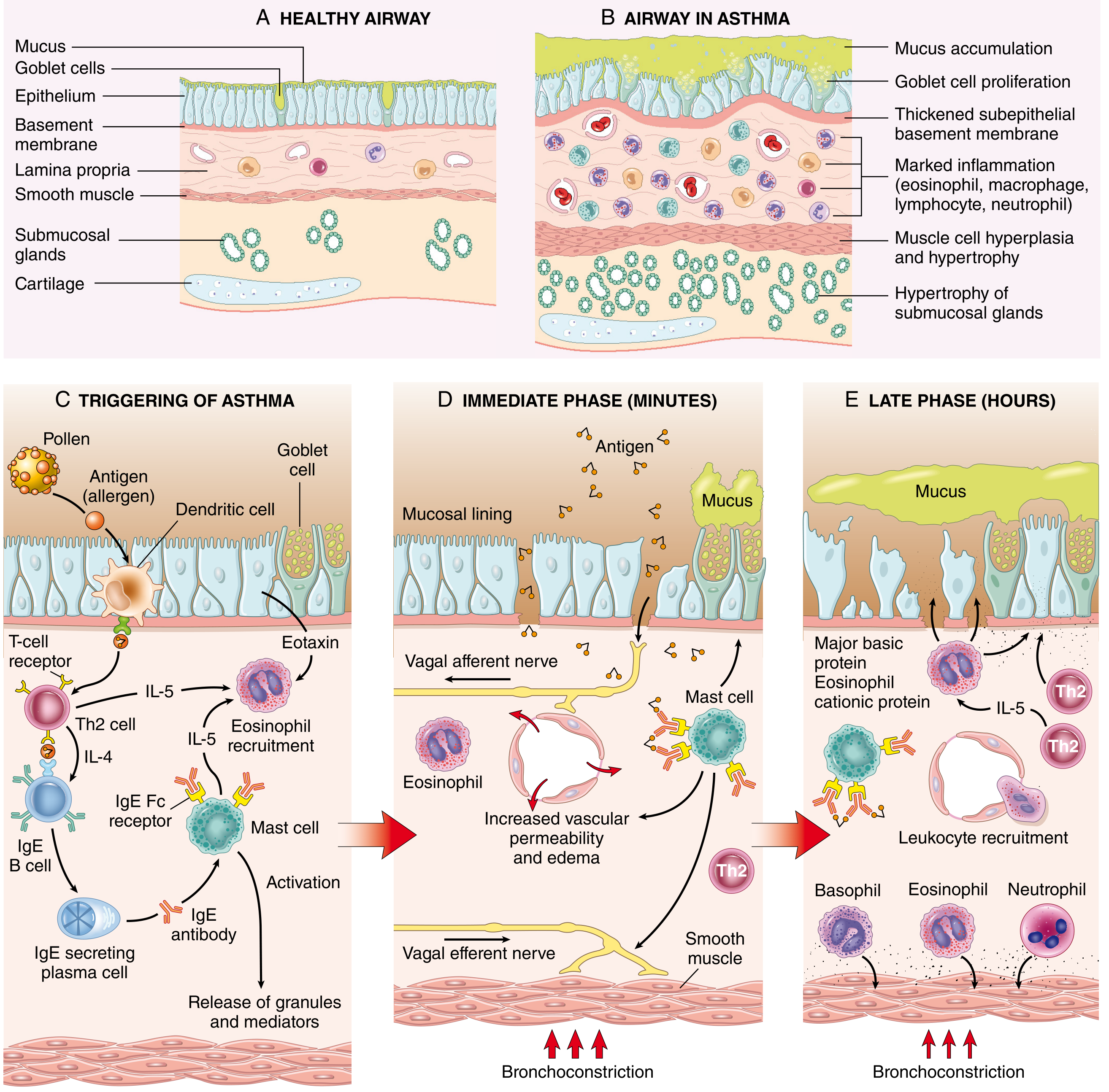
Fig. 15.10 - Robbins Pathology: (A) Normal airway vs. (B) asthmatic airway showing goblet cell proliferation, basement membrane thickening, inflammation, and smooth muscle hypertrophy; (C) Th2 sensitization; (D) immediate-phase reaction; (E) late-phase reaction.
2. Two-Phase Inflammatory Response
Immediate Phase (within minutes)
- Allergen re-exposure cross-links IgE on mast cells → mast cell degranulation
- Release of: histamine, serotonin, prostaglandin D2, leukotrienes (LTC4, LTD4, LTE4), tryptase, chymase
- Results in: bronchospasm, vascular permeability, mucus secretion
- Vagal efferent reflexes amplify bronchoconstriction
Late Phase (2-6 hours later, lasting 24-48 hours)
- Influx of eosinophils, neutrophils, basophils, Th2 cells, DCs
- Eosinophil products (major basic protein, eosinophil cationic protein) damage the epithelium and perpetuate hyperreactivity
3. Central Role of Mast Cells and Basophils
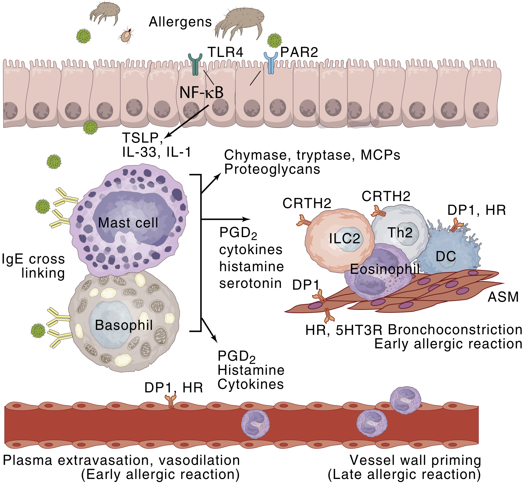
Fig. 60.6 (Murray & Nadel's): Allergen triggers IgE cross-linking on mast cells → release of chymase, tryptase, PGD2, histamine, serotonin → early bronchoconstriction and vessel wall priming for leukocyte extravasation.
4. T2 Immunity and Airway Remodeling
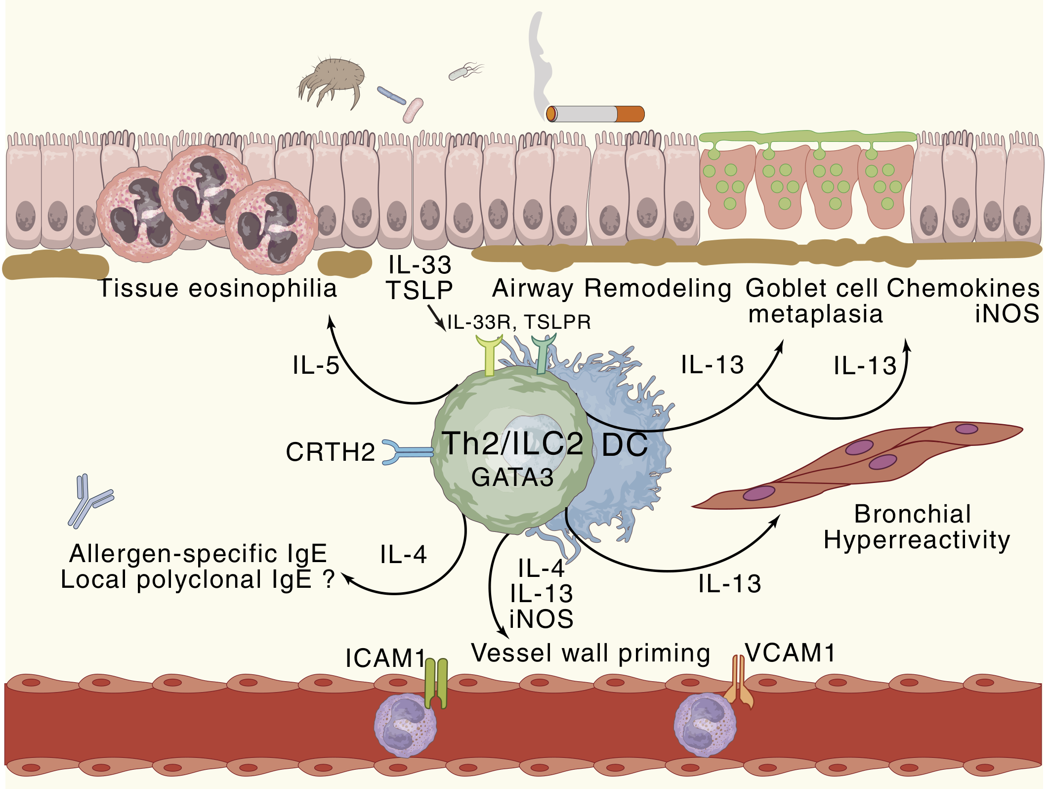
Fig. 60.5 (Murray & Nadel's): IL-5 drives tissue eosinophilia. IL-13 drives goblet cell metaplasia, iNOS, airway remodeling, and bronchial hyperreactivity. IL-4 drives IgE production.
ILC2s (Group 2 innate lymphoid cells) are activated by epithelial cytokines (TSLP, IL-33) independently of allergen recognition - they share GATA3 expression and cytokine production with Th2 cells, making them especially important in severe/late-onset asthma.
5. Mucus Plugging Mechanism
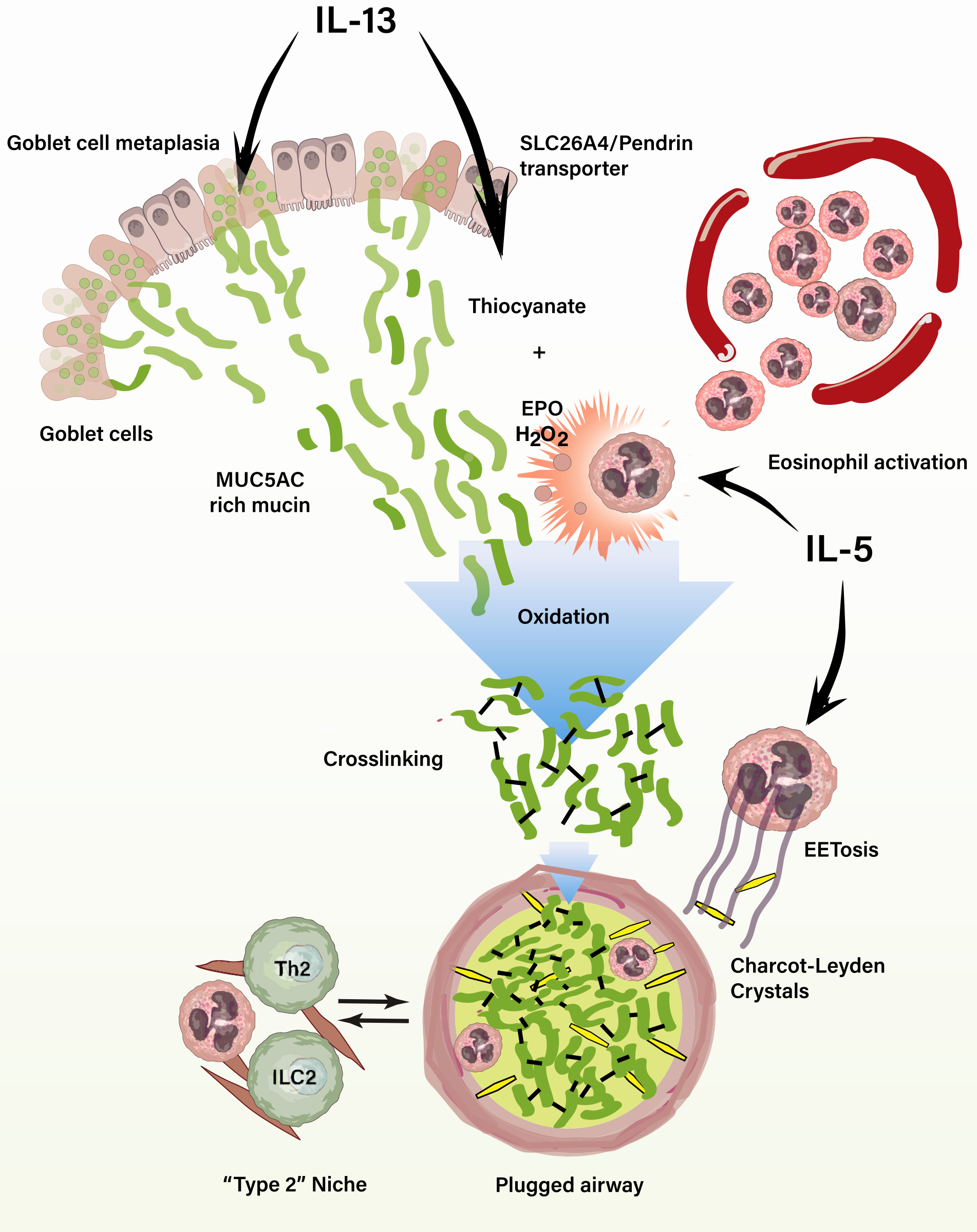
Fig. 60.7 (Murray & Nadel's): IL-13 drives goblet cell metaplasia and MUC5AC-rich mucin overproduction. IL-5 activates eosinophils, which release eosinophil peroxidase (EPO). EPO + thiocyanate + H₂O₂ cross-links mucin polymers → stiff, inspissated gel → plugged airway. Charcot-Leyden crystals form from galectin-10 released during EETosis.
6. Gross and Histological Morphology (MORPHOLOGY)
Gross Findings (Fatal Cases - Status Asthmaticus)
- Overinflated lungs with air trapping
- Small areas of atelectasis
- Thick, tenacious mucus plugs occluding bronchi and bronchioles
- Plugs contain shed epithelium forming Curschmann spirals
Histological / Microscopic Findings
Acute findings:
- Numerous eosinophils in mucus and wall
- Charcot-Leyden crystals (bipyramidal crystals of galectin-10 from eosinophil granules)
- Curschmann spirals (whorls of shed epithelium in mucus plugs)
Airway Remodeling (chronic):
- Thickening of the airway wall overall
- Sub-basement membrane fibrosis - deposition of type I and III collagens beneath the epithelial basement membrane (a hallmark finding)
- Goblet cell metaplasia - increased number of goblet cells replacing ciliated cells
- Hypertrophy of submucosal glands
- Smooth muscle hypertrophy and hyperplasia of bronchial wall
- Increased vascularity (angiogenesis)
Acute airflow obstruction = bronchoconstriction + edema + mucus plugging. Airway remodeling contributes to chronic irreversible obstruction.
Histological Biopsy (Robbins & Kumar Basic Pathology, Fig. 11.11)
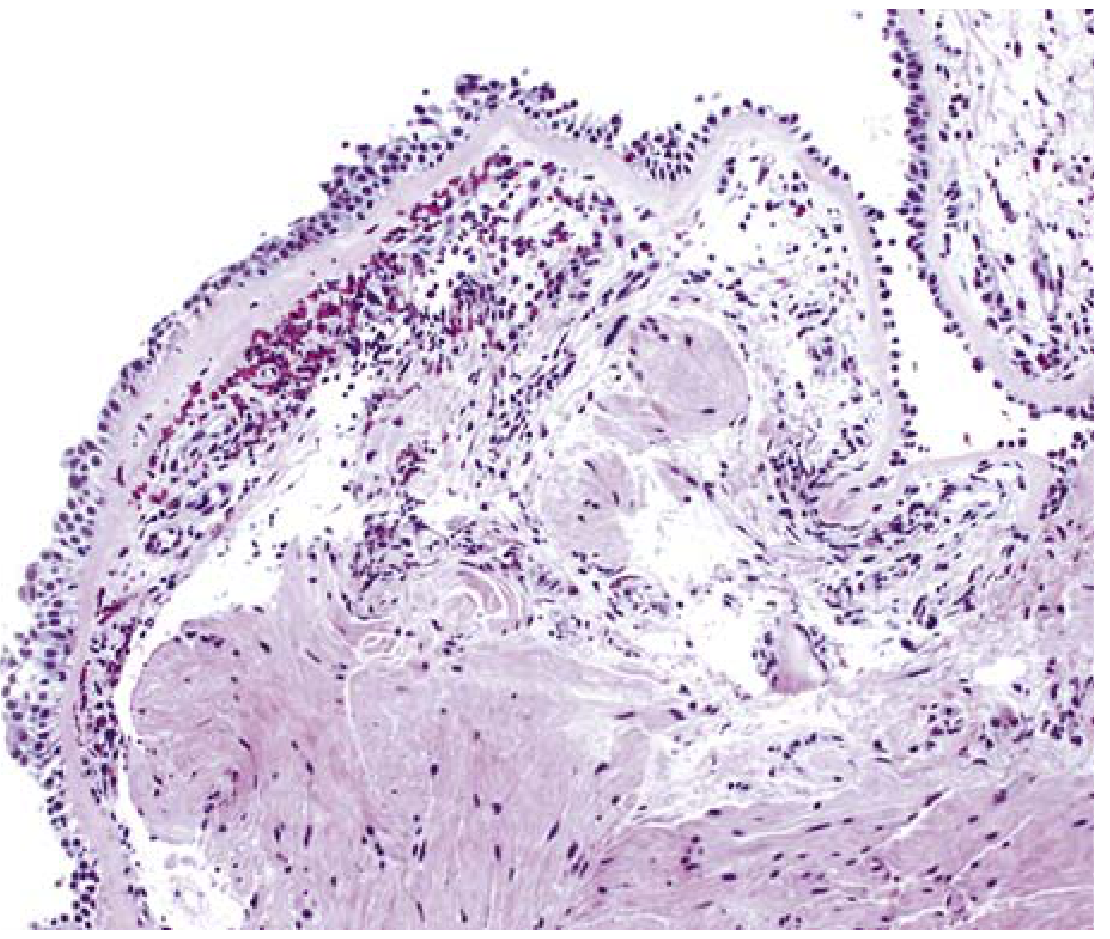
Bronchial biopsy specimen: sub-basement membrane fibrosis (pale acellular zone beneath epithelium), dense eosinophilic inflammation in the lamina propria, and smooth muscle hyperplasia in the lower layers.
7. Subtypes and Their Pathological Basis
| Subtype | Key Pathologic Feature |
|---|---|
| Atopic (extrinsic) | Th2/IgE-mediated; eosinophilic; responds to steroids |
| Non-atopic (intrinsic) | Triggered by respiratory infections; less IgE involvement |
| Aspirin-sensitive | Leukotriene overproduction (COX-1 inhibition shifts arachidonic acid to 5-LOX); adult-onset with nasal polyps |
| Occupational | Sensitization to workplace antigens (fumes, organic dusts, isocyanates) |
| T2-low / neutrophilic | Less eosinophilic; steroid-resistant; associated with severe/late-onset disease |
8. Key Pathologic Features - Summary Table
| Feature | Description |
|---|---|
| Cells | Eosinophils, mast cells, Th2 lymphocytes, ILC2s, basophils |
| Key cytokines | IL-4, IL-5, IL-13, TSLP, IL-33 |
| Sputum findings | Curschmann spirals, Charcot-Leyden crystals, eosinophils |
| Basement membrane | Sub-BM fibrosis (type I and III collagen) |
| Smooth muscle | Hypertrophy + hyperplasia |
| Mucus glands | Goblet cell metaplasia, submucosal gland hypertrophy |
| Vasculature | Increased (angiogenesis) |
| Fatal cases | Overinflation, mucus plugging, atelectasis |
Sources: Robbins, Cotran & Kumar Pathologic Basis of Disease (10th ed.), pp. 638-640; Robbins & Kumar Basic Pathology, pp. 442-456; Murray & Nadel's Textbook of Respiratory Medicine, Chapters 60-61 (Figs. 60.5-60.7).
RSV
Asking for Preferences
What aspect of RSV would you like to focus on?
Full form
RSV = Respiratory Syncytial Virus
It is a common RNA virus (family Pneumoviridae) that primarily causes respiratory tract infections. It is the leading cause of bronchiolitis and viral pneumonia in infants and young children worldwide, and can also cause serious illness in the elderly and immunocompromised.
Would you like a detailed overview of RSV - its pathology, clinical features, or treatment?
Bronchiectasis pathology with pictures
Reading File
This is a broad visual topic. Let me search the library in parallel across multiple sources.
Finding Sources
Finding Sources
Excellent. I'll read the three best sources in parallel: Robbins full pathology, Robbins basic pathology, and Murray & Nadel's (which likely has images), plus the radiology text.
Reading File
Reading File
Reading File
Excellent content found. I can see image URLs in the Robbins Basic Pathology text (Fig. 11.12 - gross specimen). Let me read more of that section to get more image URLs, and fetch the key images in parallel.
Reading File
Reading File
Loading Image
Now let me fetch the gross specimen image, the chest X-ray, and the CT scan images in parallel, plus read more of the radiology CT section.
Loading Image
Loading Image
Reading File
Excellent material. Let me now fetch the CT scan images and also get the Robbins full pathology content on pathogenesis and morphology.
Loading Image
Loading Image
Reading File
I now have comprehensive content and excellent images. Let me get the Robbins full pathology gross image (Fig 15.12) URL from the text I just read.
Reading File
I now have all the content needed. Let me fetch the Robbins full pathology gross image to display it.
Loading Image
I now have everything needed for a thorough, well-illustrated response. Let me compile the full answer.
Bronchiectasis - Pathology with Pictures
Bronchiectasis is the permanent, irreversible dilation of bronchi and bronchioles caused by destruction of smooth muscle and elastic tissue from chronic or severe infection and inflammation. It is always secondary to an underlying condition - it is never a primary disorder.
1. Definition and Key Concept
"Destruction of smooth muscle and elastic tissue by inflammation stemming from persistent or severe infections leads to permanent dilation of bronchi and bronchioles."
- Robbins, Cotran & Kumar Pathologic Basis of Disease
2. Causes / Predisposing Conditions
| Category | Examples |
|---|---|
| Congenital/Hereditary | Cystic fibrosis (most common hereditary cause), primary ciliary dyskinesia (Kartagener syndrome), immunodeficiency states |
| Post-infectious | Severe necrotizing pneumonia (S. aureus, Klebsiella, Pseudomonas, TB), viral (including post-SARS-CoV-2 reported) |
| Bronchial obstruction | Tumors, foreign bodies, mucus impaction |
| Immune/Inflammatory | Rheumatoid arthritis, SLE, IBD, allergic bronchopulmonary aspergillosis (ABPA) |
| Post-transplant | Chronic rejection (lung), graft-versus-host disease (HSCT) |
| Idiopathic | Up to 50% of cases |
3. Pathogenesis
Two intertwined processes form a vicious cycle:
Obstruction → Infection → Inflammation → Wall destruction → Dilation → More obstruction
- Obstruction (from foreign body, tumor, thick secretions) impairs mucociliary clearance
- Stasis of secretions creates a favorable environment for bacterial colonization
- Chronic bacterial infection triggers intense neutrophilic and lymphocytic inflammation
- Inflammatory mediators + bacterial toxins destroy bronchial smooth muscle and elastic tissue
- Loss of structural support → permanent dilation
- Peribronchial fibrosis develops, causing traction on airways (traction bronchiectasis)
Special mechanisms:
- Cystic fibrosis: Defective CFTR → thick viscous secretions → impaired mucociliary clearance → chronic Pseudomonas/Staphylococcus colonization → wall destruction
- Primary ciliary dyskinesia: Dynein arm mutations → non-functioning cilia → stasis → recurrent infection
- ABPA: Th2 hyperimmune response to Aspergillus fumigatus → high IgE, eosinophilic inflammation, mucus plugs → bronchiectasis
4. Morphology (Gross Pathology)
Distribution
- Usually affects lower lobes bilaterally, particularly vertical air passages
- Most severe in distal bronchi and bronchioles
- Localized when due to tumor or foreign body aspiration
Gross Findings
- Airways dilated up to 4× normal diameter
- Dilated bronchi visible almost to the pleural surface (in health, bronchioles cannot be seen by eye beyond 2-3 cm from pleura)
- Cut surface shows cystic spaces filled with mucopurulent, foul-smelling secretions
Gross Specimen - Bronchiectasis in Cystic Fibrosis (Robbins Pathology, Fig. 15.12)
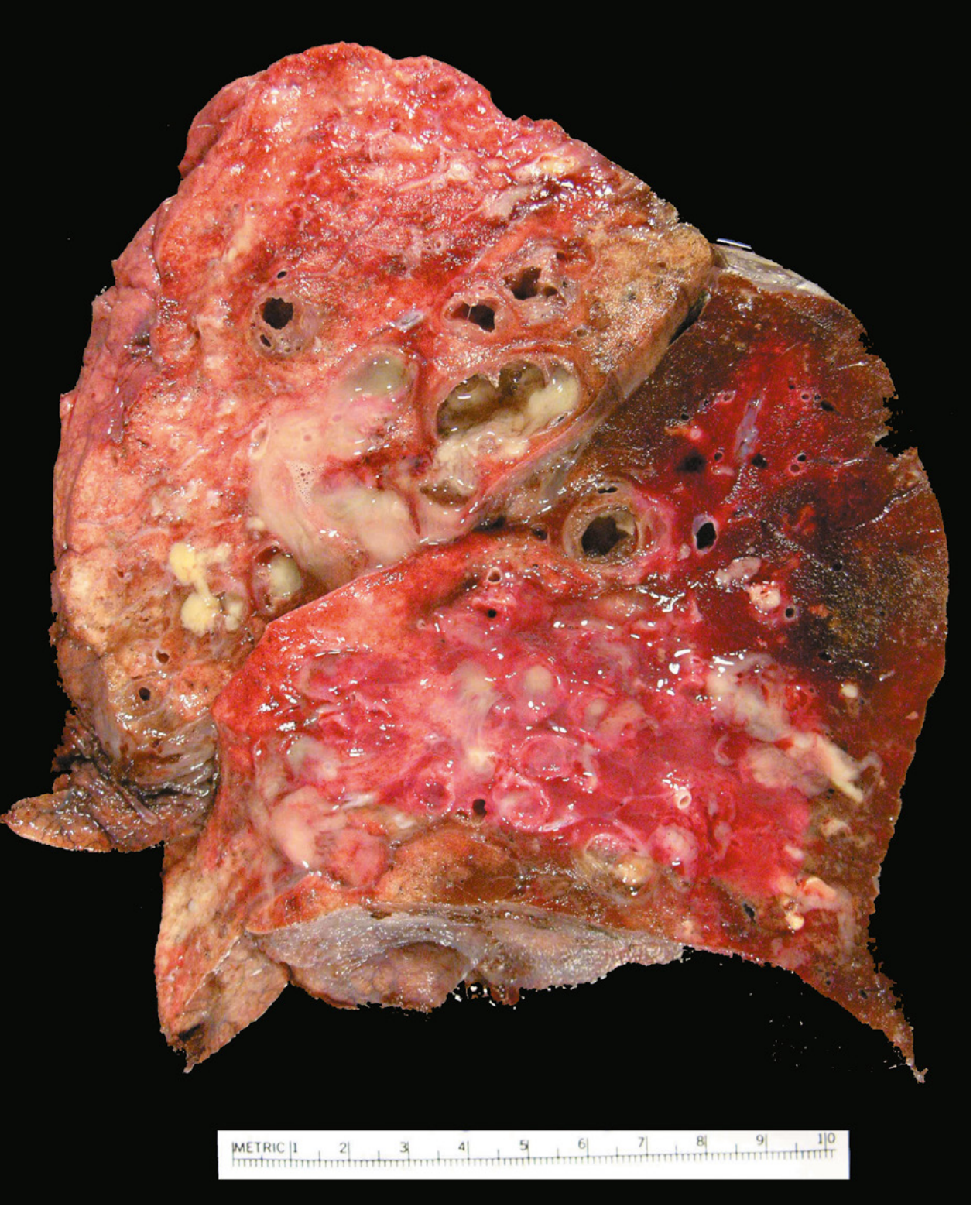
Cut surface of lung showing markedly distended peripheral bronchi filled with mucopurulent secretions, extending almost to the pleural surface.
5. Histological Findings (Microscopy)
The histology varies with activity and chronicity:
Active/Acute Phase
- Intense acute and chronic inflammatory exudate within bronchial and bronchiolar walls
- Desquamation (sloughing) of the lining epithelium
- Extensive ulceration of the mucosa
- Organisms: H. influenzae (~50%), P. aeruginosa (12-30%), nontuberculous mycobacteria
Chronic Phase
- Squamous metaplasia of remaining epithelium (further impairs mucociliary clearance)
- Fibrosis of bronchial and bronchiolar walls
- Peribronchiolar fibrosis
- In severe cases: necrosis → abscess cavity formation
- Progressive obliteration of bronchiolar lumens (bronchiolitis obliterans)
Histological Biopsy (Robbins Basic Pathology, Fig. 11.11)
Bronchial biopsy showing sub-basement membrane fibrosis, eosinophilic inflammation in the lamina propria, and smooth muscle hyperplasia.
6. Morphological Subtypes (by Severity)
| Subtype | Appearance | CT Findings |
|---|---|---|
| Cylindrical | Uniform, regular dilation (mildest) | Parallel tram-track lines; lack of tapering |
| Varicose | Non-uniform, serpiginous, beaded lumen | Beaded/irregular configuration |
| Cystic (saccular) | Large cyst-like spaces (most severe) | Clusters of cysts, often with air-fluid levels |
7. Radiological Findings
Chest X-Ray
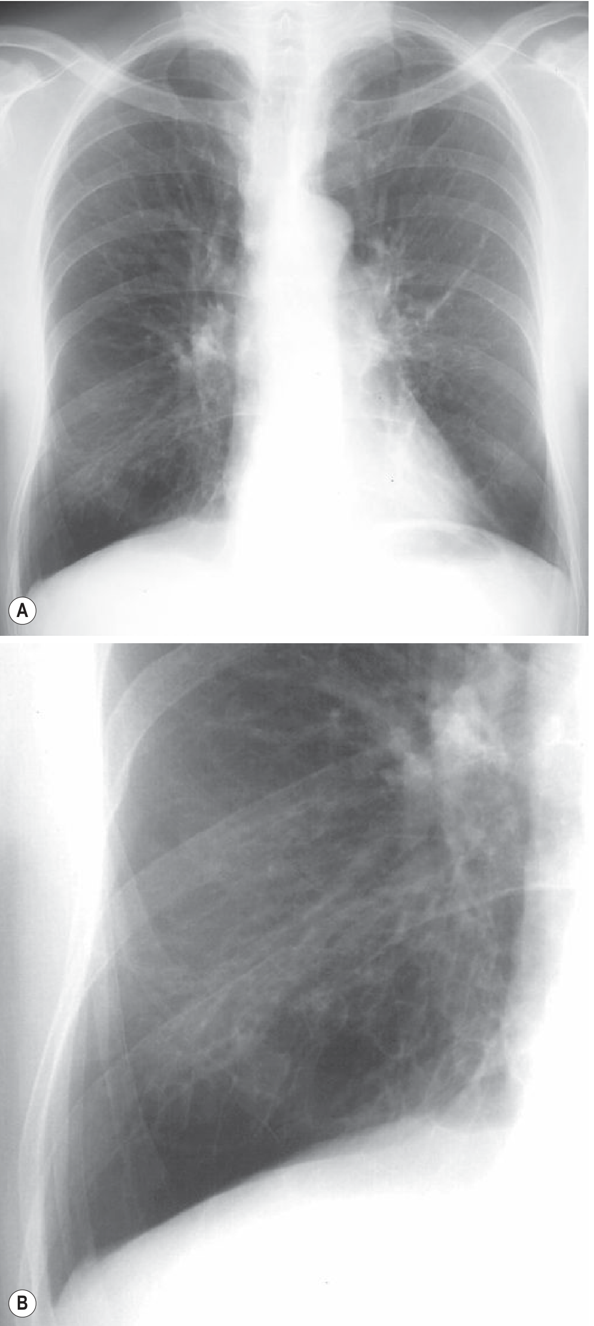
Fig. 6.15 (Grainger & Allison): (A) PA chest X-ray showing oligemia in lung bases with blood flow redistribution and slight overinflation. (B) Targeted image shows tramlines and ring opacities reflecting dilated, wall-thickened bronchi.
CXR findings:
- Tramlines - thickened parallel bronchial walls viewed en face
- Ring opacities - dilated bronchi seen end-on
- Tubular/ovoid opacities (mucus-filled bronchi)
- In cystic bronchiectasis: thin-walled ring shadows with air-fluid levels
- Overinflation (widespread disease, e.g., cystic fibrosis)
CT / HRCT (Gold Standard)
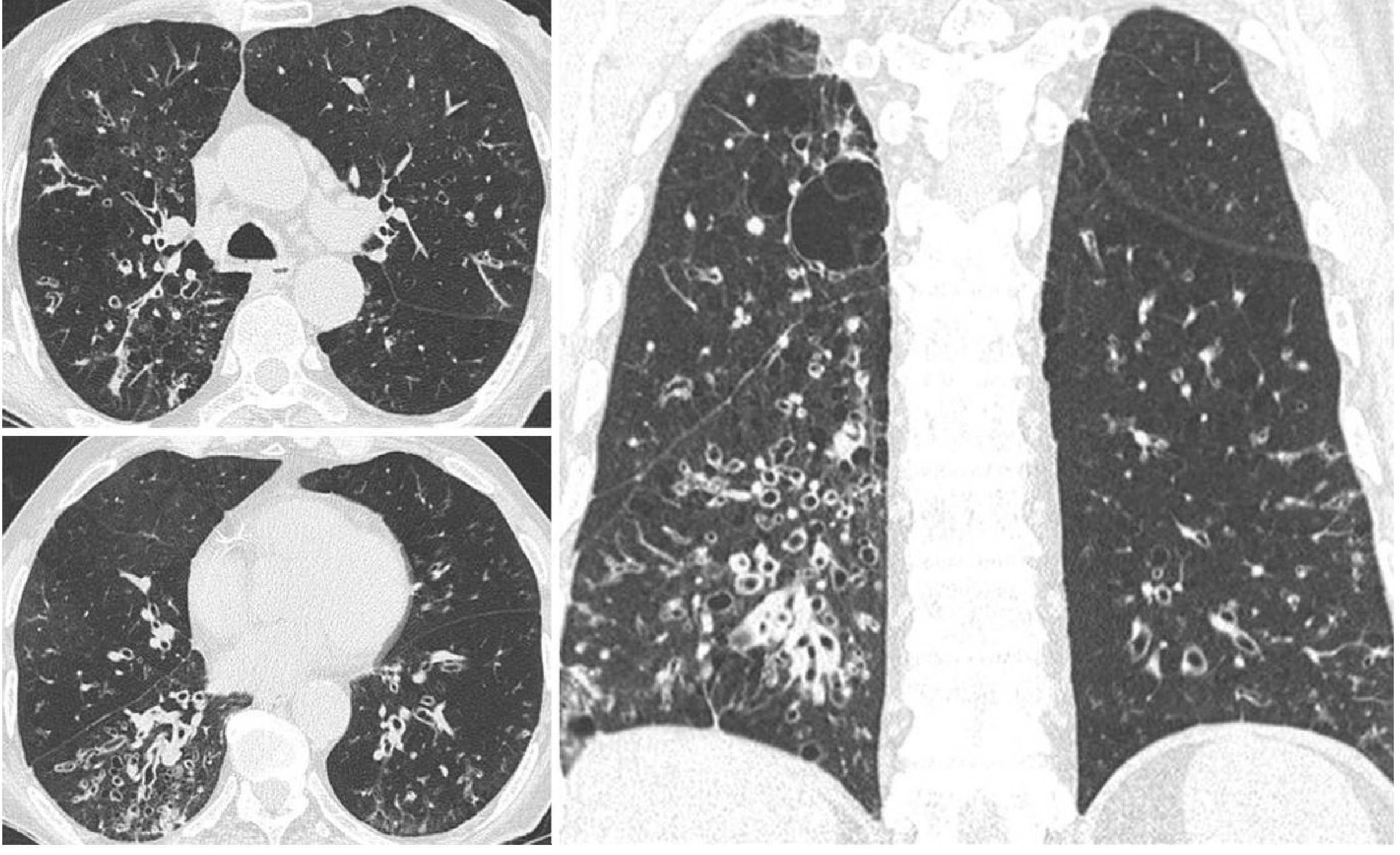
Fig. 6.17 (Grainger & Allison): Post-infectious bronchiectasis. Bilateral cylindrical bronchiectasis, right upper and lower lobes, with wall thickening and mucoid impactions.
HRCT signs:
- Lack of tapering of bronchial lumina (the cardinal sign)
- Signet ring sign: bronchial internal diameter > adjacent pulmonary artery diameter
- Bronchi visible within 1 cm of the costal pleura (normally not possible)
- Mucus-filled dilated bronchi (glove-finger, V- or Y-shaped densities)
- Tree-in-bud sign: centrilobular nodules and linear branching opacities = infectious bronchiolitis
- Air-fluid levels in dependent portions of dilated bronchi
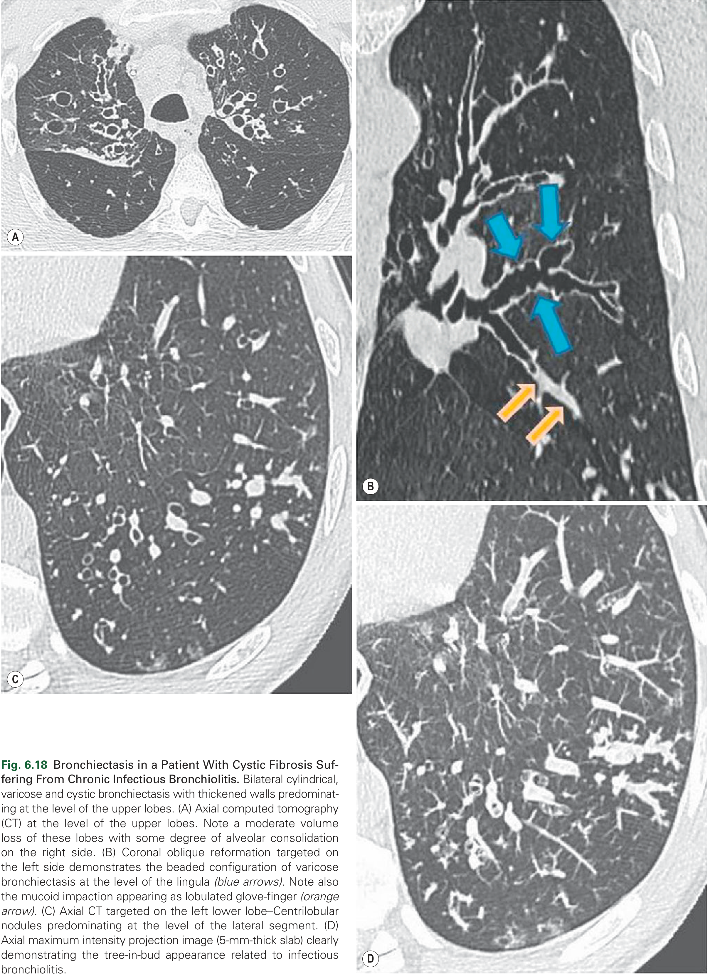
Fig. 6.18 (Grainger & Allison): Bronchiectasis in cystic fibrosis with infectious bronchiolitis. (A) Axial CT - bilateral cylindrical, varicose, and cystic bronchiectasis at upper lobes. (B) Coronal reformation - beaded configuration of varicose bronchiectasis (blue arrows) and mucoid impaction (orange arrows). (C-D) Centrilobular nodules and tree-in-bud sign.
8. Clinical Features
| Feature | Description |
|---|---|
| Cough | Severe, persistent, productive |
| Sputum | Copious, mucopurulent, foul-smelling; may be blood-streaked |
| Hemoptysis | Common; occasionally massive |
| Dyspnea | With extensive disease |
| Rhinosinusitis | Frequently co-exists |
| Episodic exacerbations | Triggered by RTIs; worse on waking (positional drainage of pus) |
Complications (now less common with modern antibiotics)
- Obstructive respiratory failure → hypoxemia, hypercapnia
- Cor pulmonale (pulmonary hypertension from chronic hypoxia)
- Brain abscess (hematogenous spread)
- Amyloidosis (AA type, chronic inflammation)
9. Summary Table
| Feature | Description |
|---|---|
| Definition | Permanent irreversible bronchial dilation |
| Mechanism | Obstruction + chronic infection → wall destruction |
| Distribution | Lower lobes, bilateral; severe distally |
| Gross | Cystic spaces, purulent secretions, extends to pleura |
| Histology | Acute/chronic inflammation, ulceration, squamous metaplasia, fibrosis |
| Organisms | H. influenzae, P. aeruginosa, NTM |
| CXR | Tramlines, ring opacities |
| CT (cardinal sign) | Lack of bronchial tapering + signet ring sign |
| Complications | Cor pulmonale, hemoptysis, abscess, amyloidosis |
Sources: Robbins, Cotran & Kumar Pathologic Basis of Disease (10th ed.), pp. 642-643; Robbins & Kumar Basic Pathology, pp. 466-509; Grainger & Allison's Diagnostic Radiology (Figs. 6.15-6.18).
This is a shared conversation. Sign in to Orris to start your own chat.