
Leiomyosarcomas
I'll start by reading the medical library skill, then search for comprehensive information on leiomyosarcomas.
Reading File
Now let me search the library and PubMed in parallel for comprehensive leiomyosarcoma coverage.
Finding Sources
Searching PubMed
leiomyosarcoma
Excellent results. Now let me read the most comprehensive sources in parallel - Robbins Pathology (core pathology), Sabiston Surgery, and Andrews' Dermatology.
Reading File
Reading File
Reading File
Now let me pull the Robbins Big (Pathologic Basis of Disease) and the Fitzpatrick entries, plus fetch the histology image:
Reading File
Reading File
Finding Sources
Loading Image
Now let me fetch the leiomyosarcoma gross/histo image and the FNCLCC grading table, plus the Gynecology staging section:
Loading Image
Reading File
Reading File
Let me now get the uterine leiomyosarcoma section from Berek & Novak and the Fitzpatrick dermatology section:
Finding Sources
Reading File
Now let me get the FIGO staging detail for uterine LMS and the Berek & Novak LMS section specifically:
Reading File
I now have comprehensive information. Let me compile the full answer.
Leiomyosarcomas
Leiomyosarcoma (LMS) is a malignant neoplasm of smooth muscle origin. It is the second most common subtype of soft tissue sarcoma (STS) after liposarcoma, and a particularly aggressive malignancy with a propensity for hematogenous spread and late recurrence.
1. Epidemiology
- Peak incidence is in the 6th and 7th decades of life.
- Retroperitoneal and uterine LMS are more common in females; other sites show a male predominance.
- Predisposing risk factors include prior radiation exposure and immunosuppression (particularly Epstein-Barr virus-related tumor promotion in post-transplant patients).
- Leiomyoma (the benign counterpart) is NOT a precursor to leiomyosarcoma.
- Sabiston Textbook of Surgery, p. 2975
2. Sites of Origin
LMS can originate from virtually any anatomic location. Major sites include:
| Site | Notes |
|---|---|
| Uterus / Myometrium | Most common visceral site; most frequent uterine sarcoma |
| Retroperitoneum | Often large, bulky, frequently unresectable |
| Extremities / Trunk | ~25% of cases; better prognosis |
| Inferior Vena Cava (IVC) | Particularly deadly; arises from great vessel wall |
| GI tract / Esophagus | Rare; associated with poor survival |
| Skin (dermis / subcutis) | Superficial cutaneous form; ~2% of STS |
- Robbins, Cotran & Kumar Pathologic Basis of Disease; Sabiston Textbook of Surgery
3. Pathogenesis & Molecular Biology
- Arise from smooth muscle cells or their precursors (e.g., myometrial cells or endometrial stromal precursor cells for uterine LMS).
- Possess complex, highly variable karyotypes with numerous chromosomal deletions and instability - no single pathognomonic marker.
- Key molecular alterations include mutations in TP53, RB, and PTEN (tumor suppressor genes).
- A subset (~30%) contains MED12 mutations, a genetic aberration otherwise largely unique to uterine smooth muscle tumors (both leiomyomas and LMS).
- Robbins & Kumar Basic Pathology, p. 679-683; Robbins, Cotran & Kumar PBD, p. 3047
4. Morphology
Gross Pathology
- Typically soft, fleshy, hemorrhagic, necrotic masses.
- Uterine LMS grows as either:
- Bulky masses invading the uterine wall, or
- Polypoid masses projecting into the uterine lumen.
- Deep soft tissue LMS are firm, rubbery, or fleshy tumors, often well-circumscribed with a cystic/necrotic center.
Histology
Eosinophilic spindle cells with blunt-ended, hyperchromatic nuclei arranged in interweaving fascicles. Diagnosis requires evaluation of three features:
| Feature | Significance |
|---|---|
| Tumor necrosis | Coagulative (geographic) necrosis is diagnostic |
| Nuclear/cytologic atypia | Marked pleomorphism vs. leiomyoma |
| Mitotic activity | ≥10 mitoses/10 HPF = malignancy (if atypia or necrosis present: ≥4/10 HPF may suffice) |
- Cases impossible to classify definitively are called STUMP (Smooth muscle tumor of uncertain malignant potential).
Histology image - uterine leiomyosarcoma:
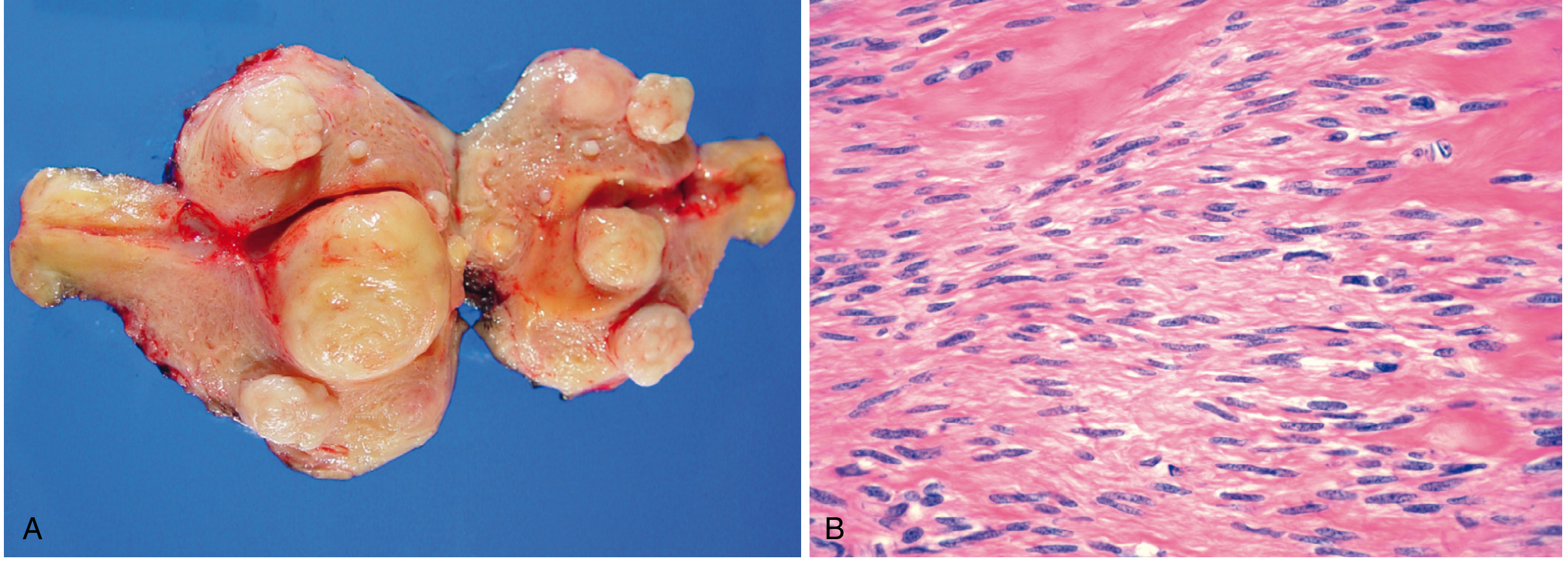
(A) Gross specimen showing hemorrhagic, necrotic uterine mass. (B) Microscopically bland spindle cells - well-differentiated variant.
Histology image - high-grade LMS with mitoses:
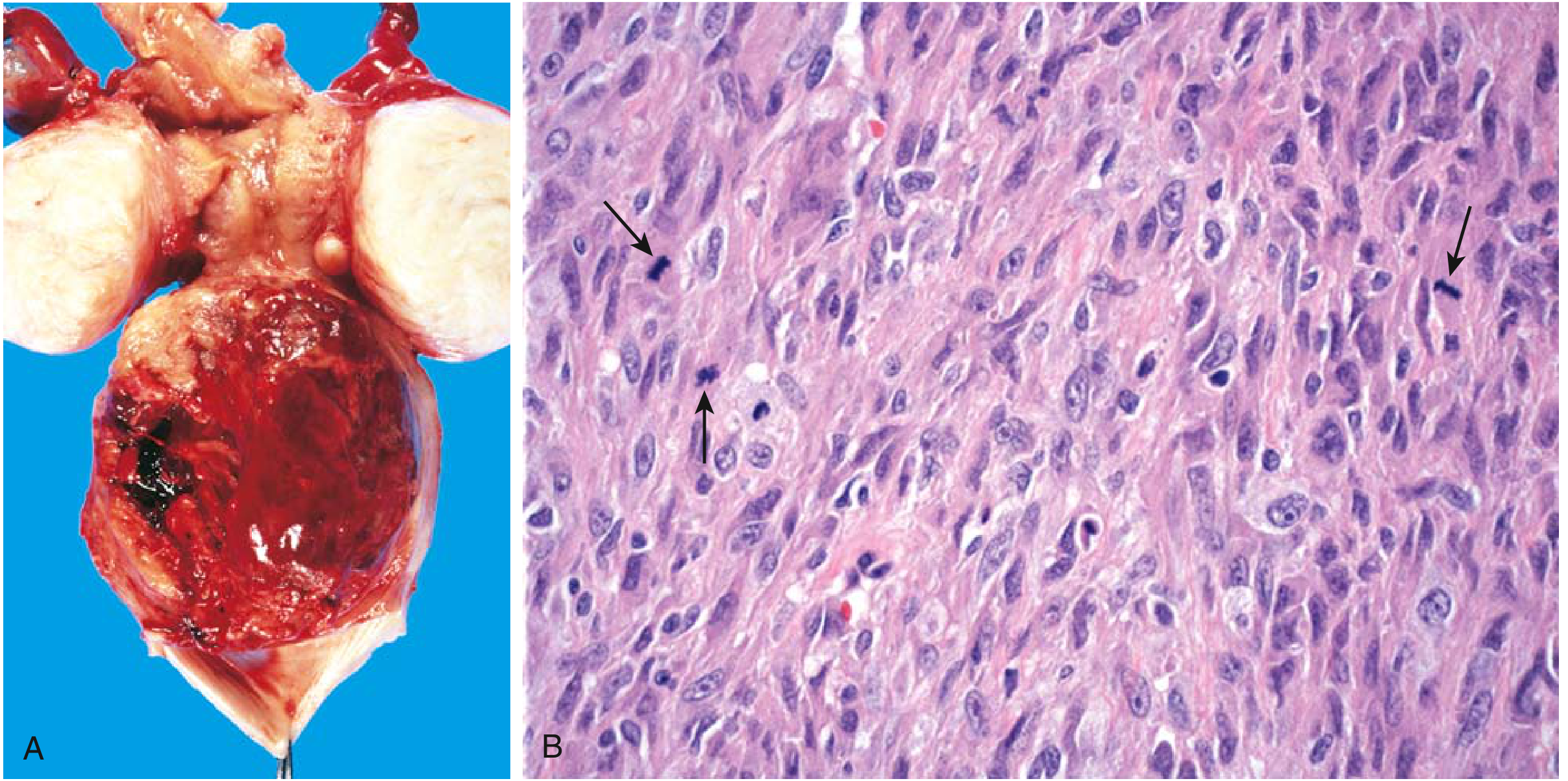
(A) Gross hemorrhagic uterine tumor. (B) High-power view showing anaplastic spindle cells with visible mitotic figures (arrows) - characteristic of high-grade LMS.
- Robbins, Cotran & Kumar PBD, p. 3052-3054; Robbins & Kumar Basic Pathology, p. 688
5. Immunohistochemistry
These tumors stain positive for smooth muscle markers, which are key to distinguishing LMS from other spindle cell/pleomorphic tumors:
- Desmin ✓
- Smooth muscle actin (SMA) ✓
- h-Caldesmon ✓
- Calponin ✓
- Sabiston Textbook of Surgery; Andrews' Diseases of the Skin, p. 1585
6. Grading: FNCLCC System
The FNCLCC (French Federation) grading system is the most widely used for STS and incorporates three parameters: Tumor differentiation score, Mitotic count, and Necrosis score.
Tumor differentiation score for LMS:
| Score | LMS Subtype |
|---|---|
| 1 | Well-differentiated leiomyosarcoma |
| 2 | Conventional leiomyosarcoma |
| 3 | Poorly differentiated / pleomorphic leiomyosarcoma |
Final FNCLCC grade: G1 = low grade, G2/G3 = high grade (most LMS are G2-G3).
- Mulholland and Greenfield's Surgery, p. 2817 (FNCLCC Table)
7. Staging
Soft Tissue Sarcoma (AJCC 8th edition) - Trunk & Extremity LMS
Staging incorporates T (tumor size), N (nodal status), M (metastasis), and G (histologic grade).
Uterine LMS: FIGO Staging (2009)
| Stage | Description |
|---|---|
| I | Tumor limited to uterus |
| IA | ≤5 cm |
| IB | >5 cm |
| II | Tumor extends beyond uterus, within pelvis |
| IIA | Adnexal involvement |
| IIB | Pelvic tissue involvement |
| III | Abdominal spread |
| IV | Bladder/rectum invasion or distant metastasis |
- Berek & Novak's Gynecology; SCHWARTZ'S PRINCIPLES OF SURGERY
8. Clinical Features
- Presentation: Painless firm mass (deep LMS); abdominal symptoms in retroperitoneal tumors; abnormal uterine bleeding or pelvic mass in uterine LMS.
- Some uterine LMS are found incidentally at hysterectomy performed for presumed leiomyoma.
- Metastasis: Predominantly hematogenous - most common sites are lung, liver, bone, and brain; abdominal dissemination also occurs.
- Lymph node metastases are uncommon.
9. Cutaneous/Superficial LMS (Special Form)
- Accounts for ~2% of all STS.
- Two subtypes differ significantly in prognosis:
| Type | Origin | Prognosis |
|---|---|---|
| Dermal (cutaneous) | Arrector pili or genital dartoic muscle | Good; metastases rare |
| Subcutaneous | Subcutaneous smooth muscle / vascular | Hematogenous metastases in ~35%; fatal in ~1/3 |
- Association with Reed syndrome (hereditary leiomyomatosis and renal cell cancer - HLRCC) has been reported; caused by fumarate hydratase (FH) gene mutations.
- Histologically: dense cellularity, nuclear pleomorphism, numerous mitoses, disarray of smooth muscle bundles.
- Treatment: wide local excision with 1-cm margin; Mohs surgery to limit recurrence (~15% recurrence rate).
- Andrews' Diseases of the Skin, p. 1583-1587; Fitzpatrick's Dermatology, p. 1882
10. Treatment
Surgery (Primary Modality)
- Macroscopically complete resection is the cornerstone of treatment for all LMS.
- Uterine LMS: Total abdominal hysterectomy (TAH) with bilateral salpingo-oophorectomy (BSO). Lymphadenectomy is not routinely required.
- Retroperitoneal LMS involving IVC: Options include tumor resection with IVC ligation or reconstruction (patch/interposition graft). For infrahepatic IVC involvement, ligation without reconstruction may be acceptable if collaterals are preserved.
Adjuvant Therapy
- Routine adjuvant chemotherapy or radiation after resection is not currently recommended (subject of ongoing trials, including the EORTC STRASS-2 study).
- Neoadjuvant chemotherapy may be considered for large retroperitoneal tumors.
Systemic Treatment for Advanced/Metastatic Disease
Active regimens include:
- Doxorubicin (backbone agent)
- Ifosfamide
- Gemcitabine + Docetaxel (particularly effective in uterine LMS)
- Trabectedin (approved in Europe for LMS specifically)
Note: Olaratumab (anti-PDGFRα monoclonal antibody) combined with doxorubicin initially showed promise in Phase II but failed to improve overall survival in the Phase III ANNOUNCE trial and has been withdrawn from the market.
- Sabiston Textbook of Surgery, p. 2979-2983
11. Prognosis
| Location/Type | 5-Year Survival |
|---|---|
| Overall uterine LMS | ~40% |
| Anaplastic (high-grade) uterine LMS | 10-15% |
| Cutaneous (dermal) LMS | Excellent |
| Subcutaneous LMS | ~65% (35% metastasis rate) |
| Retroperitoneal LMS | Poor; frequently unresectable |
| IVC leiomyosarcoma | Particularly poor |
- More than one-half of all LMS eventually metastasize hematogenously.
- Retroperitoneal tumors are often diagnosed late due to large size and silent presentation.
- Robbins, Cotran & Kumar PBD, p. 3059; Robbins & Kumar Basic Pathology, p. 690
Recent Literature (2024-2026)
- A 2025 systematic review and meta-analysis on spinal leiomyosarcoma (PMID 40368091, World Neurosurg) examined surgical outcomes in primary/metastatic spinal LMS - a rare but increasingly recognized metastatic site.
- A 2025 systematic review on retroperitoneal sarcoma (PMID 40465137, Curr Oncol Rep) covers current management strategies for retroperitoneal LMS and liposarcoma.
- A 2024 systematic review on Mohs surgery for superficial LMS (PMID 40526951, Dermatol Online J) reports ~15% recurrence with Mohs vs. higher rates with wide local excision, supporting Mohs as the preferred approach for cutaneous LMS.
Summary Mnemonic - LMS key features:
Large necrotic mass → Mitoses + atypia → SMA/Desmin positive → surgery first → hematogenous spread to Lung, Marrow, Spinal sites.
Uterine leiomyosarcomas
Reading the medical library skill first, then launching parallel searches.
Reading File
Now launching parallel searches across the library and PubMed.
Finding Sources
Finding Sources
Searching PubMed
"uterine leiomyosarcoma"[MeSH Terms]
Good. Now let me pull the key dedicated sections from Berek & Novak (gynecology), Schwartz, Robbins, and the FIGO staging table simultaneously.
Reading File
Reading File
Reading File
Searching PubMed
leiomyosarcoma uterus treatment
Excellent content found. Let me now fetch the key LMS histology image and read the rest of the Berek & Novak LMS section (chemotherapy, recurrence, FIGO staging table).
Loading Image
Reading File
Finding Sources
Let me fetch the Berek & Novak histology image, the FIGO staging table, and variants section:
Loading Image
Reading File
Reading File
I now have comprehensive, detailed information from Berek & Novak's Gynecology, Robbins PBD, and Schwartz's Surgery. Let me compile the full focused answer on uterine leiomyosarcoma.
Uterine Leiomyosarcoma (ULMS)
Uterine leiomyosarcoma is the most common uterine sarcoma, accounting for 35-40% of all uterine sarcomas. It is a malignant neoplasm of smooth muscle origin arising from the myometrium - one of the most aggressive gynecologic malignancies.
1. Epidemiology & Risk Factors
- Median age at diagnosis: 54 years (range 43-63); can occur in women in their 20s
- More common in African American women, who also have a poorer prognosis
- Premenopausal women have better survival than postmenopausal women
- No strong relationship to parity
- Prior pelvic radiation in ~4% of cases
- The incidence of sarcomatous transformation within a benign leiomyoma is only 0.13-0.81% - most ULMS arise de novo, not from pre-existing leiomyomas
- Hereditary risk: Hereditary leiomyomatosis and renal cell cancer (HLRCC) syndrome - caused by fumarate hydratase (FH) gene mutations - confers an unknown but elevated risk; also linked to hereditary retinoblastoma (RB gene mutations)
- Berek & Novak's Gynecology, p. 1862; Fitzpatrick's Dermatology
2. Pathogenesis & Molecular Biology
ULMS arises from myometrium or endometrial stromal precursor cells - not from malignant transformation of leiomyoma:
- Complex, variable karyotypes with chromosomal deletions and instability
- Key mutations: TP53, RB (retinoblastoma), PTEN (tumor suppressor)
- A subset contains MED12 mutations - an aberration otherwise largely unique to uterine smooth muscle tumors (shared with leiomyoma)
- Unlike endometrial carcinoma, ULMS has no association with unopposed estrogen exposure
- Robbins, Cotran & Kumar Pathologic Basis of Disease, p. 3047
3. Clinical Presentation
Symptoms are usually of short duration (mean 6 months) and non-specific:
| Symptom | Frequency |
|---|---|
| Abnormal uterine / vaginal bleeding | Most common |
| Pelvic pain or pressure | Common |
| Awareness of abdominal mass | Common |
| Asymptomatic (found incidentally at hysterectomy) | Minority |
- Key clinical clue: Pelvic pain + rapidly enlarging uterus, especially in a postmenopausal woman - should raise strong suspicion for ULMS.
- Physical examination reveals a pelvic mass.
- Endometrial biopsy establishes diagnosis in up to one-third of cases (if submucosal).
4. Imaging
- MRI (especially contrast-enhanced) is superior to ultrasound or CT in differentiating ULMS and STUMP from leiomyoma.
- Differentiating ULMS from degenerated leiomyoma can still be challenging on preoperative imaging - biopsy should be considered when diagnosis is uncertain.
- Diffusion-weighted MRI has been studied specifically for this distinction.
- Berek & Novak's Gynecology, p. 1868
5. Pathology
Gross Morphology
ULMS grows in two patterns:
- Bulky, fleshy, hemorrhagic, necrotic masses invading the uterine wall
- Polypoid masses projecting into the uterine lumen
Gross/histology - Robbins PBD (Fig. 22.28):
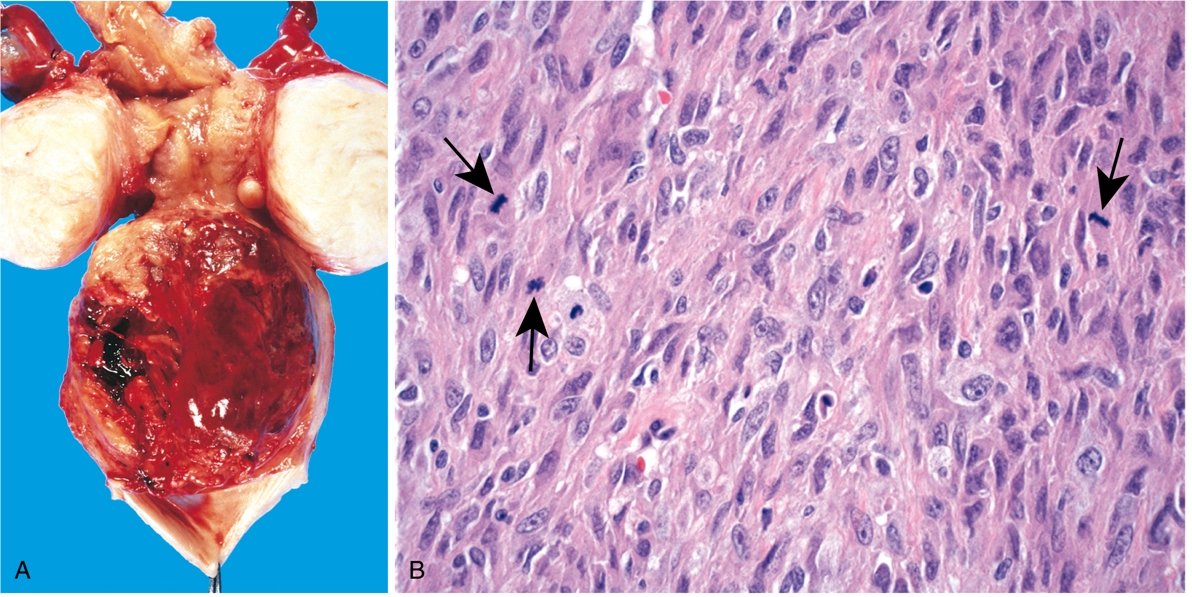
(A) A large hemorrhagic tumor mass distending the lower corpus, flanked by two leiomyomas. (B) Irregular hyperchromatic tumor cells with numerous mitotic figures (arrows).
Microscopic Criteria for Malignancy
Three cardinal histologic features must be assessed:
| Feature | Malignant Threshold |
|---|---|
| Mitotic index | ≥10 MF/10 HPF = frankly malignant; 5-10 MF/10 HPF = STUMP |
| Cytologic atypia | Severe nuclear pleomorphism; if epithelioid cells present, ≥4 MF/10 HPF sufficient |
| Coagulative tumor cell necrosis | Geographic/coagulative necrosis (vs. hyaline degeneration in leiomyoma) |
Additional features: infiltrating borders, vascular invasion.
Caveat: Mitotically active leiomyomas occur in young/pregnant women - caution required before diagnosing malignancy in this group.
Histology - Berek & Novak (Fig. 37-7):
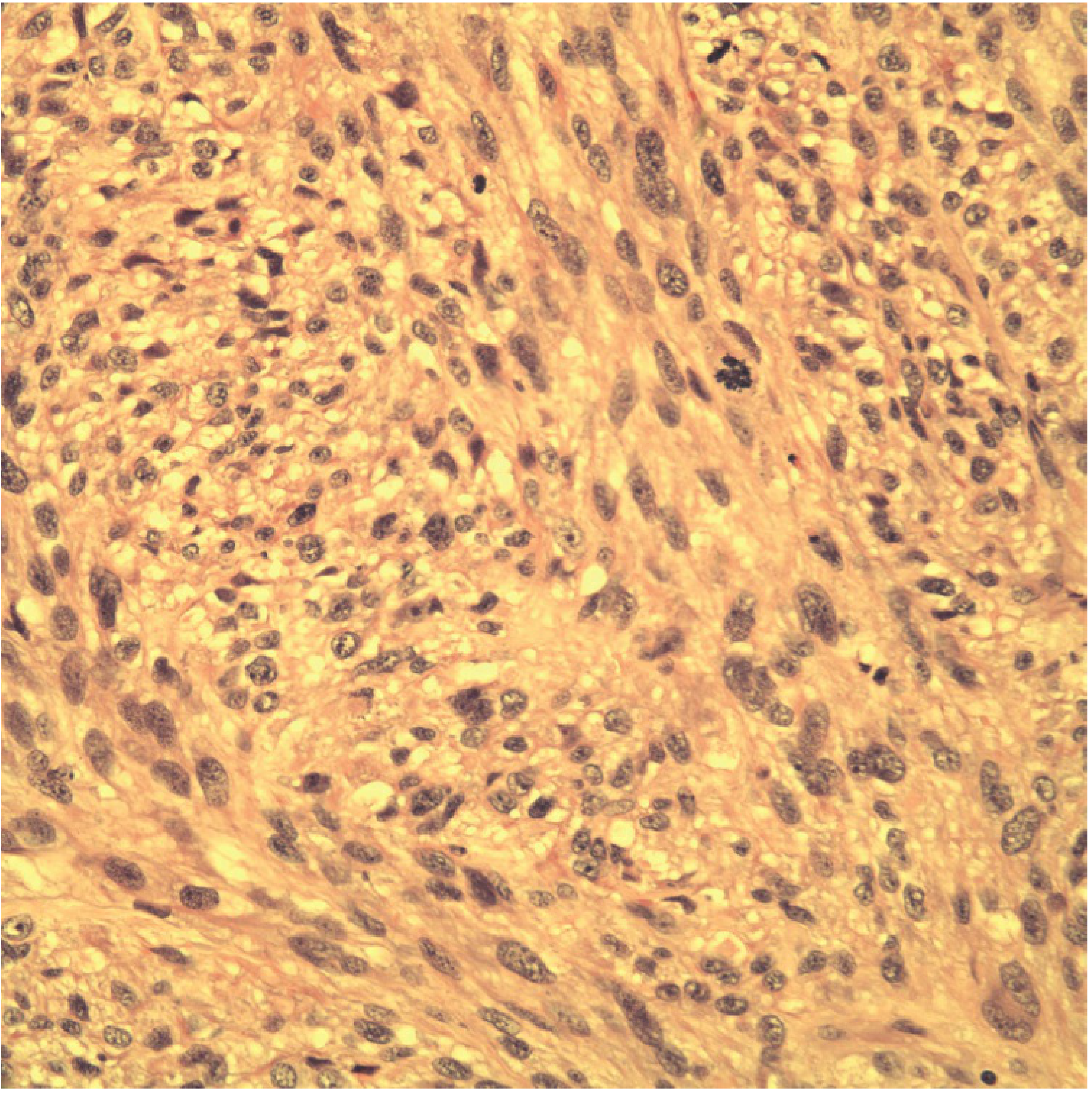
Interlacing bundles of spindle cells with fibrillar cytoplasm, irregular hyperchromatic nuclei, and multiple mitotic figures.
Immunohistochemistry
Positive for smooth muscle markers:
- Desmin ✓
- Smooth muscle actin (SMA) ✓
- h-Caldesmon ✓
6. FIGO Staging for Uterine LMS (2009)
| Stage | Definition | Substages |
|---|---|---|
| I | Tumor limited to uterus | IA: ≤5 cm; IB: >5 cm |
| II | Extends beyond uterus, within pelvis | IIA: adnexal involvement; IIB: other pelvic tissues |
| III | Invades abdominal tissues | IIIA: 1 site; IIIB: >1 site; IIIC: pelvic/para-aortic lymph nodes |
| IV | Distant disease | IVA: bladder/rectum; IVB: distant metastasis |
- Berek & Novak's Gynecology; Prat J. FIGO staging for uterine sarcomas. Int J Gynecol Obstet 2009;104:177-178
7. Pattern of Spread
Spread occurs by:
- Direct myometrial invasion → pelvic structures
- Lymphovascular invasion → pelvic blood vessels and lymphatics → intra-abdominal dissemination
- Hematogenous spread (most important) → lungs (most common distant site), bone, brain
- Lymph node metastasis is rare (<5% at diagnosis) in early-stage disease.
Prognostic factors at time of surgery:
- Tumors >5 cm → poorer prognosis
- Infiltrating (vs. pushing) borders → poorer prognosis
- Extension beyond uterus → poorer prognosis
8. Treatment
Surgery - Cornerstone of Management
- Total hysterectomy is the standard for uterine-confined disease.
- Bilateral salpingo-oophorectomy (BSO):
- Mandatory in postmenopausal women
- Mandatory with gross extrauterine disease
- Ovarian preservation acceptable in premenopausal women with early-stage disease - does NOT increase recurrence risk
- Lymphadenectomy: NOT routinely recommended (no survival advantage given low nodal metastasis rate)
- Cytoreductive surgery / pulmonary metastasectomy may improve survival in advanced disease
⚠️ Morcellation Warning
Intraoperative power morcellation is contraindicated - it increases risk of dissemination and worsens prognosis if the uterus harbors undiagnosed ULMS. The FDA has strongly discouraged power morcellators since 2014.
- Berek & Novak's Gynecology, p. 1872; Schwartz's Principles of Surgery
Radiation Therapy
- Not recommended as adjuvant therapy.
- An EORTC phase III trial comparing adjuvant pelvic radiation vs. observation in early-stage ULMS showed no difference in overall survival or recurrence rates.
- SEER database data also does not support adjuvant radiation.
Chemotherapy
| Setting | Evidence |
|---|---|
| Adjuvant (early-stage) | NOT recommended - fails to improve survival or reduce the 40-70% recurrence risk |
| Advanced / Metastatic | Indicated; increases survival |
Active regimens for advanced/metastatic ULMS:
| Regimen | Response Rate | Notes |
|---|---|---|
| Gemcitabine + Docetaxel | ~53% | Particularly active in uterine LMS |
| Doxorubicin + Ifosfamide ± Dacarbazine | ~30% | Standard STS backbone |
| Doxorubicin (single agent) | 15-25% | Most active single agent |
| Trabectedin | 15-25% | FDA/EMA approved for LMS |
| Pazopanib | 15-25% | Tyrosine kinase inhibitor |
| Eribulin | 15-25% | Active in LMS specifically |
| Liposomal doxorubicin, temozolomide, dacarbazine | Modest activity |
Note on Olaratumab: A Phase II trial showed improved OS with doxorubicin + olaratumab (OS 26.5 vs. 14.7 months). However, the Phase III ANNOUNCE trial failed to confirm overall survival benefit → olaratumab was withdrawn from the market.
Median time to progression with most regimens is <6 months; median survival with advanced disease is <2 years.
- Berek & Novak's Gynecology, p. 1892; Schwartz's Principles of Surgery, p. 2257
9. Prognosis
| Stage / Feature | Approximate 5-year Survival |
|---|---|
| Overall | 20-63% (mean ~47%) |
| Stage I (well-differentiated) | Best; young premenopausal women do best |
| Anaplastic / high-grade | 10-15% |
| Advanced / metastatic | Very poor |
- More than half of all ULMS eventually metastasize hematogenously.
- Recurrence risk: 40-70% even after complete resection of early-stage disease.
- Late recurrences are characteristic, particularly in epithelioid and myxoid variants.
10. Clinicopathologic Variants
Five special variants of uterine smooth muscle tumors:
| Variant | Key Features | Behavior |
|---|---|---|
| Myxoid LMS | Gelatinous gross appearance; myxomatous stroma; low mitotic rate (0-2 MF/10 HPF); extensively infiltrates despite circumscribed appearance | Aggressive despite low mitoses; poor prognosis |
| Epithelioid LMS | Large fleshy masses with hemorrhage; round/epithelioid cells in sheets/nests; must distinguish from trophoblastic tumors | Aggressive; prone to late recurrences |
| Intravenous leiomyomatosis | Histologically benign smooth muscle growing into venous channels/IVC; worm-like projections | Generally excellent prognosis; can extend to heart → fatal |
| Benign metastasizing leiomyoma | Histologically benign uterine SM tumor with "benign" pulmonary/nodal metastases | Indolent; responds to antiestrogen/oophorectomy |
| Disseminated peritoneal leiomyomatosis | Benign SM nodules throughout peritoneum; seen in pregnancy/OCP use; grossly alarming but histologically benign | Favorable outcome |
11. STUMP (Smooth Muscle Tumor of Uncertain Malignant Potential)
- Applied when tumors cannot be definitively classified as benign or malignant
- Mitotic index: 5-10 MF/10 HPF with or without atypia
- Less predictable behavior - requires close follow-up
- Treatment: hysterectomy; individualized adjuvant decisions
Recent PubMed Evidence (2023-2026)
- A 2025 study on uterine leiomyosarcoma serial recurrences (PMID 41027098, Gynecol Oncol) provides NIH-funded molecular analysis of disease progression at each recurrence.
- A 2025 review on future targeted therapies for LMS (PMID 40410055, Hematol Oncol Clin North Am) covers emerging targets including CDK4/6 inhibitors, PARP inhibitors, and immunotherapy.
- A 2024 review on targeted therapy for leiomyosarcoma (PMID 38473300, Cancers) highlights that uterine LMS is uniquely chemosensitive among LMS subtypes, and discusses emerging targets.
Key Summary Points:
- ULMS = most common uterine sarcoma; arises de novo from myometrium
- Diagnosed by: mitoses ≥10/10 HPF + cytologic atypia + coagulative necrosis
- Surgery is the only curative treatment; avoid morcellation
- Adjuvant radiation and chemotherapy do NOT improve early-stage outcomes
- Gemcitabine + docetaxel is the most active regimen for advanced disease
- Overall prognosis is poor - 5-year survival ~47%, with 40-70% recurrence risk
This is a shared conversation. Sign in to Orris to start your own chat.