
Abdominal muscle detail
abdominal muscles anatomy diagram layers
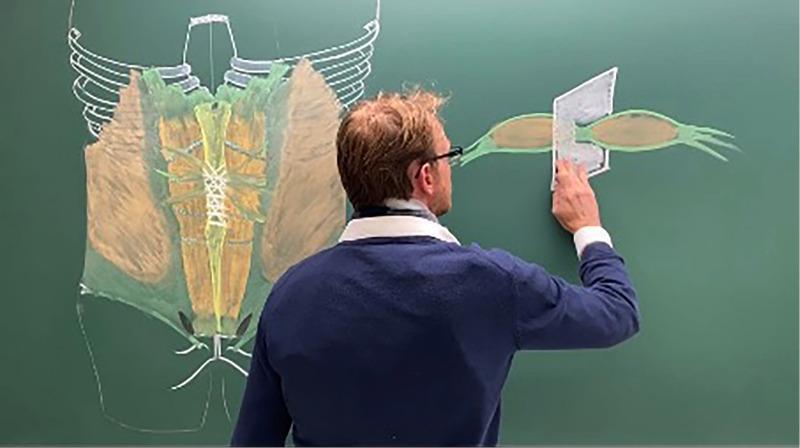
This educational image depicts a medical professional presenting anatomical diagrams of the anterior abdominal wall on a green chalkboard. The primary diagram on the left illustrates the musculoskeletal anatomy of the trunk, featuring a white outline of the inferior rib cage, sternum, and pelvis. The muscular layers are color-coded: the rectus abdominis is shown in yellow/tan along the midline, divided by the linea alba, with horizontal tendinous inscriptions clearly marked. Lateral to this, the external and internal oblique muscles are represented in brown, while green shading indicates the surrounding fascial planes, including the transversalis fascia. The right side of the board features a detailed cross-sectional or isolated diagram of a muscle belly (likely rectus abdominis or an oblique), highlighting its fusiform shape, green fascial envelope, and tendinous distal attachments. A physical cutout representing a surgical mesh or anatomical plane is positioned over this muscle to demonstrate Abdominal Wall Reconstruction (AWR) techniques or hernia repair principles. This visual is used for postgraduate surgical education, emphasizing applied anatomy in groin and incisional hernia surgery.
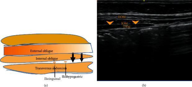
This dual-panel image features an anatomical diagram (a) and a corresponding ultrasound scan (b) demonstrating the regional anatomy for ilioinguinal and iliohypogastric nerve blocks. The diagram illustrates the layered musculature of the lateral abdominal wall, identifying the external oblique, internal oblique, and transversus abdominis muscles. It highlights the ilioinguinal and iliohypogastric nerves situated in the fascial plane between the internal oblique and transversus abdominis muscles. The ultrasound scan provides a clinical diagnostic view of the same region, showing the three muscle layers as distinct hypoechoic bands separated by bright, hyperechoic fascial planes. Specifically, the external oblique muscle (EOM) is the most superficial, the internal oblique muscle (IOM) is the intermediate layer, and the transversus abdominis muscle (TAM) is the deepest layer. Arrows on both panels indicate the target site for local anesthetic infiltration between the IOM and TAM, essential for effective regional anesthesia in the inguinal and pelvic regions.
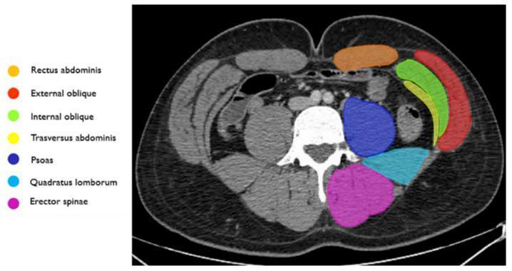
This diagnostic image is an axial CT scan of the abdomen, specifically annotated to demonstrate the anatomy of the abdominal wall musculature. The cross-section provides a clear view of the bilateral symmetry and layered arrangement of the core muscles. Anteriorly, the rectus abdominis is highlighted in orange. Moving laterally, the three distinct layers of the lateral abdominal wall are identified: the external oblique (red) as the most superficial layer, the internal oblique (green) in the middle, and the transversus abdominis (yellow) as the deep layer. In the posterior compartment, the psoas muscle (dark blue) is shown adjacent to the vertebral body. Posterior and lateral to the psoas is the quadratus lumborum (light blue), while the erector spinae group (magenta) is situated most posteriorly along the spine. This anatomical diagram serves as a key reference for identifying muscle boundaries in clinical imaging, essential for diagnosing abdominal wall hernias, hematomas, or planning surgical and interventional procedures like embolization for active bleeding.
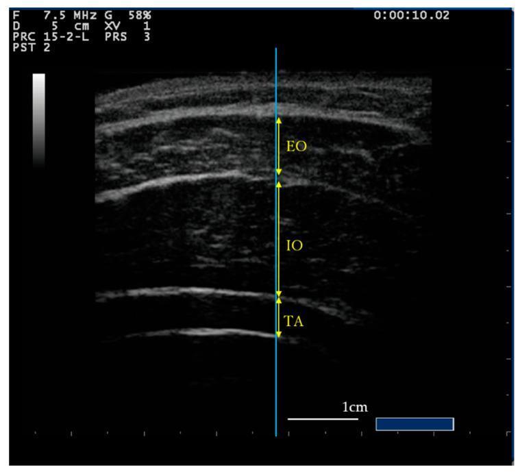
This diagnostic ultrasound image demonstrates the transverse cross-sectional anatomy of the lateral abdominal wall muscles. Three distinct layers are visible: the most superficial External Oblique (EO), the middle Internal Oblique (IO), and the deepest layer, the Transverse Abdominis (TA). The muscle bodies are characterized by relatively hypoechoic (darker) echotexture with internal speckling, separated by bright, hyperechoic fascial planes that define their boundaries. A vertical blue reference line intersects the layers to ensure consistent measurement. Yellow double-headed arrows indicate the specific thickness measurements for each of the three muscles. A 1cm scale bar is provided in the bottom right corner for quantitative assessment. This imaging modality is typically used in sports medicine and physical therapy to assess core muscle activation, hypertrophy, or atrophy through real-time visualization of muscle thickness changes during exercises like the Abdominal Drawing-In Maneuver (ADIM) or abdominal bracing.
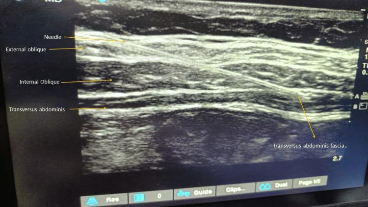
This diagnostic ultrasound image demonstrates the layered anatomy of the lateral abdominal wall, utilized for an ultrasound-guided transversus abdominis plane (TAP) block. The image clearly identifies three distinct muscle layers from superficial to deep: the external oblique, internal oblique, and transversus abdominis muscles. Each muscle layer appears as a relatively hypoechoic band separated by thin, bright hyperechoic fascial planes. A hyperechoic, linear needle is visualized using an 'in-plane' technique, traversing through the external and internal oblique muscles. The needle tip is positioned toward the fascial plane between the internal oblique and the transversus abdominis muscle, which is the target for local anesthetic deposition to block the thoracolumbar nerves (T7-L1). This imaging modality is essential in anesthesiology for providing regional analgesia following abdominal surgeries, ensuring accurate needle placement and reducing procedural complications.
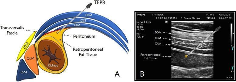
This dual-panel medical figure illustrates the Transversalis Fascia Plane Block (TFPB) through an anatomical diagram and a corresponding ultrasound image. Panel A is a cross-sectional anatomical diagram depicting the abdominal wall and retroperitoneal space. It identifies the three lateral abdominal muscle layers: external oblique (EOM), internal oblique (IOM), and transversus abdominis (TAM). Posteriorly, the latissimus dorsi (LDM), quadratus lumborum (QLM), and erector spinae (ESM) muscles are shown. The needle trajectory is illustrated passing through the three abdominal muscles to reach the target plane between the transversalis fascia and the retroperitoneal fat tissue overlying the kidney and peritoneum. Panel B is a high-frequency linear ultrasound scan demonstrating the clinical application. The ultrasound shows the characteristic stratified appearance of the EOM, IOM, and TAM as hypoechoic bands separated by hyperechoic fascial planes. An echogenic needle is visualized using an in-plane technique, with its tip positioned in the hypoechoic retroperitoneal fat layer deep to the TAM. This visual educational resource is designed for anesthesia and pain management training, focusing on regional nerve block landmarks and ultrasound-guided needle placement.
Abdominal Muscles - Detailed Overview
1. Anterolateral Muscles (Flat Muscles)
External Oblique
- Origin: Outer surface of ribs 5-12 (muscular slips from the outer surfaces of the lower 8 ribs)
- Insertion: Outer (lateral) lip of the iliac crest; aponeurosis ending in the linea alba (forming the anterior layer of the rectus sheath); lower border of the aponeurosis forms the inguinal ligament
- Innervation: Intercostal nerves T5-T11, subcostal nerve (T12), iliohypogastric nerve
- Fiber direction: Downward and medially (like hands in pockets)
- Actions:
- Unilateral: Lateral flexion to same side; rotation of trunk to opposite side
- Bilateral: Trunk flexion, stabilizes pelvis, aids expiration, compresses abdomen
Internal Oblique
- Origin: Deep layer of thoracolumbar fascia; intermediate/inner lip of iliac crest; anterior superior iliac spine (ASIS); lateral 2/3 of inguinal ligament
- Insertion: Lower borders of ribs 10-12; anterior and posterior layers of rectus sheath; linea alba; pubic crest and pectineal line
- Innervation: Intercostal nerves T8-T11, subcostal nerve (T12), iliohypogastric nerve, ilioinguinal nerve; cremaster muscle via genital branch of genitofemoral nerve
- Fiber direction: Upward and medially (perpendicular to external oblique)
- Actions:
- Unilateral: Lateral flexion and rotation of trunk to same side
- Bilateral: Trunk flexion, stabilizes pelvis, aids expiration, compresses abdomen
- Clinical note: Its inferior border forms the roof of the inguinal canal and continues onto the spermatic cord as the cremaster muscle in males.
Transversus Abdominis
- Origin: Inner surfaces of costal cartilages 7-12; deep layer of thoracolumbar fascia; inner lip of iliac crest and ASIS; lateral part of inguinal ligament
- Insertion: Posterior layer of rectus sheath; linea alba; pubic crest
- Innervation: Intercostal nerves T5-T11, subcostal nerve (T12), iliohypogastric, ilioinguinal, and genitofemoral nerves
- Fiber direction: Horizontal (transverse)
- Actions:
- Unilateral: Rotates trunk to same side
- Bilateral: Compresses abdomen, active in expiration
- Clinical note: The deepest of the three flat muscles. The plane between it and the internal oblique is where the transversus abdominis plane (TAP) block is performed for regional anesthesia.
2. Anterior (Straight/Strap) Muscles
Rectus Abdominis
- Origin: Pubic crest, pubic tubercle, and pubic symphysis
- Insertion: Costal cartilages of ribs 5-7; xiphoid process
- Innervation: Anterior rami of T7-T12 (lower six thoracic spinal nerves)
- Actions: Compresses abdominal contents; flexes vertebral column; tenses abdominal wall
- Key features:
- Enclosed within the rectus sheath (formed by aponeuroses of the three flat muscles)
- Crossed by 3-4 tendinous intersections (transverse fibrous bands) - these are visible as the "six pack" in athletes
- The linea alba runs between the two recti
Pyramidalis
- Origin: Front of pubis and pubic symphysis
- Insertion: Linea alba (superior and medially)
- Innervation: Anterior ramus of T12
- Action: Tenses the linea alba
- Note: Small triangular muscle; may be absent in some individuals
3. Posterior (Deep) Abdominal Muscles
Quadratus Lumborum
- Lies on the posterior abdominal wall
- Connects the 12th rib to the iliac crest
- Lateral flexion of trunk, fixes 12th rib during inspiration
Psoas Major
- Technically a hip muscle but runs along the posterior abdominal wall
- Described with the lower limb muscles
CT Cross-Section - Muscle Layers in Vivo
Summary Table
| Muscle | Origin | Insertion | Innervation | Key Function |
|---|---|---|---|---|
| External oblique | Outer surface, ribs 5-12 | Iliac crest; linea alba (via aponeurosis) | T7-T12 | Rotation to opposite side |
| Internal oblique | Thoracolumbar fascia; iliac crest; inguinal lig. | Ribs 10-12; linea alba | T7-T12, L1 | Rotation to same side |
| Transversus abdominis | Costal cartilages 7-12; thoracolumbar fascia; iliac crest | Linea alba; pubic crest | T7-T12, L1 | Abdominal compression |
| Rectus abdominis | Pubic crest/symphysis | Costal cartilages 5-7; xiphoid | T7-T12 | Trunk flexion |
| Pyramidalis | Pubis/pubic symphysis | Linea alba | T12 | Tenses linea alba |
Functions of the Abdominal Wall as a Unit
- Abdominal press - All muscles contract together to raise intra-abdominal pressure, which is essential for defecation, micturition, vomiting, and the expulsive phase of labor.
- Spinal stabilization - The raised intra-abdominal pressure acts like an inflated ball, stiffening the trunk and reducing intervertebral disc stress by up to 50% in the upper lumbar spine during heavy lifting.
- Trunk movement - Flexion (bilateral contraction), lateral flexion, and rotation depending on which muscles contract.
- Respiration - All flat muscles assist in expiration by compressing the thoracic cavity.
The Rectus Sheath
-
Above the arcuate line: External oblique aponeurosis forms the anterior layer; internal oblique splits to form both layers; transversus abdominis forms the posterior layer.
-
Below the arcuate line: All three aponeuroses pass anterior to the rectus abdominis, leaving only transversalis fascia posteriorly.
-
Gray's Anatomy for Students, Table 4.1, p. 344
-
THIEME Atlas of Anatomy - General Anatomy and Musculoskeletal System, pp. 173-174, 197