
Explain osteosarcoma
Reading File
Finding Sources
Finding Sources
Searching PubMed
osteosarcoma
Searching the Web
osteosarcoma National Cancer Institute treatment patient diagnosis current
Searching Images
osteosarcoma radiograph metaphysis sunburst periosteal reaction
Reading File
Reading File
Reading File
query_images_db
Reading Web Page
https://www.cancer.gov/types/bone/patient/osteosarcoma-treat…
Osteosarcoma
Osteosarcoma is a malignant primary bone tumor in which cancer cells produce osteoid, the immature matrix of bone. It is the most common primary malignant bone tumor in children and adolescents, excluding blood-cell tumors. - Robbins & Kumar Basic Pathology, p. 782
Who is affected?
It has two age peaks:
- Adolescents, especially during the growth spurt
- Older adults, usually when there is an underlying risk factor such as Paget disease of bone, previous radiotherapy, or a bone infarct
It occurs slightly more often in males. In younger patients, it usually develops in the metaphysis of long bones, most often near the knee: the distal femur or proximal tibia. - Robbins & Kumar Basic Pathology, p. 782
Symptoms
Typical features are:
- Progressive, persistent bone pain, sometimes worse at night
- Local swelling or a growing mass
- Reduced movement of a nearby joint
- Limping, when the leg is involved
- Occasionally, a pathological fracture through weakened bone
Imaging features
An initial plain X-ray may show an aggressive, mixed bone-destroying (lytic) and bone-forming (sclerotic) lesion, often extending through the cortex into soft tissues.
A classic but non-specific sign is the Codman triangle: triangular reactive bone formed when the growing tumor elevates the periosteum. MRI is used to define local extent and surgical planning; chest CT is important because the lungs are the commonest site of spread.
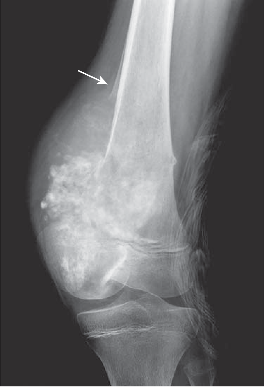
Distal femoral osteosarcoma with periosteal elevation producing a Codman triangle. - Robbins & Kumar Basic Pathology, p. 782
Diagnosis and staging
Diagnosis requires a carefully planned core-needle or open biopsy, ideally performed by the specialist sarcoma team that will provide definitive surgery. Poorly placed biopsies can complicate limb-sparing surgery.
Staging generally includes:
- X-ray and MRI of the affected bone
- CT of the chest for lung metastases
- Assessment for skeletal metastases, often with bone scan or PET/CT depending on local protocol
- Histology confirming malignant cells making osteoid
Types
The usual type is conventional high-grade intramedullary osteosarcoma, accounting for about three-quarters of cases. Other variants include telangiectatic, low-grade central, small-cell, parosteal, periosteal, and high-grade surface osteosarcoma. - Grainger & Allison’s Diagnostic Radiology, p. 1048
Causes and risk factors
Most cases are sporadic, but recognized associations include:
- Rapid bone growth during adolescence
- RB1 mutations, including hereditary retinoblastoma
- TP53 mutations, including Li-Fraumeni syndrome
- Prior radiation treatment
- Paget disease of bone
- Bone infarction
Osteosarcoma commonly has complex genetic changes, with alterations in RB and TP53 among the important abnormalities. - Robbins & Kumar Basic Pathology, p. 782
Treatment
Treatment should be delivered by a multidisciplinary sarcoma team. For most high-grade conventional tumors, it combines:
- Chemotherapy before surgery to treat microscopic systemic disease and assess tumor response.
- Wide surgical removal of the tumor, preferably with limb-sparing reconstruction when safe. Amputation is used when adequate cancer clearance and a functional limb cannot be achieved.
- Postoperative chemotherapy.
In children and adolescents, a common backbone is MAP chemotherapy: high-dose methotrexate, doxorubicin, and cisplatin, although regimens vary by age, country, tumor site, and clinical circumstances. Osteosarcoma is relatively resistant to radiotherapy; radiation is mainly considered when surgery cannot fully control local disease.
If pulmonary metastases can be completely removed, surgery for lung nodules may be part of curative-intent treatment. The NCI treatment summary notes that spread is most often to the lungs and outlines multimodal treatment for localized and metastatic disease.
Prognosis
Outcome depends mainly on:
- Whether metastases are present at diagnosis
- Ability to completely remove the primary tumor and all visible metastases
- Tumor location and size
- Histologic response to preoperative chemotherapy
- Development of recurrence, especially early recurrence
Localized, completely resectable disease has a substantially better outlook than metastatic or recurrent osteosarcoma. Lung-only metastases generally have a better outlook than spread to bone or multiple organ sites.
This is a shared conversation. Sign in to Orris to start your own chat.