
So i have biochemistry viva tomorrow and the topic is gylcogenolysis give best explanation and all notes with all question related to it
Reading File
Now let me search the medical library for glycogenolysis content in parallel.
Finding Sources
Finding Sources
Now let me read the core sections from Lippincott and Harper's in parallel:
Reading File
Reading File
Reading File
Reading File
Reading File
Reading File
Finding Sources
Reading File
Reading File
Now let me fetch the key diagrams from Lippincott for visual support:
Loading Image
Loading Image
I now have everything I need to write a comprehensive viva preparation guide. Let me compile the full notes.
🧬 Glycogenolysis - Complete Viva Notes
What is Glycogenolysis?
Glycogenolysis is the breakdown (degradation) of stored glycogen to release glucose. It is NOT a reversal of glycogenesis - it uses a completely separate set of cytosolic enzymes. The primary product from α(1→4) bonds is glucose 1-phosphate, and free glucose is released from each α(1→6) branch point.
- Occurs mainly in liver (to maintain blood glucose) and skeletal muscle (for energy during exercise)
- Liver glycogen: responds to fasting/feeding
- Muscle glycogen: responds to exercise/contraction
Step-by-Step Enzymatic Reactions
Step 1 - Glycogen Phosphorylase (Chain Shortening)
- Cleaves α(1→4) glycosidic bonds from the nonreducing ends by phosphorolysis (using inorganic phosphate, Pi)
- Product: Glucose 1-phosphate (not free glucose - no ATP wasted)
- Stops when 4 glucosyl units remain at each branch point → structure called limit dextrin
- Phosphorylase cannot act on branch points - it stops 4 residues away
- Requires pyridoxal phosphate (PLP, vitamin B6) as a coenzyme
Reaction: Glycogen (n residues) + Pi → Glycogen (n-1 residues) + Glucose 1-phosphate
Step 2 - Debranching Enzyme (Branch Removal)
The debranching enzyme is a bifunctional single protein with two distinct enzymatic activities:
Activity 1 - 4:4 Transferase (oligo-α(1→4)→α(1→4)-glucantransferase):
- Transfers the outer 3 of 4 glucosyl residues from the branch to a nonreducing end of another chain
- Breaks an α(1→4) bond and makes a new α(1→4) bond (does NOT yield free glucose or glucose 1-P)
Activity 2 - α(1→6)-Glucosidase (amylo-α(1→6)-glucosidase):
- Hydrolyzes the remaining 1 glucosyl residue attached at the α(1→6) branch point
- Releases free glucose (not glucose 1-phosphate!)
- This is the ONLY step that produces free glucose in glycogenolysis
Remember: ~8:1 ratio of glucose 1-P to free glucose (since branches occur every ~8-12 residues)
Step 3 - Phosphoglucomutase
- Converts Glucose 1-phosphate → Glucose 6-phosphate
- Requires glucose 1,6-bisphosphate as intermediate cofactor
Step 4 - Fate of Glucose 6-Phosphate
| Tissue | What happens | Why |
|---|---|---|
| Liver | Glucose 6-phosphatase removes Pi → free glucose → enters blood | Liver has glucose 6-phosphatase |
| Muscle | Enters glycolysis directly as glucose 6-P | Muscle lacks glucose 6-phosphatase |
Key exam point: Muscle glycogenolysis cannot raise blood glucose because muscle lacks glucose 6-phosphatase.
Regulation of Glycogenolysis
Regulation occurs at two levels: (1) covalent (hormonal) and (2) allosteric.
Diagram: Hormonal (Covalent) Regulation Cascade
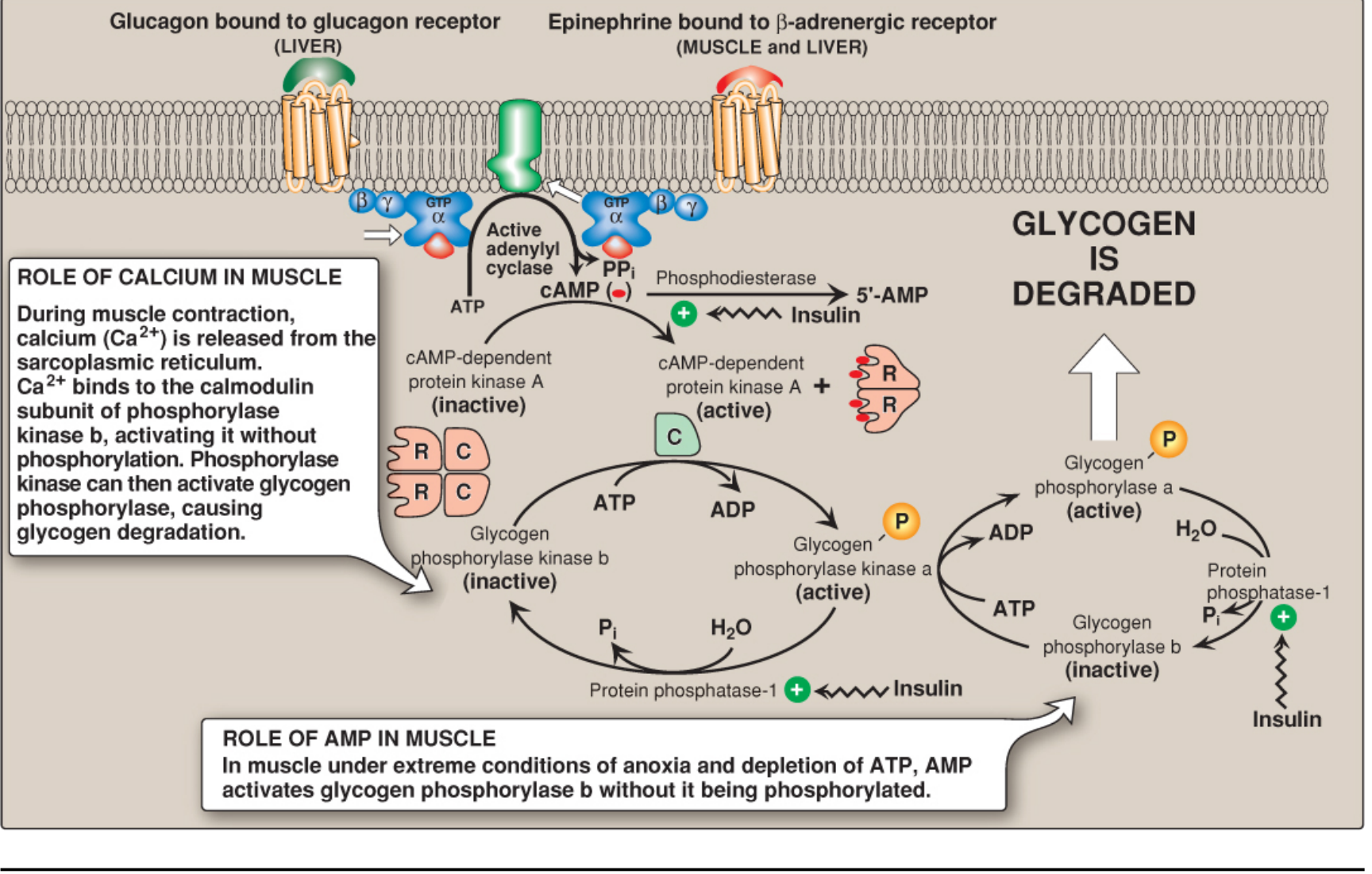
A. Covalent (Hormonal) Regulation - The Cascade
Hormonal triggers: Glucagon (liver), Epinephrine (liver + muscle)
Cascade (memorize this sequence):
Glucagon / Epinephrine
↓ (binds GPCR)
Adenylyl cyclase activated → ATP → cAMP ↑
↓
PKA (Protein Kinase A) activated [inactive R₂C₂ → active 2C after cAMP binds R subunits]
↓ (phosphorylates)
Phosphorylase kinase b (inactive) → Phosphorylase kinase a (active)
↓ (phosphorylates)
Glycogen phosphorylase b (inactive) → Glycogen phosphorylase a (active)
↓
GLYCOGENOLYSIS ↑
Amplification cascade: A few hormone molecules → millions of glucose 1-P molecules. Each step amplifies the signal enormously.
PKA also simultaneously:
- Phosphorylates glycogen synthase → inactivates it (stops glycogenesis)
- Phosphorylates inhibitor-1 → inhibits protein phosphatase-1 (prevents inactivation of phosphorylase)
Insulin counteracts glycogenolysis:
- Activates phosphodiesterase → breaks down cAMP
- Activates protein phosphatase-1 → dephosphorylates and inactivates phosphorylase a → phosphorylase b (inactive)
B. Allosteric Regulation
| Allosteric effector | Effect on phosphorylase | Tissue |
|---|---|---|
| AMP | Activates phosphorylase b (without phosphorylation) | Muscle only |
| ATP, Glucose 6-P, Glucose | Inhibits phosphorylase a | Muscle (ATP, G6P); Liver (glucose too) |
| Ca²⁺ (via calmodulin) | Activates phosphorylase kinase b (without phosphorylation) | Muscle + Liver |
Calcium's Role in Muscle:
- Nerve stimulation → Ca²⁺ released from sarcoplasmic reticulum
- Ca²⁺ binds to calmodulin (δ subunit) of phosphorylase kinase
- Activates phosphorylase kinase b without phosphorylation
- This links muscle contraction directly to glycogenolysis
AMP's Role in Muscle:
- During extreme anoxia/ATP depletion: AMP ↑
- AMP directly activates glycogen phosphorylase b (bypasses the whole cascade)
C. Glycogenolysis in Liver Can Be cAMP-Independent
- α₁-adrenergic receptor stimulation by norepinephrine (or epinephrine) → IP₃ pathway → Ca²⁺ mobilization from ER → Ca²⁺/calmodulin activates phosphorylase kinase
- Also activated by vasopressin, oxytocin, and angiotensin II (via Ca²⁺ or PIP₂ pathway)
- (Harper's Illustrated Biochemistry, 32nd Ed)
Liver vs. Muscle Glycogenolysis - Key Differences
| Feature | Liver | Muscle |
|---|---|---|
| Purpose | Maintain blood glucose | Fuel for contraction |
| Hormone trigger | Glucagon (primary), epinephrine | Epinephrine, neural Ca²⁺ |
| Glucose 6-phosphatase | Present → free glucose released | Absent → G6P stays in cell |
| Raises blood glucose? | YES | NO |
| AMP activates phosphorylase b? | No | Yes (myophosphorylase) |
| cAMP-independent pathway? | Yes (α₁-adrenergic/Ca²⁺) | No |
Glycogen Storage Diseases (GSDs) - Viva Favorites!
The diagram below shows the key GSD types and where they block glycogen degradation:
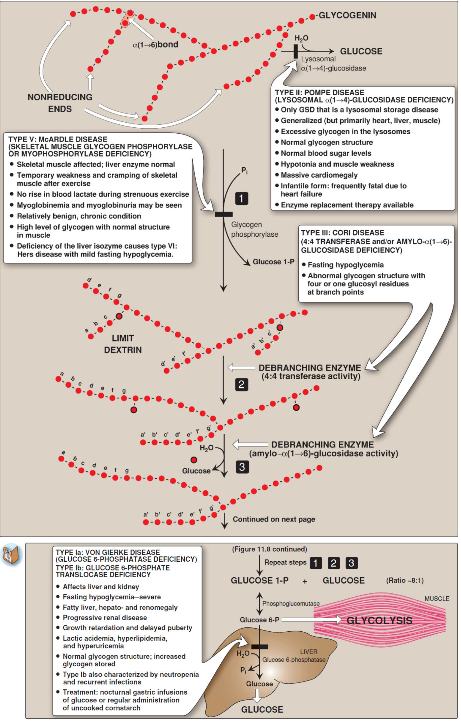
| Type | Name | Enzyme Deficient | Tissue | Key Features |
|---|---|---|---|---|
| I (Ia) | Von Gierke disease | Glucose 6-phosphatase | Liver + Kidney | Severe fasting hypoglycemia, hepatomegaly + renomegaly, lactic acidemia, hyperlipidemia, hyperuricemia, fatty liver |
| I (Ib) | Von Gierke Ib | G6P translocase | Liver + Kidney | Same as Ia + neutropenia + recurrent infections |
| II | Pompe disease | Lysosomal acid α-glucosidase (acid maltase) | ALL organs (heart, muscle, liver) | ONLY lysosomal GSD; massive cardiomegaly; hypotonia; infantile form often fatal by 2 years; enzyme replacement therapy (ERT) available |
| III | Cori disease (Forbes) | Debranching enzyme (4:4 transferase + α-1,6-glucosidase) | Liver + Muscle | Fasting hypoglycemia; abnormal glycogen (short outer chains, many branches) |
| IV | Andersen disease | Branching enzyme (enzyme of SYNTHESIS) | Liver | Liver cirrhosis - fatal in early childhood; glycogen has long unbranched chains |
| V | McArdle disease | Muscle phosphorylase (myophosphorylase) | Muscle | Muscle cramps + weakness on exercise; myoglobinuria; NO rise in blood lactate on exercise; liver enzyme normal; relatively benign |
| VI | Hers disease | Liver phosphorylase | Liver | Mild fasting hypoglycemia; hepatomegaly; benign |
| IX | Phosphorylase kinase deficiency | Phosphorylase kinase | Liver (X-linked) or Muscle | Similar to Hers disease |
Most GSDs are autosomal recessive. Exceptions: Type IX (phosphorylase kinase - X-linked), Danon disease (X-linked).
Likely Viva Questions & Model Answers
Basic Mechanism
Q: What is the first enzyme of glycogenolysis?
A: Glycogen phosphorylase. It cleaves α(1→4) glycosidic bonds by phosphorolysis, producing glucose 1-phosphate.
Q: Why is the product glucose 1-phosphate and not free glucose?
A: Phosphorolysis uses inorganic phosphate (Pi) instead of water. This traps glucose in the cell as glucose 1-phosphate (charged, cannot cross membranes) and conserves the energy of the glycosidic bond. No ATP is consumed.
Q: What is a limit dextrin?
A: The residual glycogen structure after phosphorylase has cleaved all available α(1→4) bonds, leaving 4 glucosyl residues on each chain before a branch point. Phosphorylase cannot degrade it further.
Q: What are the two activities of the debranching enzyme?
A: (1) 4:4 transferase - transfers 3 of 4 residues from a branch to another chain; (2) amylo-α(1→6)-glucosidase - hydrolyzes the remaining α(1→6)-linked residue to yield free glucose.
Q: What is the ratio of glucose 1-phosphate to free glucose in glycogenolysis?
A: Approximately 8:1 (or 9:1). The ratio reflects the average chain length between branch points.
Q: What coenzyme does glycogen phosphorylase require?
A: Pyridoxal phosphate (PLP), a derivative of vitamin B6. It acts as an acid-base catalyst.
Regulation
Q: How does glucagon stimulate glycogenolysis?
A: Glucagon binds GPCR → Gs protein activates adenylyl cyclase → ATP → cAMP → PKA activated → phosphorylates phosphorylase kinase b → active phosphorylase kinase a → phosphorylates glycogen phosphorylase b → active phosphorylase a → glycogenolysis.
Q: What is the difference between phosphorylase a and phosphorylase b?
A: Phosphorylase a is the phosphorylated, active form. Phosphorylase b is the dephosphorylated, inactive form (though it can be activated allosterically by AMP in muscle).
Q: How does calcium activate glycogenolysis in muscle?
A: During neural stimulation, Ca²⁺ is released from the sarcoplasmic reticulum. Ca²⁺ binds to calmodulin (the δ-subunit of phosphorylase kinase b), activating it without phosphorylation. Activated phosphorylase kinase then phosphorylates glycogen phosphorylase b → active a form.
Q: How does AMP activate glycogenolysis in muscle?
A: Under extreme anoxia/ATP depletion, AMP accumulates and directly activates glycogen phosphorylase b allosterically without requiring phosphorylation, bypassing the entire hormonal cascade.
Q: How does insulin inhibit glycogenolysis?
A: Insulin activates phosphodiesterase (degrades cAMP), activates protein phosphatase-1 (dephosphorylates and inactivates phosphorylase a), and increases glucose uptake → raises glucose 6-phosphate (allosteric inhibitor of phosphorylase a).
Q: Can glycogenolysis in liver be activated without cAMP?
A: Yes. Epinephrine/norepinephrine acting via α₁-adrenergic receptors activates the IP₃/Ca²⁺ pathway → Ca²⁺/calmodulin activates phosphorylase kinase independent of cAMP. Also activated by vasopressin, oxytocin, and angiotensin II.
Q: Why does epinephrine cause glycogenolysis in both liver and muscle but glucagon only in liver?
A: Because glucagon receptors are present only on hepatocytes, not on muscle cells. Epinephrine binds β-adrenergic receptors on both liver and muscle (via cAMP pathway).
Clinical / Applied
Q: Why can't muscle glycogenolysis raise blood glucose?
A: Muscle lacks glucose 6-phosphatase. Therefore, glucose 6-phosphate cannot be converted to free glucose and must be used within the muscle via glycolysis.
Q: What is McArdle disease? What is its key biochemical feature?
A: Type V GSD - deficiency of muscle phosphorylase (myophosphorylase). Key feature: failure of blood lactate to rise during exercise (because muscle cannot break down glycogen → no glucose → no glycolysis → no lactate).
Q: What is Von Gierke disease?
A: Type I GSD - deficiency of glucose 6-phosphatase (Type Ia) or its translocase (Ib). Both glycogenolysis and gluconeogenesis are impaired (both produce glucose 6-P which cannot be released as free glucose). Features: severe fasting hypoglycemia, massive hepatomegaly, lactic acidemia, hyperlipidemia, hyperuricemia.
Q: Which is the only lysosomal GSD?
A: Pompe disease (Type II GSD) - deficiency of lysosomal acid α-glucosidase (acid maltase). It is the only GSD involving lysosomes.
Q: What disease results from branching enzyme deficiency?
A: Andersen disease (Type IV GSD) - a disease of glycogen SYNTHESIS (not breakdown). The abnormal unbranched glycogen deposited triggers liver cirrhosis and is fatal in early childhood.
Q: What is the signal amplification cascade and why is it important?
A: The multi-step cascade (hormone → cAMP → PKA → phosphorylase kinase → phosphorylase) amplifies the signal at each step. A few hormone molecules binding their receptors ultimately activate many phosphorylase molecules, allowing a rapid, large release of glucose.
Quick Memorization Mnemonics
GSD types for liver GSDs (hypoglycemia): Type I (G6Pase), Type III (debrancher), Type VI (liver phosphorylase), Type IX (phosphorylase kinase), Type 0 (glycogen synthase)
Phosphorylase "b" = inactive, "a" = active (b is "bad"/inactive, a is "active")
Glycogen synthase is opposite: "a" form = active (dephosphorylated), "b" form = inactive (phosphorylated)
Debranching enzyme activities:
- 4:4 transferase = moves 3 residues
- α-1,6-glucosidase = releases free glucose
Blood lactate test distinguishes:
- McArdle (no rise) vs. normal (lactate rises on exercise)
Summary Flowchart
GLYCOGEN
|
| ① Glycogen phosphorylase (cleaves α-1,4 bonds)
| Coenzyme: PLP (Vit B6)
↓
LIMIT DEXTRIN (4 residues before branch)
|
| ② Debranching Enzyme: 4:4 transferase activity
| (transfers 3 residues to adjacent chain)
|
| ③ Debranching Enzyme: α-1,6-glucosidase activity
| (releases 1 free glucose from branch point)
↓
GLUCOSE 1-PHOSPHATE (major product, ~8/9 of total)
+ FREE GLUCOSE (~1/9 of total, from branch points)
|
| Phosphoglucomutase
↓
GLUCOSE 6-PHOSPHATE
| |
LIVER MUSCLE
| |
G6Pase Glycolysis
↓
FREE GLUCOSE → Blood
Sources: Lippincott Illustrated Reviews: Biochemistry, 8th Ed; Harper's Illustrated Biochemistry, 32nd Ed; Basic Medical Biochemistry: A Clinical Approach, 6th Ed; Robbins & Kumar Basic Pathology
Good luck on your viva tomorrow! Know the enzyme sequence cold, be ready to explain regulation at both covalent and allosteric levels, and be prepared to link any GSD to its deficient enzyme, product accumulation, and clinical features.
This is a shared conversation. Sign in to Orris to start your own chat.