
Blood coagulation
blood coagulation cascade clotting factors diagram
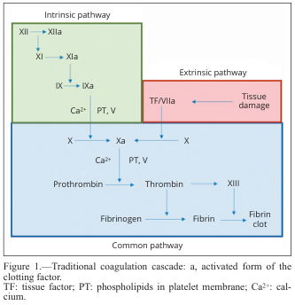
Summary : This diagram illustrates the traditional coagulation cascade, detailing the sequence of events and factors involved in blood clotting via the intrinsic, extrinsic, and common pathways. flowchart: # Pathways : • Intrinsic pathway (green box): Initiated by factor XII, proceeds through XI, IX, and VIII, with activation steps indicated by "a" (e.g., XIIa, XIa, IXa). • Extrinsic pathway (red box): Initiated by tissue damage, which activates tissue factor (TF) and factor VIIa. • Common pathway (blue box): Both intrinsic and extrinsic pathways converge at factor X, leading to the activation of prothrombin to thrombin, which then converts fibrinogen to fibrin, forming a fibrin clot. # Nodes : • XII → XIIa (Intrinsic) • XI → XIa (Intrinsic) • IX → IXa (Intrinsic) • Ca²⁺, PT, V (Intrinsic, Common) • TF/VIIa (Extrinsic) • Tissue damage (Extrinsic) • X → Xa (Common) • Prothrombin → Thrombin (Common) • Fibrinogen → Fibrin (Common) • XIII (Common) • Fibrin clot (Common) # Connectors : • Arrows indicate the direction of activation or conversion between factors. • Intrinsic and extrinsic pathways both feed into the activation of factor X in the common pathway. • Ca²⁺ and PT, V are required cofactors at specific steps. # Layout : • Three main colored sections: green (intrinsic, top left), red (extrinsic, top right), blue (common, bottom). • Flow proceeds from top (initiation) to bottom (clot formation). • Arrows show sequential activation and convergence of pathways. # Analysis : • The diagram clearly separates the intrinsic and extrinsic initiation mechanisms, both converging on the common pathway at factor X. • The cascade is highly sequential, with each activated factor catalyzing the next step. • Calcium ions (Ca²⁺) and phospholipids (PT) are essential cofactors at multiple steps. • The final outcome is the formation of a stable fibrin clot, highlighting the coordinated nature of the coagulation process.
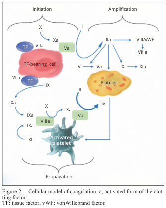
Summary : This figure presents a cellular model of blood coagulation, illustrating the sequential activation of clotting factors and the roles of different cell types (TF-bearing cell, platelet, activated platelet) in the initiation, amplification, and propagation phases of coagulation. flowchart: # Phases of Coagulation : • Initiation, Amplification, and Propagation are the three main phases depicted. ## Nodes : • TF-bearing cell (red, with "TF" label) • Platelet (yellow, labeled "Platelet") • Activated platelet (blue-green, labeled "Activated platelet") • Clotting factors: X, Xa, VIIa, IX, IXa, XI, XIa, II, IIa, V, Va, VIII, VIIIa, vWF (von Willebrand factor) • Green rectangles represent activated forms of clotting factors (e.g., Va, VIIIa). ## Connectors : • Arrows indicate activation and conversion steps between clotting factors. • Initiation: TF-bearing cell activates VIIa, which then activates X to Xa (with Va), and IX to IXa. • Amplification: II is converted to IIa, which activates V to Va, VIII/vWF to VIIIa, and XI to XIa on the platelet. • Propagation: Activated platelet facilitates further activation of X to Xa (with Va), IX to IXa (with VIIIa), and XI to XIa, leading to more IIa production. ## Layout : • The diagram is divided into three horizontal sections: Initiation (left), Amplification (center), Propagation (bottom). • Arrows flow from TF-bearing cell to platelet, then to activated platelet, showing the progression of the coagulation cascade. ## Design Encodings : • Cell types are color-coded: TF-bearing cell (red), Platelet (yellow), Activated platelet (blue-green). • Activated clotting factors are shown in green rectangles. • Blue arrows indicate activation pathways. • Factor names and their activated forms are clearly labeled. # Analysis : • The figure visually demonstrates the stepwise activation of clotting factors, beginning with tissue factor (TF) exposure, progressing through platelet activation, and culminating in a robust propagation phase on the activated platelet surface. • The amplification phase shows multiple feedback loops, with IIa (thrombin) activating several upstream factors, ensuring rapid and localized clot formation. • The propagation phase highlights the central role of the activated platelet in sustaining and amplifying the coagulation response.
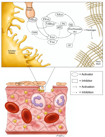
Summary : This figure illustrates the process of blood coagulation, focusing on the interactions between coagulation factors, inhibitors, and platelets at a site of vascular injury, and the formation of a fibrin mesh. It combines a schematic pathway diagram (top) with a cross-sectional tissue illustration (bottom), and includes a legend explaining the symbols used. process diagram and medical illustration: # Schematic Pathway Diagram (Top) : • Shows the cascade of coagulation factors involved in blood clotting. • Key factors depicted: VII(a), IX(a), VIII(a), XI(a), X(a), V(a), (Pro)Thrombin (IIa). • Inhibitors shown: TFPI, APC, PS, TM, AT. • Tissue Factor is present at the site of injury, activating VII(a). • Activated Platelet is shown as the starting point for the cascade. • Fibrinogen is converted to Fibrin Mesh via (Pro)Thrombin (IIa). • Solid arrows indicate activation; dashed arrows indicate inhibition. • Legend clarifies: ovals = Activator, rectangles = Inhibitor, solid line = Activation, dashed line = Inhibition. # Cross-sectional Tissue Illustration (Bottom) : • Depicts a blood vessel wall with endothelial cells, red blood cells, white blood cells, and platelets. • Shows platelets adhering to the vessel wall at the site of injury. • Fibrin mesh is forming at the injury site, trapping blood cells. • Endothelial cells line the vessel lumen. • Platelets are shown in yellow, red blood cells in red, white blood cells in purple. # Key Nodes and Interactions : • Activators: VII(a), IX(a), VIII(a), XI(a), X(a), V(a), (Pro)Thrombin (IIa). • Inhibitors: TFPI, APC, PS, TM, AT. • Activation pathways: Tissue Factor → VII(a); VII(a) → IX(a); IX(a) + VIII(a) → X(a); X(a) + V(a) → (Pro)Thrombin (IIa); (Pro)Thrombin (IIa) → Fibrinogen → Fibrin Mesh. • Inhibition pathways: TFPI inhibits VII(a); APC/PS inhibits VIII(a) and V(a); TM/AT inhibits (Pro)Thrombin (IIa). # Legend : • Oval shape = Activator. • Rectangle shape = Inhibitor. • Solid line = Activation. • Dashed line = Inhibition. # Analysis : • The figure visually integrates the molecular coagulation cascade with its anatomical context at a vascular injury site. • It highlights the balance between activation and inhibition in clot formation. • The schematic clarifies how multiple factors and inhibitors interact to regulate thrombin generation and fibrin mesh formation. • The tissue illustration contextualizes the process, showing how platelets and fibrin mesh physically contribute to clotting at the site of vessel damage.
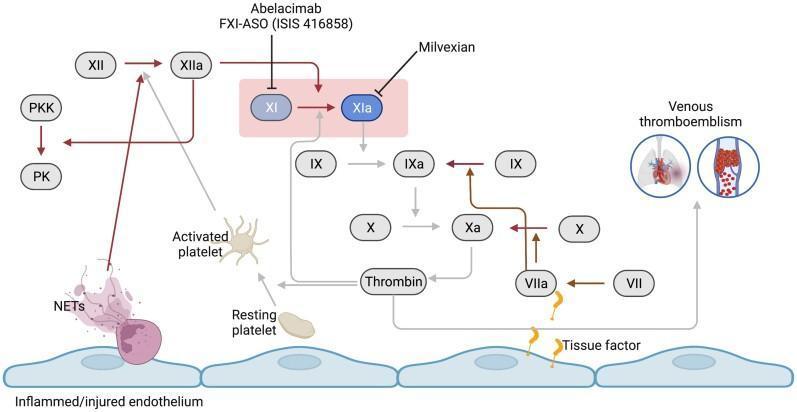
This pathophysiology diagram illustrates the coagulation cascade focusing on the central role of Factor XI (FXI) in venous thromboembolism. The visual depicts an inflamed/injured endothelial layer at the base, leading to the activation of the contact pathway (via FXII and NETs) and the extrinsic pathway (via Tissue Factor and FVII). Key clotting factors, including FXII, FXI, FIX, FX, and Thrombin, are shown as oval nodes interconnected by arrows representing biochemical activation. A highlighted pink box focuses on the conversion of FXI to FXIa, noting pharmacologic inhibitors like Abelacimab, FXI-ASO, and Milvexian. The diagram differentiates between resting and activated platelets and includes an icon for venous thromboembolism featuring pulmonary embolism and deep vein thrombosis. Dark brown arrows indicate pathways unaffected by FXI inhibition, while lighter grey arrows signify the attenuated thrombin formation pathway. This educational material is designed for medical students and clinicians studying hematology, pharmacology, and cardiovascular medicine.
Blood Coagulation
1. Overview of Hemostasis Events
- Vascular constriction - smooth muscle contraction reduces blood flow immediately
- Platelet plug formation - platelets adhere and aggregate at the injury site
- Blood clot formation - the coagulation cascade generates a fibrin mesh
- Fibrous tissue ingrowth - permanent vessel repair
2. Vascular Constriction
- Local myogenic spasm
- Autacoid factors from traumatized tissues and platelets
- Nervous reflexes (pain impulses)
3. Platelet Plug Formation
- Platelets adhere via GP Ib receptor (binds vWF) and GP Ia receptor (binds collagen)
- Degranulation releases ADP (induces aggregation) and serotonin (aggregation + vasoconstriction)
- TXA2 is synthesized from arachidonic acid and amplifies platelet activation
- Conformational change in αIIbβIII (GP IIb/IIIa) integrin enables fibrinogen binding, cross-linking adjacent platelets into a platelet plug
4. The Coagulation Cascade
- Formation of prothrombin activator (via intrinsic or extrinsic pathway)
- Prothrombin activator + Ca²⁺ → thrombin
- Thrombin converts fibrinogen → fibrin mesh
Coagulation Cascade Diagram
Extrinsic Pathway (faster, tissue injury)
- Activates factor X → Xa (directly)
- Activates factor IX → IXa
Intrinsic Pathway (slower, contact activation)
Common Pathway
5. Conversion of Fibrinogen to Fibrin
- Fibrinogen is a large plasma protein (MW ~340,000) produced by the liver (100-700 mg/dL in plasma)
- Thrombin cleaves fibrinogen, releasing fibrinopeptides → fibrin monomers
- Fibrin monomers polymerize into fibrin fibers (initially held by weak hydrogen bonds)
- Factor XIIIa (activated by thrombin, also called fibrin-stabilizing factor) forms covalent cross-links between fibrin strands, creating a strong, stable 3D meshwork
- The clot traps blood cells, platelets, and plasma, and adheres to damaged vessel surfaces
Cellular Model of Coagulation
| Phase | Location | Key Events |
|---|---|---|
| Initiation | TF-bearing cell | VIIa-TF activates X and IX |
| Amplification | Platelet surface | Thrombin activates V, VIII, XI |
| Propagation | Activated platelet | Massive thrombin burst via IXa/VIIIa and Xa/Va complexes |
6. Clotting Factors (Summary Table)
| Factor | Common Name | Pathway |
|---|---|---|
| I | Fibrinogen | Common |
| II | Prothrombin | Common |
| III | Tissue thromboplastin (TF) | Extrinsic |
| IV | Calcium (Ca²⁺) | All |
| V | Proaccelerin | Common |
| VII | Proconvertin | Extrinsic |
| VIII | Antihemophilic factor A | Intrinsic |
| IX | Antihemophilic factor B (Christmas factor) | Intrinsic |
| X | Stuart-Prower factor | Common |
| XI | PTA (plasma thromboplastin antecedent) | Intrinsic |
| XII | Hageman factor (contact factor) | Intrinsic |
| XIII | Fibrin-stabilizing factor | Common |
7. Natural Anticoagulants (Inhibitors of Coagulation)
| Inhibitor | Mechanism |
|---|---|
| Antithrombin III (AT-III) | Binds and inactivates thrombin and factors IX-XII; greatly enhanced by heparin |
| Protein C | Activated by thrombomodulin-thrombin complex; inactivates factors Va and VIIIa |
| Protein S | Cofactor for protein C |
| TFPI (Tissue Factor Pathway Inhibitor) | Inhibits VIIa-TF complex, limiting extrinsic pathway |
| Fibrin fibers | Adsorb 85-90% of generated thrombin, preventing clot spread |
| Endothelial PGI2 and NO | Inhibit platelet aggregation and vasoconstriction |
8. Coagulation with Activators and Inhibitors
9. Clinical Relevance
- Vitamin K deficiency or liver disease → reduced prothrombin and fibrinogen → bleeding tendency
- Hemophilia A - factor VIII deficiency (intrinsic pathway defect) → deep tissue/joint bleeding
- Von Willebrand disease - defective platelet adhesion (primary hemostasis defect) → mucosal/surface bleeding
- DVT/PE - pathological venous clot formation; fibrin-rich "red thrombus"
- Arterial thrombosis - platelet-rich "white thrombus" in high-shear arterial flow
- Heparin - potentiates AT-III (inactivates thrombin + factors IXa-XIIa)
- Warfarin - inhibits vitamin K-dependent factor synthesis (II, VII, IX, X)
- Direct oral anticoagulants (DOACs) - dabigatran (thrombin inhibitor), rivaroxaban/apixaban (Xa inhibitors)
- Antiplatelet drugs - aspirin (inhibits TXA2), clopidogrel (ADP receptor blocker), abciximab (GP IIb/IIIa inhibitor)