
Feline esophagus ?
The "feline esophagus" is a radiological finding where the esophagus shows a transverse striped pattern resembling a cat's esophagus, caused by transient mucosal folds. It is most commonly seen on barium swallow studies and is associated with gastroesophageal reflux or eosinophilic esophagitis.
Searching sources...
Reading File
Finding Sources
Searching Images
feline esophagus barium swallow transverse folds radiology
Searching PubMed
"feline esophagus" eosinophilic esophagitis
Now let me read the relevant textbook passages for more detail:
Reading File
Reading File
Let me fetch the endoscopic image referenced in Robbins:
Loading Image
Finding Sources
Now let me also fetch the endoscopic image from the Robbins figure caption that specifically shows the feline/ringed appearance:
Finding Sources
Reading File
I have comprehensive information now. Let me also get the endoscopic image showing the feline esophagus appearance from the Robbins figure:
Reading File
Here is a complete synthesis of the topic:
Feline Esophagus
What It Is
"Feline esophagus" (also called ringed esophagus or trachealization) is an endoscopic/radiologic finding where the esophageal mucosa shows stacked circular rings or concentric transverse ridges, resembling the striped pattern of a cat's tail or the rings of a trachea. The term refers to the supposed endoscopic resemblance to a striped cat's coat.
- Robbins Pathologic Basis of Disease, p. 707: "the esophageal mucosa can resemble stacked circular rings (referred to as feline esophagus because of supposed endoscopic resemblance to a striped cat's tail) with strictures and linear furrows."
Primary Association: Eosinophilic Esophagitis (EoE)
Feline esophagus is the hallmark endoscopic feature of Eosinophilic Esophagitis (EoE). It is one of the most characteristic findings alongside linear furrows and white exudates (eosinophilic abscesses).
Endoscopic Findings in EoE
| Feature | Description |
|---|---|
| Feline/ringed esophagus | Stacked circular mucosal rings (fixed or transient) |
| Trachealization | Same as above - rings resemble a trachea |
| Linear furrows | Vertical longitudinal lines |
| White exudates/plaques | Whitish papules = eosinophilic abscesses |
| Edema/pallor | Decreased vascular markings |
| Strictures | Narrowing of esophageal lumen |
| Narrow-caliber esophagus | Long-segment narrowing (small-caliber esophagus) |
| Crepe-paper mucosa | Mucosal fragility |
Important: Endoscopic findings are normal in at least one-third of EoE patients - normal endoscopy does not exclude the diagnosis. - Sleisenger & Fordtran's GI and Liver Disease
Histology of EoE (the diagnostic standard)
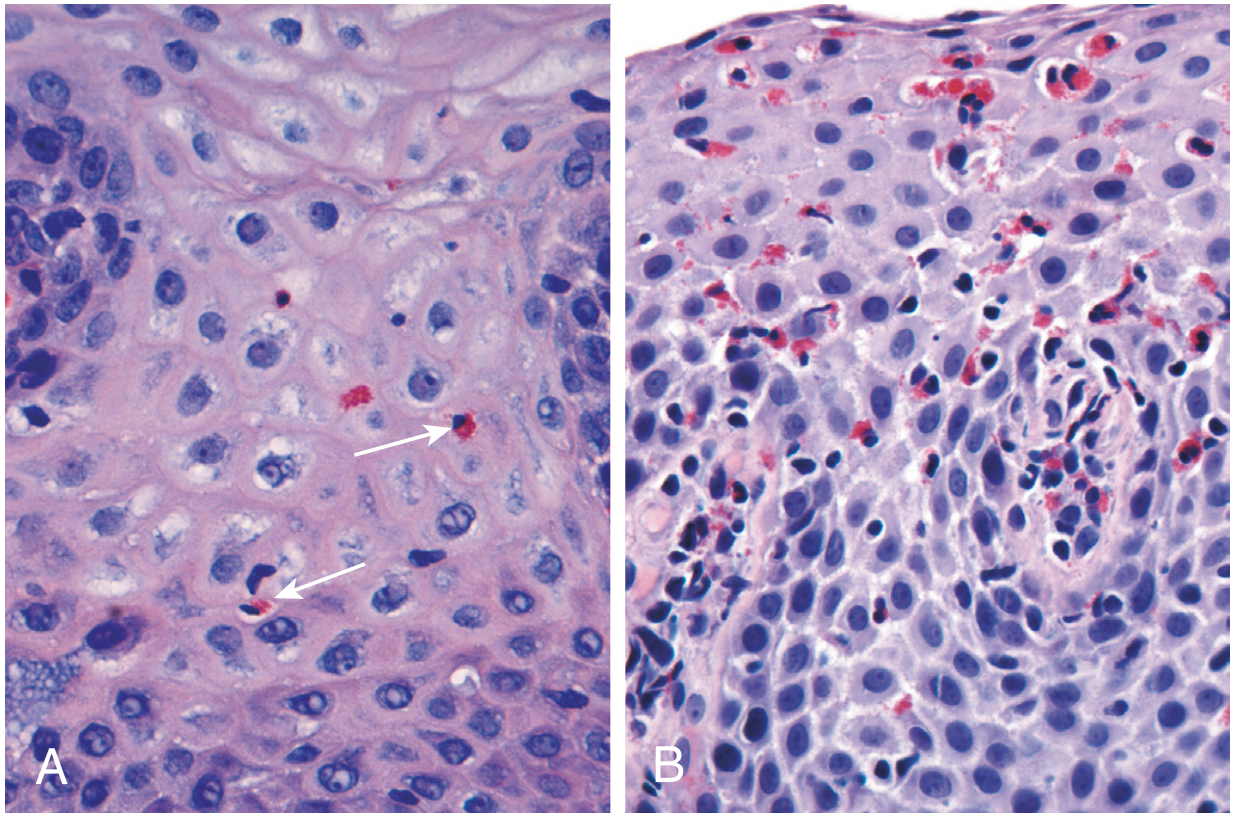
Fig: (A) Reflux esophagitis - scattered intraepithelial eosinophils with mild basal zone expansion. (B) EoE - numerous intraepithelial eosinophils (dense sheets).
The cardinal histologic feature is:
- >15 eosinophils per high-power field (x40) - large numbers of intraepithelial eosinophils, predominantly superficial, forming clusters and sheets
- Intercellular edema
- Basal hyperplasia and papillary lengthening
Sensitivity of biopsy: a single specimen has only 55% sensitivity; taking 5 biopsy specimens increases sensitivity to 100% (due to patchy distribution). - Sleisenger & Fordtran
Rings vs. Trachealization: Key Distinction
From the Clinical GI Endoscopy textbook, rings in EoE are classified and must be distinguished from transient plications (which can mimic feline esophagus). Rings are best appreciated when the esophagus is fully insufflated during endoscopy.
- Fixed rings (fibrostenotic) - also called concentric rings, corrugated esophagus, trachealization - graded by severity
- Transient rings - may be peristalsis-related; easily confused with true feline/ringed esophagus
Differential Diagnosis of Feline Esophagus
While most strongly associated with EoE, feline esophagus can also appear in:
- GERD - overlap exists; ringed appearance can occur with chronic reflux
- Transient mucosal folds - peristaltic contractions can transiently produce a striped/ringed pattern on fluoroscopy (original radiologic description)
- Hiatal hernia-related changes
Pathogenesis of EoE
EoE is an immune-mediated (Th2-driven) allergic disorder:
- Associated with atopy (atopic dermatitis, allergic rhinitis, asthma)
- Food antigens (especially cow's milk) trigger IgE- and non-IgE-mediated eosinophilic infiltration
- Mast cells and IL-4/IL-13 signaling are implicated
- Incidence has increased over the last 20 years in parallel with other Th2-mediated allergic disorders
Clinical Features of EoE
| Feature | Children | Adults |
|---|---|---|
| Presentation | Reflux, vomiting, food refusal, failure to thrive | Dysphagia, food impaction, chest pain |
| Demographics | Young children, predominantly male | Can occur at any age |
| Atopy | Common | Common |
Diagnosis of EoE
- Suggestive history (dysphagia, food impaction)
- Esophageal eosinophilic infiltrate on biopsy (>15 eos/hpf)
- Exclusion of GERD (normal pH monitoring or failure to respond to high-dose PPIs)
- Multiple biopsies (at least 5) for adequate sensitivity
Treatment
-
Dietary: Empiric elimination of cow's milk and dairy (first-line dietary approach); guided elimination based on skin patch/IgE testing
-
Pharmacologic: PPIs, topical corticosteroids (swallowed budesonide/fluticasone)
-
Biologic: IL-4/IL-13 receptor antagonists (dupilumab) are beneficial
-
Endoscopic: Dilation of strictures - must be done in small increments to reduce risk of mucosal tears and perforation; does not reduce inflammation
-
Robbins, Cotran & Kumar Pathologic Basis of Disease; Schwartz's Principles of Surgery, 11th ed.; Sleisenger & Fordtran's GI and Liver Disease
This is a shared conversation. Sign in to Orris to start your own chat.