
Hernia kia Hoti hain
hernia kya hoti hai types causes symptoms in Urdu Hindi
types of hernia diagram inguinal umbilical
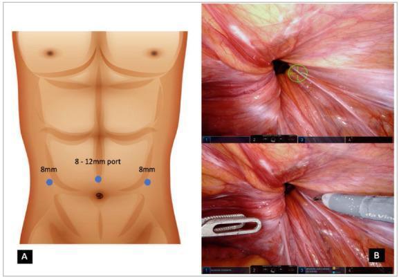
Educational medical composite illustrating robotic inguinal hernia repair. Panel A is an anatomical diagram showing abdominal port placement on a male torso; it highlights a central supraumbilical port (8-12 mm) flanked laterally by two 8 mm robotic ports, essential for the da Vinci surgical system setup. Panel B displays a robotic intraoperative view of the internal inguinal region. The image shows the inguinal defect as a dark, circular opening within the reddish-pink peritoneal tissue. Distinct anatomical landmarks such as the medial umbilical ligament and the radiating striated texture of the abdominal wall are visible. Robotic surgical instruments, including a dissector and a grasping tool, are shown targeting the defect area for dissection and preparation of the preperitoneal space. This visual material demonstrates the surgical approach and anatomical orientation required for robotic-assisted transabdominal preperitoneal (rTAPP) hernia repair.
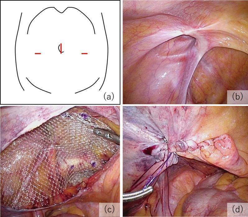
A multi-panel educational figure illustrating the key procedural steps of a Transabdominal Preperitoneal (TAPP) inguinal hernia repair. (a) Schematic diagram of the abdomen showing the laparoscopic port arrangement: a 12 mm umbilical port and two 5 mm lateral ports. (b) Intraoperative laparoscopic photograph showing the internal inguinal anatomy and the hernial orifice, classified as EHS-L2 (European Hernia Society classification for a medium indirect lateral hernia). (c) Surgical view demonstrating mesh fixation; a semi-transparent, macroporous polypropylene mesh is positioned in the preperitoneal space to cover the defect and is secured with visible surgical tacks. (d) Final procedural step showing the closure of the peritoneal flap using continuous suturing to exclude the mesh from the peritoneal cavity, preventing contact with abdominal viscera. The series emphasizes proper anatomical orientation, mesh placement, and peritoneal closure in minimally invasive hernia surgery.
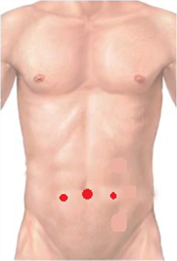
This medical illustration depicts the standard trocar port placement for laparoscopic inguinal hernia repair on a male torso. The diagram uses red circular markers to indicate surgical entry points. A primary 10 mm trocar port is positioned centrally at the umbilicus. Two secondary 5 mm working ports are located bilaterally in the lower abdominal quadrants. These lateral ports are situated along the midclavicular lines, approximately 1 cm inferior to the horizontal umbilical line. This configuration is typical for providing optimal triangulation and visualization of the preperitoneal space (including Retzius and Bogros spaces) during minimally invasive abdominal surgery. The anatomical focus is on the lower abdominal wall and the geometric relationship between the umbilicus and midclavicular landmarks for surgical access.
hernia abdominal wall anatomy diagram showing defect bulge
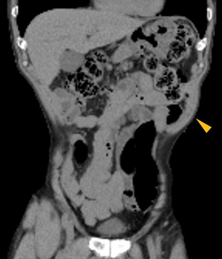
A diagnostic image showing a coronal view of a non-contrast abdominal CT scan. The image displays the anatomy of the torso, including the liver in the right upper quadrant, the spleen in the left upper quadrant, and loops of the small and large intestines containing gas and fecal material. A yellow arrowhead points to a focal area of the left lateral abdominal wall where there is a distinct outward bulging. This area is characterized by thinning and relaxation of the abdominal wall musculature (oblique and transversus abdominis muscles). Notably, there is no evidence of a fascial defect, hernia sac, or protrusion of intra-abdominal contents into the bulge, a finding characteristic of an abdominal pseudohernia. This clinical entity is often associated with neuropathic muscle paralysis, such as that caused by herpes zoster reactivation or post-surgical nerve injury. The image serves as a clinical reference for differentiating pseudohernia from true incisional or ventral hernias based on the preservation of anatomical continuity despite muscle laxity.
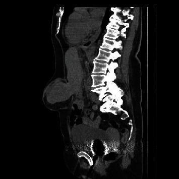
This diagnostic image is a non-contrast sagittal CT scan of the abdomen and pelvis. The image clearly displays the thoracolumbar vertebral column and sacrum as posterior landmarks. The primary finding is a large ventral abdominal wall defect at the supra-umbilical level, involving the linea alba. A significant portion of solid abdominal organ tissue, consistent with liver segment III, is seen herniating through the defect into a large epigastric hernia sac. The herniated liver tissue appears homogenous with density similar to the intra-abdominal liver parenchyma, contrasting with the lower-density subcutaneous adipose tissue. There is a visible bulge on the anterior abdominal wall corresponding to the hernia sac. This clinical imaging demonstrates a complex epigastric hernia with visceral content, highlighting the potential for liver herniation in severe ventral abdominal wall defects. The image is intended for educational purposes regarding diagnostic radiology and surgical anatomy of abdominal wall hernias.
🫁 Hernia - Mukammal Maloomat
Hernia Kya Hoti Hai?
- Reducible hoti hai - wapas apni jagah jaa sakti hai, ya
- Incarcerated hoti hai - wapas nahi ja sakti
Hernia Ki Iqsam (Types)
| Naam | Jagah | Khaasiyat |
|---|---|---|
| Inguinal Hernia | Groin / Kamar ke nichle hisse mein | Sabse zyada common - aant ya charbi inguinal canal mein ghus jaati hai |
| Femoral Hernia | Groin ke niche, upper thigh mein | Auraton mein zyada, strangulation ka risk zyada (40%) |
| Umbilical Hernia | Naaf (belly button) ke paas | Naaf ke paas ek ubhaar banta hai |
| Incisional Hernia | Operation ke nishaanaat (scar) ke paas | Pahle ki surgery ke baad |
| Hiatal Hernia | Pait (stomach) ka chest mein jaana | Diaphragm ki kamzori se |
| Internal Hernia | Andar ki taraf | Baahar se nazar nahi aata |
Asbaab (Causes) - Kyun Hoti Hai?
- Janm se kamzori (congenital)
- Umar ke saath muscles kamzor hona
- Pehle ki surgery ya zakhm
- Wazan uthana (heavy lifting)
- Khansi ya chheenk (chronic cough)
- Mote hona (obesity)
- Qabz aur zor lagana
- Hamla (pregnancy)
- Dhoorpaan (smoking)
Alamaat (Symptoms)
- Ek jagah ubhaar (bulge) - laitne se ghata, uthne se badhta hai
- Us jagah dard ya bhari bhaari feeling
- Khaney ke baad takleef
- Kabhi kabhi koi alamat nahi bhi hoti
⚠️ Emergency Alamaat (Foran Doctor ke paas jaao)
- Ubhaar andar nahi ja raha
- Tez dard
- Ukaai/ultiyaan
- Bukhaar
- Ubhaar ki jagah lal ho jaana
Teekra Khatarnaak Kaun Si Hernia Hai?
- Femoral Hernia: 40% cases mein presentation ke waqt already strangulated hoti hai
- Umbilical Hernia: 60% cases mein emergency surgery mein strangulation milti hai
- Incisional Hernia: Sirf 2% mein strangulation hoti hai (nisbatan safe)
Ilaj (Treatment)
- Open Hernia Repair - Kaat kar jaal (mesh) lagana
- Laparoscopic Repair - Keyhole surgery (TAPP / TEP) - chote teedon se
Kise Zyada Khatra Hai?
- Pehle se hernia ho
- Mardaangi (males mein inguinal hernia zyada)
- Wazni kaam karne wale
- Mote afraad
- Dhuwan peene wale
- Pait ki surgery ho chuki ho
- Family history ho
Zaroori Baat: Agar koi ubhaar nazar aaye ya dard ho, apne doctor se zaroor miley. Hernia apne aap theek nahi hoti aur waqt ke saath badh sakti hai.
What are the causes of enlargement of prostate gland
causes of prostate gland enlargement BPH pathophysiology 2024
benign prostatic hyperplasia BPH prostate anatomy zones diagram
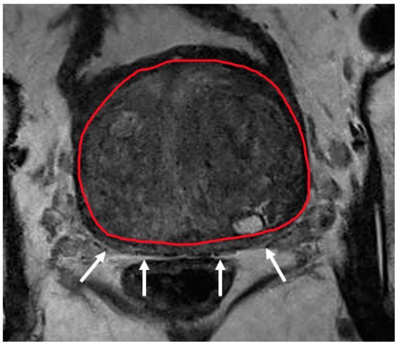
This diagnostic axial MRI scan (T2-weighted) of the prostate illustrates severe benign prostatic hyperplasia (BPH) with transition zone hypertrophy. The transition zone is segmented with a red outline, occupying the vast majority of the prostatic volume and exhibiting a heterogeneous, predominantly medium-gray signal intensity. Due to this significant expansion, the peripheral zone is severely compressed and displaced posteriorly, visible only as a thin, crescent-shaped sliver of lower signal intensity, indicated by white arrows. This image demonstrates a common challenge in prostatic zonal segmentation and radiological assessment: the effacement of anatomical landmarks between the transition and peripheral zones. The anatomical region focuses on the mid-gland level of the prostate. Educational focus includes understanding prostatic zonal anatomy, the pathophysiological effect of transition zone expansion on surrounding tissues, and the clinical imaging characteristics of severe hypertrophy in the context of urological diagnostics and radiotherapy planning.
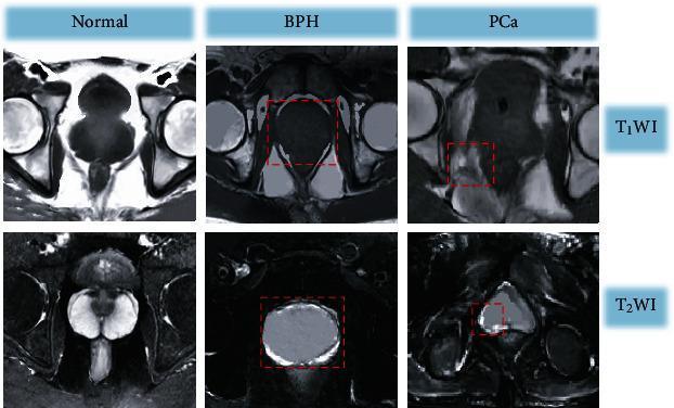
A comparative diagnostic MRI panel displaying axial views of the pelvic region to distinguish between Normal, Benign Prostatic Hyperplasia (BPH), and Prostate Cancer (PCa). The top row contains T1-weighted images (T1WI), while the bottom row shows T2-weighted images (T2WI). In the T1WI sequence, all three conditions present relatively homogeneous isosignals, making it difficult to differentiate the internal anatomy of the prostate. Red dashed boxes highlight regions of interest. In the T2WI sequence, significant diagnostic features are visible: the normal prostate displays a symmetrical high-signal crescent-shaped peripheral zone; BPH shows marked enlargement of the central and transitional zones while maintaining the peripheral high-signal crescent; and the PCa image demonstrates a loss of normal architecture with restricted low-signal areas in the peripheral zone and mixed signal intensity in the central region, indicative of neoplastic infiltration. These images serve as an educational comparison of signal intensity changes and morphological alterations in common prostatic pathologies.
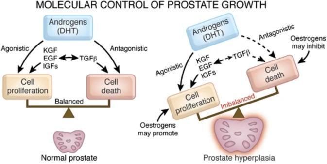
This medical schematic illustrates the pathophysiology of benign prostatic hyperplasia (BPH) compared to a normal prostate, focusing on the molecular signaling pathways that regulate tissue growth. The diagram is divided into two comparative sections: 'Normal prostate' and 'Prostate hyperplasia.' In a normal prostate, a balanced scale shows homeostatic equilibrium between cell proliferation and cell death. Androgens, primarily dihydrotestosterone (DHT), exert agonistic effects via growth factors (KGF, EGF, IGFs) to promote proliferation, and antagonistic effects via TGF-beta to promote apoptosis. In the prostate hyperplasia model, the scale is tilted toward proliferation, indicating an 'imbalanced' state. This pathological state is characterized by increased agonistic signaling and potential hormonal shifts, where estrogens may further promote cell proliferation and inhibit cell death. The illustration uses flow arrows to define the relationships between hormonal inputs (DHT, Oestrogens), intermediary growth factors (KGF, EGF, IGFs, TGF-beta), and the resulting physiological outcomes within the prostatic stroma and epithelium.
Causes of Prostate Gland Enlargement
1. Benign Prostatic Hyperplasia (BPH) - Most Common
Pathophysiology - How BPH Develops
- Testosterone in the blood is converted to DHT inside the prostate by the enzyme 5α-reductase type 2
- DHT is 10 times more potent than testosterone and binds to androgen receptors in the nucleus
- This stimulates growth factors (KGF, EGF, IGFs) that increase stromal cell proliferation and decrease epithelial cell death (apoptosis)
- The result: continuous, progressive enlargement of the gland
"BPH does not occur in males who are castrated before puberty or in males with genetic diseases that block androgen activity." - Robbins & Kumar Basic Pathology
Risk Factors for BPH
| Risk Factor | Mechanism |
|---|---|
| Age | Most important - affects ~50% of men >50 yrs, ~90% of men in their 80s |
| Androgens (DHT) | Central hormonal driver of prostate growth |
| Estrogens | With aging, testosterone falls but estrogen persists; estrogens act synergistically with DHT to further drive growth |
| Family history / Genetics | Genetic predisposition increases risk |
| Obesity / Metabolic syndrome | Increases circulating estrogens via peripheral conversion of androgens; atherosclerosis of prostatic vasculature also implicated |
| Diabetes / Insulin resistance | Higher insulin/IGF-1 levels promote prostatic cell growth |
| Sedentary lifestyle | Associated with increased BPH risk |
| Gut microbiota alterations | Emerging evidence (2024) links gut dysbiosis to prostatic inflammation |
Prostate Size in BPH
- Normal prostate: ~20 mL in young men; ~24-38 mL between ages 50-80
- In BPH: volume exceeds 30 mL and can reach 60-100 g (3-5x normal weight)
2. Prostate Cancer (Carcinoma of the Prostate)
- Arises primarily in the peripheral zone of the prostate (unlike BPH which affects the transition zone)
- Most common in men over 50 years
- Key molecular events: TMPRSS2-ETS fusion genes and PI3K/AKT pathway activation promote tumor cell growth
- Also androgen-dependent - treated by surgical or drug-induced castration
3. Prostatitis (Inflammation/Infection)
- Acute bacterial prostatitis - caused by gram-negative organisms (E. coli most common); presents with fever, dysuria, perineal pain
- Chronic bacterial prostatitis - recurrent UTIs with persistent infection
- Chronic pelvic pain syndrome (CPPS) - most common form; often no identifiable organism
- Granulomatous prostatitis - seen in TB, fungal infections, or post-BCG therapy for bladder cancer
Summary Comparison
| Cause | Zone Affected | Age Group | Key Feature |
|---|---|---|---|
| BPH | Transition zone | >50 yrs | Benign, hormonal (DHT), very common |
| Prostate Cancer | Peripheral zone | >50 yrs | Malignant, palpable on DRE, raised PSA |
| Prostatitis | Diffuse | Any age | Inflammatory, painful, usually reversible |
Symptoms of Enlargement (LUTS)
- Hesitancy (difficulty starting urination)
- Weak or interrupted stream
- Urgency and frequency
- Nocturia (waking at night to urinate)
- Incomplete bladder emptying
- Overflow incontinence / dribbling
Management of enlargement of prostate gland
BPH management guidelines 2024 alpha blockers TURP surgical treatment
TURP transurethral resection prostate surgical procedure
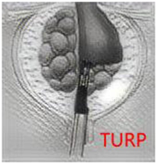
A medical illustration depicting the Transurethral Resection of the Prostate (TURP) procedure. The image shows a transverse or cross-sectional view of the prostate gland housed within its surgical capsule. The left side of the gland exhibits a nodular, bumpy texture characteristic of benign prostatic hyperplasia (BPH) with multiple distinct lobules. A metallic, cylindrical surgical instrument—a resectoscope—is positioned centrally within the prostatic urethra. The resectoscope is actively removing tissue from the right side of the gland, creating a smooth, hollowed-out surgical bed as it resects tissue from the bladder neck toward the verumontanum. The anatomical focus is the lower urinary tract, specifically the prostate gland's internal architecture during a minimally invasive urological intervention. This diagram serves as a clinical education tool to demonstrate the mechanical removal of obstructive prostatic tissue to alleviate urinary symptoms.
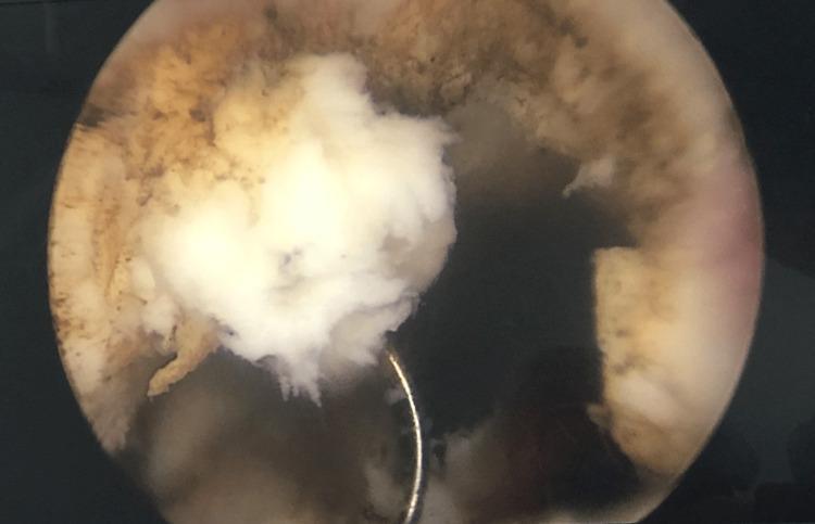
This endoscopic intraoperative image demonstrates a Transurethral Resection of the Prostate (TURP) procedure. The primary focus is a large, white, caseous-appearing mass measuring approximately 12 mm x 15 mm x 32 mm. The mass exhibits a distinct friable, 'fluffy,' and irregular texture, characteristic of tuberculous granulomatous lesions with central caseous necrosis. It is seen protruding from the prostatic urethra into the darker, open space of the bladder lumen, causing significant urinary tract obstruction. A metallic, curved surgical loop (resectoscope loop) is visible in contact with the inferior aspect of the white mass, illustrating the active resection process. The surrounding bladder neck and prostatic urethral tissue appear hyperemic with a brownish-red, rough surface texture, contrasting sharply with the pale necrotic material. This clinical photograph is used to teach the visual presentation of genitourinary tuberculosis as a cause of bladder outlet obstruction and its differentiation from typical benign prostatic hyperplasia or prostatic malignancy during endoscopy.
Management of Enlarged Prostate Gland (BPH)
- Mild (IPSS 0-7): Watchful waiting
- Moderate (IPSS 8-19): Medical therapy
- Severe (IPSS 20-35) or complications: Surgical intervention
1. Watchful Waiting (Active Surveillance)
- Regular follow-up and monitoring
- Lifestyle modifications:
- Reduce fluid intake in the evening (less nocturia)
- Avoid caffeine and alcohol
- Bladder training exercises
- Treat constipation
- Not all BPH progresses - spontaneous improvement occurs in some men
- Risk of urinary retention on watchful waiting is ~7% over 4 years
2. Medical Therapy
A. Alpha-1 Blockers (First-line, fastest relief)
| Drug | Dose | Type |
|---|---|---|
| Tamsulosin | 0.4-0.8 mg daily | α1A-selective (prostate-specific) |
| Silodosin | 8 mg daily | α1A-selective |
| Alfuzosin | 10 mg daily | α1A-selective |
| Terazosin | 5-10 mg daily | α1, long-acting |
| Doxazosin | 4-8 mg daily | α1, long-acting |
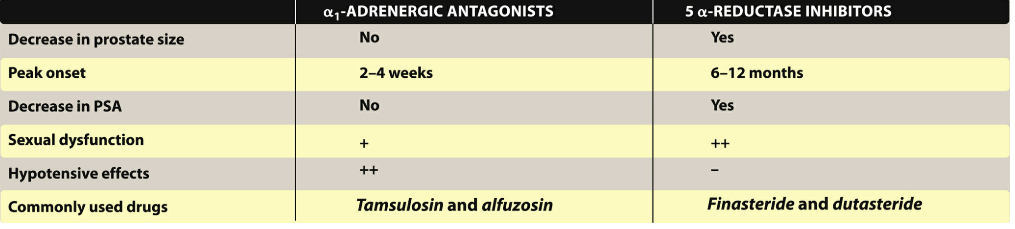
- Symptom relief starts within 2-4 weeks
- Do not reduce prostate size - only relax smooth muscle
- Side effects: orthostatic hypotension, dizziness, retrograde ejaculation, floppy iris syndrome (important before cataract surgery)
- AUA 2023 guidelines recommend any of: alfuzosin, doxazosin, silodosin, tamsulosin, or terazosin
B. 5α-Reductase Inhibitors (Shrink the prostate)
| Drug | Dose | Isoenzyme blocked |
|---|---|---|
| Finasteride | 5 mg daily | Type 2 only |
| Dutasteride | 0.5 mg daily | Type 1 and 2 (more potent) |
- Take 6-12 months to achieve maximum effect - not for quick symptom relief
- Only effective if prostate is enlarged (>40 cm³)
- Reduce PSA by ~50% (important when interpreting PSA for cancer screening)
- Prevent disease progression and reduce risk of acute urinary retention
- Side effects: decreased libido, erectile dysfunction, decreased ejaculate, gynecomastia
- ⚠️ Teratogenic - pregnant women must not handle these tablets (male fetus genital defects)
C. Combination Therapy (Alpha-blocker + 5α-RI)
- Doxazosin + Finasteride (MTOPS trial) - reduced clinical progression
- Dutasteride + Tamsulosin (CombAT study) - available as a fixed combination (Duodart)
- Rationale: alpha-blocker gives rapid symptom relief while 5α-RI shrinks the gland over months
D. Tadalafil (PDE-5 Inhibitor)
- Tadalafil 5 mg daily is the only PDE-5 inhibitor approved for BPH
- Relaxes smooth muscle in prostate and bladder via increased cGMP/nitric oxide
- Useful in men who also have erectile dysfunction (ED) - treats both conditions simultaneously
3. Surgical Management
- Urinary retention unresponsive to medical therapy
- Recurrent urinary tract infections
- Recurrent gross hematuria
- Bladder stones
- Renal insufficiency from obstruction
- Large bladder diverticula
- Failure of medical therapy
Standard Surgery
- A resectoscope is passed through the urethra and a wire loop shaves away prostatic tissue under direct vision
- Performed under regional or general anaesthesia
- Most effective surgical treatment
- Morbidity ~18%, mortality ~0.23%
- Retreatment rate ~17-18% at 5 years
Minimally Invasive Surgical Alternatives (MIST)
| Procedure | Description |
|---|---|
| HoLEP (Holmium Laser Enucleation) | Laser enucleates prostatic lobes; effective for large glands |
| GreenLight PVP (Photoselective Vaporization) | Laser vaporizes tissue; less bleeding |
| TUIP (Transurethral Incision) | Incisions in bladder neck; for small glands |
| Rezum (Water Vapor Therapy) | Steam injected into transition zone causes necrosis; day procedure; preserves ejaculation |
| UroLift (Prostatic Urethral Lift) | Implants hold lateral lobes apart; no cutting or heat; preserves sexual function |
| Robotic Waterjet (Aquablation) | Robotic water jet ablation; retreatment rate <5% at 5 years; preserves ejaculatory function better than TURP |
Open Prostatectomy
- For very large prostates (>80-100 g)
- Suprapubic or retropubic approach
- Higher morbidity but effective for massive glands
4. Management of Complications
| Complication | Management |
|---|---|
| Acute Urinary Retention | Urinary catheterization (Foley), then trial without catheter (TWOC) with alpha-blocker; TURP if fails |
| Recurrent UTI | Treat infection, then definitive surgical relief of obstruction |
| Renal failure (hydronephrosis) | Urgent catheterization, nephrology consult |
| Bladder stones | Cystolithotripsy + TURP |
Summary Algorithm
BPH Diagnosed
     |
IPSS Score
     |
 ┌───┴───┐
Mild    Moderate/Severe
(0-7)    (8-35)
  |          |
Watchful   Medical Therapy
Waiting    (Alpha-blocker ± 5α-RI)
               |
          Not improving /
          Complications
               |
          SURGICAL (TURP / MIST)

Ye medicine de hain doctor ny
Citralka syrup uses urinary alkalinizer sodium citrate potassium citrate
📋 Prescription ki Tafseel
1. 💊 Tab. Cyrocin 250mg (Ciprofloxacin)
- Antibiotic (fluoroquinolone family)
- Peshab ki nali aur masana ki infection (UTI) khatam karne ke liye
- Ciprofloxacin peshab mein bohot achi concentration haasil karta hai isliye UTI mein bohot effective hai
- Poora course khatam karein - khud se band na karein
- Khali pait na lein - khane ke saath lein
- Paani khub peeyein (rozana 8-10 gilaas)
2. 💊 Tab. Etoricox 60mg (Etoricoxib)
- COX-2 selective Anti-inflammatory / Painkiller (NSAID)
- Peshab ki nali, masane ya prostate ki sojan aur dard kam karta hai
- Burning sensation aur takleef mein fauri aaraam deta hai
- Khane ke baad lein - khali pait na lein (pait mein taklif ho sakti hai)
- Dil ke marzon, kidney problems wale log doctor ko zaroor bataayein
3. 🧴 Syp. Citralka (Disodium Hydrogen Citrate)
- Urine Alkalizer - peshab ko alkaline (less acidic) banata hai
- Jab peshab zyada acidic hoti hai to jalan hoti hai - Citralka yeh pH theek karta hai
- Kidney stones dissolve karne mein aur UTI antibiotics ka asar badhane mein madad karta hai
- Ciprofloxacin ka asar alkaline urine mein aur bhi zyada hota hai - isliye ye dono saath prescribe hue hain
- Paani mein ghol kar peeyein
- Theek aur meetha lagta hai - regular lena zaroori hai
📌 Khulasa - Kyun Yeh Combination Diya Gaya
| Dawai | Maqsad |
|---|---|
| Ciprofloxacin | Bacterial infection khatam karna |
| Etoricoxib | Dard aur sojan kam karna |
| Citralka | Peshab ki jalan kam karna + antibiotic ka asar badhana |
⚠️ Zaroori: Dawai poori miqdar mein aur poore waqt ke liye lein. Agar 3 din mein aaraam na aaye, jwar (bukhaar) aa jaaye, kamar mein dard ho ya peshab mein khoon aaye to foran doctor ko dobara dikhayein - yeh kidney infection ki alamat ho sakti hai.

Aur report ye hai
🔬 Prostate Ultrasound Report - Tafseel
📌 Prostate Gland ke Nataaij
| Measurement | Report ki Reading | Normal Hona Chahiye |
|---|---|---|
| Size | 3.1 cm × 5.3 cm × 3.8 cm | ~3 cm × 4 cm × 3 cm |
| Volume | 33 ml | 20-30 ml |
| Weight | 34 grams | 20 grams tak (upper limit) |
| Capsule | Intact (salaamat) | Intact honi chahiye |
| Hypoechoic area | Nahi mili | Nahi honi chahiye (cancer ka sign hota) |
| Calcification | Nahi | -- |
✅ Matlab: Prostate badi ho gayi hai - weight normal ki had se 14 gram zyada hai. Lekin capsule salaamat hai aur cancer ka koi nishaan nahi - yeh BPH (Benign Prostatic Hyperplasia) hai yani ek non-cancerous enlargement.
📌 Urinary Bladder (Masana) ke Nataaij
| Finding | Report | Matlab |
|---|---|---|
| Bladder wall | Thodi moti (slightly thick) | Sojan ki wajah se |
| Mass ya pathri | Nahi | Good - koi serious masla nahi |
| Pre-micturation volume | 507 ml | Peshab karne se pehle masane mein itna pani tha |
| Post-micturation volume | 73 ml (residual urine) | Peshab karne ke baad bhi 73ml reh gayi |
⚠️ Residual Urine 73ml: Yeh thoda zyada hai. Normal mein peshab ke baad masana bilkul khali ho jaata hai (< 50ml). Iska matlab prostate ki wajah se peshab poori tarah nahi nikal rahi - yeh bladder outlet obstruction ki alamat hai.
⚠️ Thick Bladder Wall: Yeh Mild Cystitis (masane ki sojan/infection) ki taraf ishara karta hai - isliye doctor ne antibiotic (Ciprofloxacin) di hai.
📌 Incidental Findings (Ittifaqan Mile Masail)
Bilateral early reducible indirect inguinal hernia
- Dono taraf (right aur left) inguinal hernia hai
- Right side: 7.4mm, Left side: 8.7mm
- "Early reducible" matlab: abhi choti hai aur wapas ja sakti hai - fouri surgery ki zaroorat nahi lekin nazar rakhen
- Hum ne pehle baat ki thi hernia ke baare mein - yeh wahi hai!
Penile shaft soft tissue edema
- Ling ke hisse mein thodi sojan hai
- Yeh UTI/infection ya local inflammation ki wajah se ho sakta hai
🏁 Doctor ki Final Conclusion (Khulasa)
| # | Finding | Matlab |
|---|---|---|
| 1 | Prostate enlarged 34g (normal 20g) | BPH - Benign Prostatic Hyperplasia |
| 2 | Bladder residual urine 73ml | Prostate ki wajah se peshab poori nahi nikal rahi |
| 3 | Thick bladder wall | Mild Cystitis (masane ki infection/sojan) |
| 4 | Bilateral inguinal hernia | Ittifaqan mili - future mein surgical consultation |
💊 Doctor ki Dawaiyon se Talluq
| Dawai | Kyun Di |
|---|---|
| Ciprofloxacin | Cystitis (masane ki infection) khatam karne ke liye |
| Etoricoxib | Dard aur sojan (masana + prostate) kam karne ke liye |
| Citralka | Peshab ki jalan kam karo + urine alkaline karo |
🔔 Aham Baat: Yeh dawaiyen sirf UTI aur infection ka ilaj karengi. Prostate ki BPH ki wajah se jo peshab poori nahi nikal rahi - us ke liye doctor agle visit mein shayad Alpha-blocker (Tamsulosin) ya koi aur BPH ki dawai bhi shuru karen. Ashraf sahab ko follow-up zaroor karwaani chahiye.
Tamsulosin bh chl rahi hai
💊 Tamsulosin - Poori Maloomat
Yeh Kya Hai?
Kaam Kaise Karta Hai? (Mechanism)
Lippincott Pharmacology ke mutabiq: "Tamsulosin and silodosin are more selective for the α1A receptor found in the prostate - they have less effect on blood pressure because they target prostate-specific receptors."
Ashraf Sahab ke Case mein Kya Faida Hoga?
| Masla | Tamsulosin ka Faida |
|---|---|
| Peshab poori tarah nahi nikal raha | Prostate relax → peshab poora nikal jayega |
| Residual urine 73ml | Kam hogi - target < 50ml |
| Peshab ki dhaar kamzor | Flow rate behtar hogi |
| Baar baar peshab ki hajat | Urgency aur frequency kam hogi |
| Raat ko uthna (Nocturia) | Behtar neend |
Khaane ka Tarika
- 0.4 mg - roz ek baar (sham ko khaane ke baad - subah nahi)
- Raat ko sone se pehle lena best hota hai - blood pressure girne ka khatra kam hota hai
- Khane ke baad lein - khali pait na lein
Side Effects (Mumkina Pareshaniyan)
| Side Effect | Kitna Common | Kya Karein |
|---|---|---|
| Chakkar aana (dizziness) | Thoda | Dhire se uthein, seedha na kharen |
| Blood pressure thoda gire | Kam (kyunki α1A selective hai) | Pareshani nahi |
| Retrograde ejaculation | Ho sakta hai | Mani peeche masane mein jaati hai - nuqsandeh nahi |
| Naak band rehna | Kabhi kabhi | -- |
| Floppy Iris Syndrome | Aankh ki surgery mein | ⚠️ Agar cataract ki surgery honi ho to doctor ko zaroor batayein ke Tamsulosin chal rahi hai |
Ab Ashraf Sahab Ki Poori Dawai List
| Dawai | Kaam | Timing |
|---|---|---|
| Ciprofloxacin 250mg | UTI/Cystitis infection khatam kare | Subah + Raat (khaane ke baad) |
| Etoricoxib 60mg | Dard + sojan kam kare | Subah ek baar (khaane ke baad) |
| Citralka Syrup | Peshab ki jalan kam kare | Subah + Raat (paani mein mila kar) |
| Tamsulosin 0.4mg | Prostate relax kare, peshab aasaan kare | Raat ko sone se pehle (khaane ke baad) |
⏱️ Kab Asar Hoga?
- Tamsulosin ka asar 2-4 haftoon mein nazar aata hai
- Peshab ki dhaar behtar hogi aur residual urine kam hogi
- Prostate size nahi ghate gi - iske liye alag dawai (Finasteride/Dutasteride) hoti hai - doctor zaroorat hogi to baad mein add kar sakte hain
✅ Khulasa: Doctor ne bilkul sahi treatment di hai - infection ke liye antibiotic, dard ke liye pain killer, jalan ke liye Citralka, aur BPH ke liye Tamsulosin. Yeh ek complete package hai. Tamsulosin lifelong bhi chal sakti hai - apni marzi se band na karein.