
Long case viva questions on peripheral arterial diseases
Reading File
Now let me search the medical library for peripheral arterial disease content in parallel.
Finding Sources
Finding Sources
Good. Now let me read the main PAD sections from the most comprehensive sources in parallel.
Reading File
Finding Sources
Finding Sources
The first file returned references, not the main content. Let me read the actual PAD content from Textbook of Family Medicine and Bailey & Love in parallel.
Reading File
Reading File
Finding Sources
Excellent content found. Let me read more from the Family Medicine textbook and Bailey & Love for the clinical details, diagnosis, and management sections.
Reading File
Reading File
Great content. Let me read more of both texts for management, investigation, and treatment details.
Reading File
Reading File
Excellent content. Let me get the treatment/management and revascularization sections now.
Reading File
Reading File
I now have very rich material. Let me get the management/treatment section from Bailey & Love and also the acute limb ischemia section to round out the viva questions.
Reading File
Finding Sources
I have enough rich content to now compile a comprehensive set of long case viva questions on peripheral arterial disease. Let me write the detailed answer.
Long Case Viva Questions on Peripheral Arterial Disease (PAD)
Below is a structured set of viva questions organized the way a long case presentation unfolds - from history to examination to investigations to management. Expected answer points are included for each question.
SECTION 1: Introduction and Epidemiology
Q1. What is peripheral arterial disease? How prevalent is it and why is it clinically significant beyond the leg?
Expected answer:
- PAD is atherosclerotic occlusive disease of the peripheral (predominantly lower limb) arteries, causing a spectrum from asymptomatic stenosis to limb-threatening ischemia.
- It is age-dependent, underdiagnosed, and undertreated. The PARTNERS program found PAD in 29% of primary care patients aged >70 or aged 50-69 with smoking/diabetes history.
- More than 50% of patients with PAD are entirely asymptomatic.
- Clinically significant because it is a powerful independent predictor of mortality - PAD patients have a ~4-fold higher risk of MI and 2-3x higher risk of stroke vs. non-PAD patients. Approximately 40% of patients with atherosclerotic vascular disease have disease in more than one vascular bed.
- Patients with critical limb ischemia (CLI) have an annual mortality rate of ~25%.
(Textbook of Family Medicine 9e)
Q2. What are the risk factors for PAD?
Expected answer:
- Modifiable: Cigarette smoking (single most important), diabetes mellitus, hypertension, hyperlipidaemia, obesity, sedentary lifestyle.
- Non-modifiable: Age (>50 years), male sex, family history, ethnicity (higher in Black populations).
- Emerging: Hyperhomocysteinaemia, elevated lipoprotein(a), chronic kidney disease (CKD), inflammatory markers (CRP).
- Smoking is particularly important - it roughly doubles the risk of PAD and is directly associated with claudication severity and amputation risk.
SECTION 2: History Taking
Q3. How would you take a history from a patient presenting with possible PAD?
Expected answer:
- Claudication history:
- Onset, site, character of pain (cramping/aching in muscle groups)
- Claudication distance (how far before pain starts) - note this varies little day to day
- Relieved by standing still within 5 minutes (differentiates from nerve compression, which requires sitting)
- Pain not present on first step (unlike osteoarthritis)
- Worsening with uphill walking, increased speed, carrying weights
- Rest pain:
- Pain in foot/calf at rest, worse at night, worse with leg elevation
- Patient hangs foot out of bed or sleeps in chair for relief (gravity effect)
- Indicates more severe ischemia than claudication
- Skin changes: Non-healing ulcers, blackened tissue (gangrene)
- Cardiovascular review: Ask about chest pain, dyspnoea, palpitations, TIA/stroke, renal impairment (co-existing atherosclerosis)
- Risk factor assessment: Smoking history (pack-years), diabetes management, hypertension and medications, lipid profile, family history
- Functional impact: How has claudication affected occupation, social, and leisure activities?
- Erectile dysfunction - may indicate aortoiliac disease (Leriche syndrome)
(Bailey and Love's Short Practice of Surgery 28e; Textbook of Family Medicine 9e)
Q4. What is Leriche syndrome? What is its triad?
Expected answer:
- Leriche syndrome results from aortoiliac occlusion.
- Classic triad: (1) Buttock and thigh claudication; (2) absence of femoral pulses bilaterally; (3) erectile dysfunction (sexual impotence) in men due to internal iliac artery insufficiency.
- The level of claudication is one anatomical level below the arterial obstruction - aortoiliac disease causes thigh and buttock claudication.
(Bailey and Love's Short Practice of Surgery 28e)
Q5. How do you differentiate claudication from other causes of leg pain on history?
Expected answer:
| Feature | Arterial Claudication | Neurogenic Claudication (spinal stenosis) | Osteoarthritis |
|---|---|---|---|
| Onset | After fixed walking distance | Variable | On movement |
| Relief | Standing still (5 min) | Sitting/leaning forward | Rest (prolonged) |
| Position | Any resting position | Must sit | Any |
| First step | Pain-free | Often pain-free | Painful |
| Uphill | Worse | Better (spinal flexion) | Variable |
| Pulses | Absent/reduced | Normal | Normal |
SECTION 3: Clinical Examination
Q6. How would you examine a patient with suspected PAD?
Expected answer:
- General inspection: Pallor, cyanosis, tar-stained fingers (smoking), xanthelasma, corneal arcus
- Inspect the limbs: Skin colour (pallor, cyanosis, dependent rubor "sunset foot"), hair loss, muscle wasting, trophic skin changes, ulcers, gangrene
- Buerger's test:
- Elevate both legs to 45 degrees for 1-2 minutes - ischaemic limb goes pale (positive)
- Then hang legs down - ischaemic limb flushes deep red-purple (reactive hyperaemia/dependent rubor = "sunset foot" sign)
- Buerger's angle = the angle at which the limb becomes pale; <20 degrees = severe ischemia
- Capillary refill time: Press heel/toe pulp for 5 seconds; normal 2-3 seconds; prolonged >10 sec in severe ischemia
- Pulses: Systematically palpate femoral, popliteal, posterior tibial, dorsalis pedis
- Compare bilateral femoral pulses
- Note: pulse distal to an arterial occlusion is usually absent; collaterals may occasionally allow palpable distal pulses even with stenosis
- Exercise (walking to claudication) will cause a distal pulse to disappear as vasodilation occurs below the obstruction
- Auscultation: Bruit over aorta, iliac, femoral arteries (indicates turbulent flow, suggests stenosis). Note: tight stenoses/complete occlusions may be silent.
- Abdomen: Palpate for aortic aneurysm (may coexist with lower limb occlusive disease)
- Venous guttering: A chronically ischaemic leg elevated will show collapsing, guttered veins
(Bailey and Love's Short Practice of Surgery 28e)
Q7. What does the site of absent pulses tell you about the level of disease?
Expected answer: (Mirrors Table 61.1 from Bailey & Love)
| Site of obstruction | Clinical findings |
|---|---|
| Aortoiliac | Claudication in buttocks, thighs, calves. Both femoral and distal pulses absent. Aortoiliac bruit. Impotence (Leriche). |
| Iliac (unilateral) | Unilateral thigh/calf (sometimes buttock) claudication. Unilateral absent femoral and distal pulses. Iliac bruit. |
| Femoropopliteal (SFA - commonest, 70%) | Unilateral calf claudication. Femoral pulse present, popliteal and distal pulses absent. |
| Distal (tibial/peroneal) | Femoral and popliteal pulses present. Ankle pulses absent. Claudication in calf and foot. |
SECTION 4: Investigations
Q8. What is the ankle-brachial index (ABI)? How is it measured and how do you interpret it?
Expected answer:
- ABI = highest ankle systolic pressure (dorsalis pedis, posterior tibial, or peroneal) / highest ipsilateral brachial systolic pressure.
- Measured using a hand-held Doppler probe and sphygmomanometer.
- Interpretation:
- 0.9-1.4: Normal
- <0.9: Hemodynamically significant arterial lesion (PAD diagnosis)
- 0.5-0.9: Claudication range
- <0.4: Critical limb-threatening ischemia (CLTI)
-
1.4: Arterial calcification/incompressibility (falsely elevated - seen in diabetes, chronic renal failure)
- A drop of >20% from resting ABI after exercise is indicative of flow-limiting arterial disease (exercise ABI is useful when resting ABI is normal but symptoms suggest PAD).
- Pitfall: In diabetics with medial calcification, vessels are non-compressible. Use toe-brachial index (TBI) instead - toe (digital) arteries are rarely affected by calcification. TBI <0.6 suggests significant arterial disease.
(Bailey and Love's Short Practice of Surgery 28e; Textbook of Family Medicine 9e)
Q9. What investigations would you order for a patient with PAD? Justify each.
Expected answer:
Bedside/Basic:
- ABI with hand-held Doppler (confirms diagnosis, grades severity)
- Blood glucose, HbA1c (diabetes screening/monitoring)
- Full blood count (anaemia worsens ischemia by reducing O2 delivery)
- Lipid profile (identify and quantify dyslipidaemia)
- Renal function (coexisting renovascular disease; relevant for contrast use)
- ECG (coexisting coronary artery disease is present in ~50%)
Vascular Imaging (when intervention is planned):
- Duplex Doppler Ultrasonography (DUS): First-line non-invasive imaging. Localises stenosis, determines degree using peak systolic velocity ratio (PSVR). PSVR ≥2.4 = obstructive disease; PSVR ≥3.0 = functionally significant. Useful for graft/stent surveillance.
- CT Angiography (CTA): Best anatomical detail, 3D reconstruction, invaluable for pre-operative planning. Limitation: iodinated contrast (risk of nephropathy, especially in diabetics), ionising radiation. Metformin must be withheld periprocedurally.
- MR Angiography (MRA): No ionising radiation, no iodinated contrast. Uses gadolinium (caution in severe CKD - nephrogenic systemic fibrosis). Good for aortoiliac and femoropopliteal segments.
- Digital Subtraction Angiography (DSA): Gold standard for arterial anatomy. Access via Seldinger technique (usually common femoral artery). Provides dynamic flow information and can be combined with endovascular treatment. Complications: haematoma, false aneurysm, thrombosis, arterial dissection, distal embolisation, renal dysfunction, allergic reaction (~5%). Reserved for when intervention is planned.
(Bailey and Love's Short Practice of Surgery 28e; Textbook of Family Medicine 9e)
Q10. What is the Seldinger technique?
Expected answer:
- A technique for gaining percutaneous arterial access.
- Steps: (1) Needle puncture of the artery (usually CFA); (2) guidewire inserted through needle; (3) needle withdrawn over wire; (4) sheath/catheter threaded over wire; (5) wire removed.
- Allows introduction of catheters, sheaths, and interventional devices without surgical cut-down.
SECTION 5: Classification
Q11. Describe the Fontaine classification of PAD.
Expected answer:
| Stage | Clinical Features |
|---|---|
| I | Asymptomatic |
| IIa | Mild claudication (claudication distance >200 m) |
| IIb | Moderate-severe claudication (claudication distance <200 m) |
| III | Ischaemic rest pain |
| IV | Ulceration or gangrene |
Q12. What is the Rutherford classification of PAD?
Expected answer:
| Grade | Category | Description |
|---|---|---|
| 0 | 0 | Asymptomatic |
| I | 1 | Mild claudication |
| I | 2 | Moderate claudication |
| I | 3 | Severe claudication |
| II | 4 | Ischaemic rest pain |
| III | 5 | Minor tissue loss (non-healing ulcer) |
| III | 6 | Major tissue loss (frank gangrene) |
Categories 4, 5, 6 = critical limb-threatening ischemia (CLTI).
Q13. What is meant by chronic limb-threatening ischaemia (CLTI)?
Expected answer:
- CLTI (previously called critical limb ischemia/CLI) refers to patients with ischaemic rest pain and/or ischaemic ulceration/gangrene (tissue loss).
- Defines the highest-risk end of the PAD spectrum with an imminently threatened limb.
- Requires urgent vascular assessment and revascularisation to prevent major amputation.
- These patients have the lowest ABIs (<0.4) and the highest annual mortality (~25%).
- Bilateral co-morbidities (coronary, cerebral, renal) are almost universally present.
(Bailey and Love's Short Practice of Surgery 28e; Textbook of Family Medicine 9e)
SECTION 6: Management
Q14. Outline the management of a patient with intermittent claudication.
Expected answer:
Conservative/Medical (most claudicants improve or stabilise with this):
- Smoking cessation - single most effective intervention; dramatically slows progression
- Supervised exercise rehabilitation - structured walking programme (30-45 min, 3x/week); increases pain-free walking distance by improving collateral flow and muscle metabolism
- Antiplatelet therapy - aspirin 75-100 mg/day or clopidogrel 75 mg/day (reduces cardiovascular events, not claudication per se)
- Statins - reduce cardiovascular events; some improvement in walking distance
- Antihypertensives - ACE inhibitors preferred (also shown to reduce cardiovascular events in HOPE trial)
- Diabetes control - tight glycaemic control
- Cilostazol (PDE-3 inhibitor) - only drug with evidence of improving claudication: increases maximal walking distance by 54% vs placebo. Contraindicated in heart failure. Approved by FDA for claudication.
- Pentoxifylline - no randomised data showing benefit over placebo; not recommended.
- Foot care - especially in diabetics to prevent ulceration
Endovascular/Surgical (for disabling claudication or CLTI):
- Indications: lifestyle-limiting claudication not responding to 3-6 months of conservative treatment, rest pain, tissue loss
- Angioplasty ± stenting (PTA): First-line for short segment lesions (TASC A/B); shorter recovery, less morbidity. Risk of restenosis from neointimal hyperplasia.
- Surgical bypass: For long-segment occlusions (TASC C/D), failed endovascular procedures; e.g., aortobifemoral bypass (aortoiliac), femoro-popliteal bypass.
- Graft material: Autologous vein (long saphenous - best patency) vs. synthetic (PTFE or Dacron - acceptable above knee, poor below knee).
(Textbook of Family Medicine 9e; Bailey and Love's Short Practice of Surgery 28e)
Q15. What is cilostazol and how does it work? When is it contraindicated?
Expected answer:
- Cilostazol is a phosphodiesterase type III (PDE-3) inhibitor. It inhibits platelet aggregation and causes arterial vasodilation by preventing breakdown of cAMP.
- Approved by the FDA for treatment of intermittent claudication (1999).
- In randomised trials comparing cilostazol 100 mg twice daily vs. pentoxifylline vs. placebo, cilostazol improved maximum walking distance by 54% vs. 30% for pentoxifylline and 34% for placebo.
- Contraindication: Heart failure of any severity (increased mortality in trials with PDE inhibitors in heart failure).
- Dose: 100 mg twice daily.
(Textbook of Family Medicine 9e)
Q16. What are the indications for revascularisation in PAD?
Expected answer:
- Lifestyle-disabling intermittent claudication not improved after 3-6 months of maximal conservative therapy
- Ischaemic rest pain (Fontaine III / Rutherford 4)
- Tissue loss - non-healing ulceration (Fontaine IV / Rutherford 5)
- Gangrene with limb salvage potential (Rutherford 6)
- In CLTI, revascularisation should be performed urgently
Q17. What is the TASC classification? How does it guide treatment?
Expected answer:
- TASC (Trans-Atlantic Inter-Society Consensus) classifies lesions by morphology to guide revascularisation strategy:
- TASC A: Short single stenoses; recommended endovascular (PTA ± stenting)
- TASC B: Multiple short stenoses or single longer stenosis; endovascular preferred
- TASC C: Multiple long stenoses or single occlusion; surgery preferred
- TASC D: Complete occlusions, long segment or aortoiliac involvement; surgery recommended
- Endovascular has lower short-term morbidity; surgery has better long-term patency for complex lesions.
SECTION 7: Acute Limb Ischaemia (ALI) - Viva Extension
Q18. What are the 6 Ps of acute limb ischemia?
Expected answer:
- Pain - sudden severe onset
- Pallor - white/pale limb
- Pulselessness - absent distal pulses
- Paraesthesia - pins and needles (early nerve ischaemia)
- Paralysis - motor deficit (late/severe; urgent sign)
- Perishing cold (Poikilothermia)
Paraesthesia and paralysis indicate irreversible ischemia is approaching and demand emergency surgical intervention.
Q19. How do you differentiate thrombosis from embolism as causes of acute limb ischemia?
Expected answer:
| Feature | Embolism | Thrombosis in-situ |
|---|---|---|
| Onset | Sudden, abrupt | Gradual, preceding claudication |
| Contralateral limb | Normal pulses | Absent/reduced (background PAD) |
| AF/cardiac disease | Often present | Less common |
| Collaterals | Absent (no pre-existing PAD) | Present (compensated) |
| Severity | More severe (no collaterals) | Less severe (collaterals) |
| Treatment | Embolectomy | Thrombolysis ± bypass |
Q20. What is the management of acute limb ischemia?
Expected answer:
- Immediate: IV heparin to prevent propagation; analgesia; urgent vascular surgical referral
- Imaging: Emergency DSA or duplex to determine level and cause
- Surgical embolectomy (Fogarty balloon catheter) - for embolic occlusion, viable limb
- Catheter-directed thrombolysis - for thrombotic occlusion or if surgical risk is high; slower, requires ICU monitoring, risk of haemorrhage
- Surgical bypass - for thrombotic occlusion with anatomical disease
- Amputation - if limb is non-viable (irreversible ischemia with fixed mottling, rigor, paralysis)
- Post-procedure: Investigate and treat the source (echocardiogram if AF/cardiac embolism; anticoagulation)
SECTION 8: Complications and Prognosis
Q21. What are the long-term complications of PAD?
Expected answer:
- Progression to rest pain and CLTI
- Amputation (major or minor) - <5% of claudicants reach amputation; much higher in CLTI
- Myocardial infarction (fourfold risk vs. non-PAD)
- Stroke (2-3x risk)
- Cardiovascular death (leading cause of death in PAD patients)
- Reduced quality of life and functional decline
Q22. What is the natural history of intermittent claudication?
Expected answer:
- Apparent "improvement" in IC over time does NOT reflect disease improvement - it often reflects functional decline (patients walk slower/shorter distances to avoid pain).
- PAD is a progressive disorder causing significant decline in quality of life.
- ~25% of claudicants deteriorate, 25% improve (often due to functional decline), 50% stable.
- Fewer than 5% will ever require amputation.
- The dominant risk is cardiovascular mortality - not limb loss.
- The lower the ABI, the greater the cardiovascular event risk.
(Textbook of Family Medicine 9e)
SECTION 9: Special Situations
Q23. How does diabetes modify PAD presentation and management?
Expected answer:
- PAD in diabetics is more distal (tibial/peroneal vessels affected preferentially), more diffuse, and often bilateral.
- Neuropathy may mask ischemic pain - patient may present with painless ulceration or gangrene without preceding claudication.
- ABI is unreliable due to medial calcification; use TBI <0.6 as threshold.
- Foot ulcers in diabetics are often of mixed ischaemic and neuropathic origin.
- Worse prognosis with higher amputation rates.
- Tight glycaemic control, meticulous foot care, podiatry referral are essential.
- Metformin must be withheld before iodinated contrast studies (CTA/DSA) due to risk of lactic acidosis.
Q24. What is the ABCD (or secondary prevention) strategy in PAD?
Expected answer:
- A - Antiplatelet agents (aspirin or clopidogrel) + ACE inhibitors
- B - Blood pressure control (target <130/80)
- C - Cholesterol (statins - target LDL <1.8 mmol/L) + Cigarette cessation
- D - Diabetes control (HbA1c target ~7%) + Diet and exercise
These reduce cardiovascular events and mortality - the primary threat in PAD - rather than just improving claudication.
KEY IMAGES TO KNOW
The following visuals are commonly discussed in viva settings:
- Aortoiliac bifurcation disease angiogram with collateralisation via lumbar and inferior mesenteric arteries
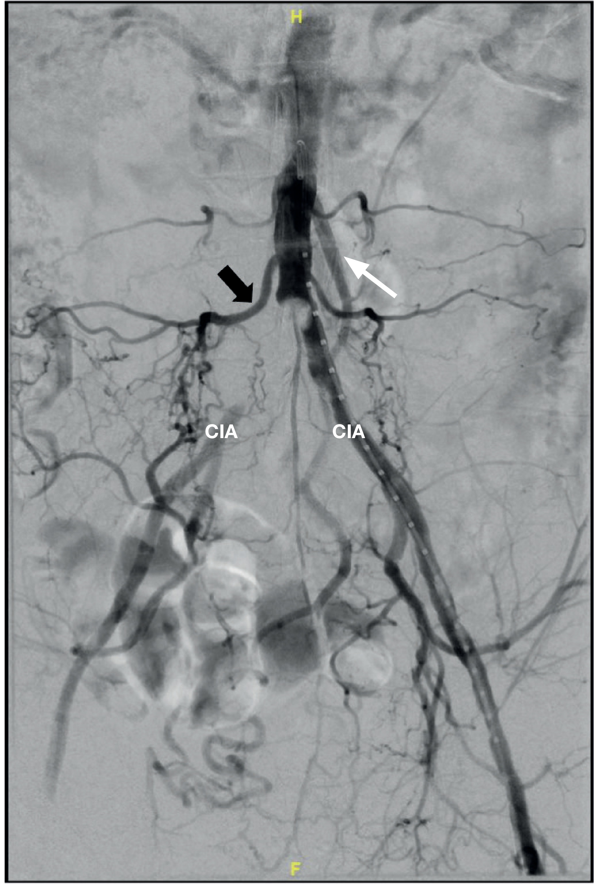
- Dependent rubor / "sunset foot" - elevation pallor, dependency rubor
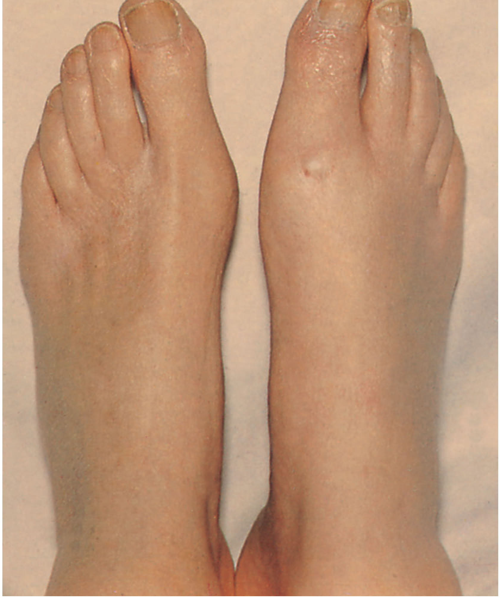
- Dry gangrene of the toes in CLTI
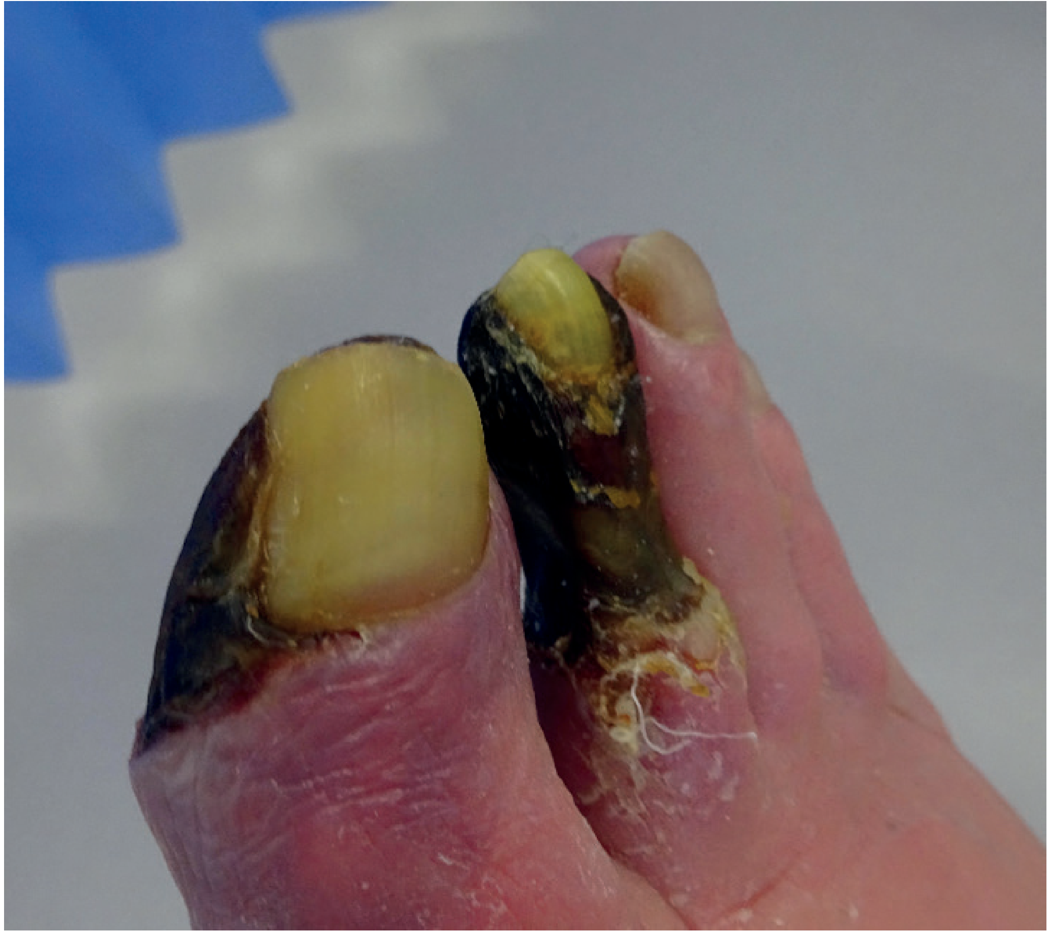
QUICK REFERENCE SUMMARY TABLE
| Topic | Key Point |
|---|---|
| Commonest artery affected | Superficial femoral artery (70% of claudication cases) |
| Claudication relief | Standing still ≤5 minutes |
| ABI normal range | 0.9-1.4 |
| ABI in claudication | 0.5-0.9 |
| ABI in CLTI | <0.4 |
| ABI falsely elevated | Diabetes/CKD (calcification) - use toe-brachial index |
| Drug for claudication | Cilostazol (PDE-3 inhibitor) - contraindicated in HF |
| Gold standard imaging | DSA |
| Most common cause of death in PAD | Cardiovascular (MI/stroke) |
| Leriche syndrome | Aortoiliac occlusion: buttock claudication + absent femoral pulses + impotence |
| CLTI annual mortality | ~25% |
Sources: Bailey and Love's Short Practice of Surgery 28th Edition; Textbook of Family Medicine 9e; Bailey and Love's Short Practice of Surgery Ch. 61.
Make a detailed Pg viva questions on pad
Reading File
Finding Sources
Finding Sources
Finding Sources
Reading File
Finding Sources
Finding Sources
Reading File
Finding Sources
Finding Sources
Finding Sources
Reading File
Reading File
Reading File
Finding Sources
Reading File
Reading File
Now I have all the content needed. Let me compile the comprehensive PG-level viva questions.
PG Viva Questions on Peripheral Arterial Disease
Compiled from Sabiston Textbook of Surgery, Schwartz's Principles of Surgery, Bailey & Love, Textbook of Family Medicine, Goldman-Cecil Medicine
PART 1: BASIC SCIENCE & PATHOPHYSIOLOGY
Q1. Describe the pathophysiology of atherosclerosis as it relates to PAD.
Answer:
- Atherosclerosis is the underlying cause of PAD in >90% of cases. It begins in childhood as fatty streaks and progresses through:
- Endothelial injury - by smoking, hypertension, hyperlipidaemia, hyperglycaemia, shear stress
- Lipid deposition - LDL enters the subintima and undergoes oxidation
- Inflammatory recruitment - monocytes migrate into the intima, differentiate into macrophages, engulf oxidized LDL to become foam cells
- Foam cell accumulation forms the fatty streak
- Smooth muscle cell migration from media to intima; SMCs produce extracellular matrix forming a fibrous cap over the lipid core - the atheromatous plaque
- Plaque progression - calcification, neovascularisation, enlargement causing lumen stenosis
- Plaque rupture/erosion - triggers acute thrombosis, can cause acute limb ischemia
- In the lower limb, atherosclerosis preferentially affects:
- Aortoiliac segment
- Superficial femoral artery at the adductor canal (most common - 70%)
- Tibial/peroneal arteries (especially in diabetics)
- The muscle group affected by claudication is characteristically one level below the arterial occlusion (because it is the most distal vascular territory that becomes ischemic under exercise demand).
Q2. What is the haemodynamic basis of intermittent claudication?
Answer:
- At rest, blood flow through a stenosed vessel may be adequate because vascular resistance in the distal capillary bed is high.
- With exercise, the metabolic demands of muscle increase massively. The capillary bed vasodilates, reducing peripheral resistance. In a normal limb, this drives increased flow.
- Across a stenosis, the pressure gradient rises as flow increases (Poiseuille's Law: resistance ∝ length/radius⁴). Flow cannot keep pace with metabolic demand.
- Anaerobic metabolism occurs in the exercising muscle → lactic acid and other metabolites accumulate → nociceptive pain stimulus → claudication.
- On resting, metabolic demands fall, lactate clears within 2-5 minutes, and pain resolves. This is the basis of the classic "stop and rest" relief.
- In rest pain (critical ischemia), perfusion pressure is inadequate even without exercise. It is exacerbated by elevation (removes gravity-assisted perfusion pressure) and relieved by dependency (gravity augments pressure in the distal arteries).
Q3. What is the significance of collateral circulation in PAD?
Answer:
- Collateral vessels are pre-existing arterioles and small arteries that expand (arteriogenesis) in response to a chronic pressure gradient across a stenosis.
- Driven by increased shear stress, growth factors (VEGF, FGF, PDGF), and monocyte infiltration.
- Collaterals develop slowly - explain why chronic occlusion is better tolerated than acute occlusion.
- Important examples:
- Inferior mesenteric artery (IMA) and lumbar arteries collateralise around aortoiliac disease
- Profunda femoris (deep femoral artery) is the most important collateral route around a superficial femoral artery (SFA) occlusion - hence it is called the "artery of the thigh"
- Geniculate arteries collateralise around a popliteal occlusion
- Clinically: a patent, non-diseased profunda is the key vessel to preserve in infrainguinal surgery - inflow to the profunda should always be assessed.
Q4. What is the role of the profunda femoris artery in lower limb ischaemia?
Answer:
- The profunda femoris artery (PFA/deep femoral artery) is the major collateral supply to the thigh and lower leg when the SFA is occluded.
- Its branches (perforators) communicate with the geniculate arteries around the knee and tibial vessels distally.
- In SFA occlusion, the limb survival often depends on the profunda - a patient with SFA occlusion but a healthy profunda may have only mild claudication.
- Profundaplasty - endarterectomy and patch widening of the profunda ostium - can improve perfusion to the limb when the SFA is occluded, especially in patients unfit for more complex bypass surgery.
- In above-knee amputation, the profunda-geniculate collaterals supply the skin flap; this is why amputation should be above the knee (not through the distal thigh where these collaterals are absent) when the profunda is compromised.
PART 2: CLINICAL ASSESSMENT
Q5. How do you clinically grade the severity of lower limb ischemia? Compare Fontaine and Rutherford classifications.
Answer:
| FONTAINE | Clinical | RUTHERFORD Grade | Category | Clinical |
|---|---|---|---|---|
| Stage I | Asymptomatic | 0 | 0 | Asymptomatic |
| Stage IIa | Mild claudication (>200 m) | I | 1 | Mild claudication |
| Stage IIb | Moderate-severe claudication (<200 m) | I | 2 | Moderate claudication |
| I | 3 | Severe claudication | ||
| Stage III | Ischaemic rest pain | II | 4 | Ischaemic rest pain |
| Stage IV | Ulceration / gangrene | III | 5 | Minor tissue loss |
| III | 6 | Major tissue loss |
- Fontaine is simpler and widely used in Europe; Rutherford is more detailed and used in research.
- Rutherford Grade III, Categories 4-6 = Chronic Limb-Threatening Ischaemia (CLTI). (Schwartz's Principles of Surgery 11e; Sabiston Textbook of Surgery)
Q6. What is Buerger's test and what does a positive test tell you?
Answer:
- Method: The patient lies supine. Both legs are elevated to 45° for 1-2 minutes. Then the patient sits up and hangs both legs down.
- Positive test (ischaemic limb):
- On elevation: the foot becomes pale/white (blood drains away and cannot be replenished against gravity due to low perfusion pressure)
- On dependency: the foot flushes deep red/purple (reactive hyperaemia = dependent rubor / "sunset foot" sign) - ischaemic vessels maximally dilate and fill slowly with blood on dependency
- Buerger's angle: The angle of leg elevation at which the foot goes pale. Normal = >90°. Significant ischaemia = <20°.
- A positive Buerger's test strongly suggests critical limb ischemia (CLTI).
Q7. What is Leriche syndrome? What is its pathological basis, clinical triad, and treatment?
Answer:
- Pathology: Atherosclerotic occlusion of the abdominal aorta at or just above the bifurcation into the iliac arteries, or bilateral common iliac artery stenosis/occlusion.
- Clinical Triad:
- Bilateral buttock and thigh claudication (sometimes calf)
- Absent or markedly diminished bilateral femoral pulses
- Erectile dysfunction/impotence in men (due to ischaemia of the internal iliac arteries supplying the corpora cavernosa)
- Other features: Atrophy of lower limb musculature, pallor of both legs, absent distal pulses bilaterally, aortoiliac bruit on auscultation
- Treatment:
- Endovascular: aortoiliac angioplasty ± kissing stents (bilateral iliac stenting)
- Surgery: Aortobifemoral bypass (gold standard for extensive AIOD) using Dacron/PTFE bifurcated graft. 5-year patency >85%.
- Extraanatomic (high-risk patients): axillofemoral bypass or femoro-femoral crossover bypass.
Q8. Describe the complete examination of a patient presenting with lower limb ischaemia.
Answer:
General:
- Signs of hyperlipidaemia (xanthelasma, xanthomata, corneal arcus)
- Smoking history (tar staining)
- BMI, blood pressure, signs of diabetes (acanthosis nigricans)
Inspection (affected limb):
- Colour: pallor, cyanosis, dependent rubor ("sunset foot")
- Trophic changes: loss of hair, shiny atrophic skin, muscle wasting
- Ulcers: site (tips of toes/pressure areas/trauma points = arterial; gaiter area = venous), depth, slough/necrosis, surrounding skin
- Gangrene: dry (mummified, demarcated - arterial) vs wet (infected, malodorous)
- Venous guttering (visible collapse of veins on leg elevation in severe ischaemia)
Palpation:
- Temperature: compare both limbs - ischaemic limb is cooler
- Capillary refill: press heel/toe pulp for 5 seconds; normal <3 sec; prolonged >10 sec = severe
- Pulses: Radial (general cardiovascular), carotid, abdominal aorta, femoral, popliteal, posterior tibial, dorsalis pedis (compare sides)
- Popliteal pulse - must be examined bimanually with knee slightly flexed; if aneurysmal pulsation - suspect popliteal aneurysm
Auscultation:
- Bruits over: aorta, renal, iliac, femoral arteries
- Continuous "machinery" murmur = arteriovenous fistula
Neurological:
- Sensation (ischaemic neuropathy) - assess in digits and forefoot
- Motor power (paralysis = irreversible ischaemia in acute setting)
Cardiovascular system:
- Cardiac rhythm (AF = embolic source), murmurs, JVP, evidence of heart failure
PART 3: INVESTIGATIONS
Q9. Describe in detail how the Ankle-Brachial Index (ABI) is measured and interpreted. What are its limitations?
Answer:
Measurement:
- Patient rests supine for 10 minutes.
- Apply BP cuff just above the ankle.
- Apply Doppler probe over dorsalis pedis then posterior tibial artery.
- Inflate cuff to suprasystolic pressure, then deflate slowly until Doppler signal returns = ankle systolic pressure.
- Record both ankle pressures and both brachial pressures.
- ABI = highest ankle pressure (DP or PT) / highest brachial pressure (left or right).
Interpretation:
| ABI | Interpretation |
|---|---|
| 1.0-1.4 | Normal |
| 0.9-1.0 | Borderline |
| <0.9 | PAD (hemodynamically significant) |
| 0.5-0.9 | Claudication range |
| 0.4-0.5 | Severe ischaemia |
| <0.4 | Critical limb-threatening ischaemia (CLTI) |
| >1.4 | Non-compressible vessels (falsely elevated) |
Exercise ABI: A drop of >20% from resting ABI after exercise (standardised treadmill - 3.2 km/h, 12% grade) = flow-limiting arterial disease. Used when resting ABI is normal but symptoms suggest PAD.
Limitations:
- Medial arterial calcification (diabetes, CKD, elderly) - vessels cannot be compressed; ABI falsely elevated >1.4. Use Toe-Brachial Index (TBI) instead (toe arteries are rarely calcified). TBI <0.6 = significant arterial disease; TBI <0.15 = CLTI.
- ABI does not localise the level of disease - use imaging for this.
- Normal resting ABI does not exclude PAD if symptoms are present (use exercise ABI).
- Operator-dependent skill required.
- Proximal disease (aortoiliac) may be missed if collaterals maintain near-normal ankle pressures at rest.
(Bailey and Love's 28e; Textbook of Family Medicine 9e)
Q10. What is a noninvasive vascular study (NVIS)? What information does it provide?
Answer:
- A NVIS is a battery of non-invasive tests to assess arterial haemodynamics:
- Segmental limb pressures - BP cuffs at thigh (proximal/distal), calf, ankle. A drop of >20 mmHg between adjacent segments suggests occlusive disease at that level.
- Pulse volume recordings (PVR) - pneumatic cuffs measure volumetric pulsation. Reduced amplitude = reduced flow. Waveform analysis: triphasic (normal) → biphasic → monophasic (severe disease).
- ABI at rest and after exercise
- Toe pressures (especially useful in diabetics)
- Provides: Physiological/haemodynamic information, approximate level of disease.
- Does not provide: Precise anatomical localisation needed for intervention (duplex, CTA, or DSA required for this).
- The NVIS also includes Doppler waveform analysis: triphasic in normal vessels (forward flow, brief reversal, forward flow); monophasic in severe disease.
- A PSVR (peak systolic velocity ratio) ≥2.4 on duplex = obstructive disease; ≥3.0 = functionally significant stenosis.
(Textbook of Family Medicine 9e)
Q11. Compare the imaging modalities used in PAD. When would you choose each?
Answer:
| Modality | Advantages | Disadvantages | Best Used For |
|---|---|---|---|
| Duplex Doppler US | Non-invasive, no contrast/radiation, cheap, portable, graft surveillance | Operator-dependent; poor for aortoiliac; limited by calcification/bowel gas | First-line imaging; femoropopliteal assessment; graft surveillance |
| CTA | Fast, wide availability, 3D reconstruction, good anatomical detail | Iodinated contrast (nephrotoxicity), radiation, calcification artefact | Pre-operative planning; aortoiliac and runoff assessment; emergency (fastest for ALI) |
| MRA | No radiation, no iodinated contrast, good soft tissue | Gadolinium (NSF in severe CKD), expensive, time-consuming, poor for calcification | Renal impairment; allergy to iodinated contrast; aortoiliac and fem-pop assessment |
| DSA (Digital Subtraction Angiography) | Gold standard for arterial anatomy; dynamic flow; can combine with intervention | Invasive, contrast + radiation, complications (~5%): haematoma, pseudoaneurysm, dissection, embolism, AKI | When intervention planned; run-off vessel assessment; failed non-invasive imaging |
Key DSA complications:
- Haematoma at access site
- False aneurysm (pseudoaneurysm)
- Arterial thrombosis/dissection
- Distal embolisation
- Contrast-induced nephropathy
- Allergic reaction to contrast
- Retroperitoneal haematoma (if high puncture above inguinal ligament)
Q12. What are the blood investigations you would order in PAD and why?
Answer:
- FBC - anaemia worsens ischaemia (reduces O₂ delivery); polycythaemia increases viscosity and thrombosis risk
- Urea, creatinine, eGFR - CKD is common co-morbidity; limits use of contrast agents; calcification causes false ABI
- Fasting blood glucose and HbA1c - diabetes accelerates PAD; affects ABI interpretation; influences wound healing
- Fasting lipid profile - identifies dyslipidaemia; guides statin dosing
- Coagulation screen - if thrombolysis or surgery planned; screens for hypercoagulable state
- Cardiac enzymes (troponin, BNP) - co-existing CAD is present in ~50%; cardiac risk stratification pre-operatively
- ECG - atrial fibrillation (embolic source in ALI); coronary disease
- Echocardiography - if embolic source suspected (thrombus in LA/LV, valvular disease, patent foramen ovale)
- Thrombophilia screen (in younger patients, non-atherosclerotic PAD): protein C, protein S, antithrombin III, antiphospholipid antibodies, factor V Leiden, homocysteine
- Inflammatory markers (CRP, ESR) - if vasculitis or Buerger's disease suspected
PART 4: MEDICAL MANAGEMENT
Q13. Discuss the evidence-based medical management of PAD. Name the landmark clinical trials.
Answer:
A - Antithrombotic/Antiplatelet therapy:
- All symptomatic PAD patients should receive antiplatelet therapy (expert consensus despite the 2022 USPSTF guidance on primary prevention in the general population).
- Aspirin (75-325 mg/day) - COX-1 inhibitor, most widely used
- Clopidogrel (75 mg/day) - P2Y12 receptor antagonist; CAPRIE trial showed clopidogrel reduced cardiovascular events more than aspirin in PAD subgroup (3.71% vs 4.86%/year). Preferred in current smokers.
- Ticagrelor - EUCLID trial showed similar benefit to clopidogrel for MACE prevention in symptomatic PAD (HR 1.02; not superior).
- Dual antiplatelet therapy (DAPT): CHARISMA trial - clopidogrel + aspirin not significantly better than aspirin alone for MACE; increased bleeding risk.
- Low-dose rivaroxaban (2.5 mg BD) + aspirin - COMPASS trial showed reduced MACE and MALE events vs aspirin alone in PAD, at cost of increased bleeding. Now guideline-recommended for selected high-risk PAD patients.
B - Blood pressure control:
- Target <130/80 mmHg
- ACE inhibitors preferred (HOPE trial: ramipril reduced cardiovascular events in PAD patients independent of BP-lowering effect)
- Beta-blockers NOT contraindicated in PAD (historical concern unsubstantiated)
C - Cholesterol (Statins):
- Mandatory for all PAD patients
- Reduce MACE, slow atherosclerosis progression
- High-intensity statin (atorvastatin 40-80 mg); target LDL <1.8 mmol/L (<70 mg/dL)
- Also shown to modestly improve claudication distance
D - Diabetes control:
- Target HbA1c ~7% (53 mmol/mol)
- Tight control prevents microvascular complications (neuropathy, retinopathy, nephropathy) but does not clearly reduce macrovascular PAD progression
E - Exercise rehabilitation:
- Supervised exercise programme: 30-45 min walking, 3x per week, minimum 3 months
- More effective than pharmacotherapy or angioplasty for claudication
- Mechanism: improved collateral function, mitochondrial enzyme activity, oxygen utilisation, muscle metabolism
F - Pharmacotherapy for claudication:
- Cilostazol (PDE-3 inhibitor): 100 mg BD - 54% improvement in maximal walking distance vs placebo; superior to pentoxifylline (30% improvement). Contraindicated in heart failure.
- Pentoxifylline - no RCT evidence of benefit over placebo; NOT recommended.
- Naftidrofuryl - 5-HT₂ antagonist; modest benefit in claudication (used in Europe)
(Sabiston Textbook of Surgery; Textbook of Family Medicine 9e)
Q14. Why are beta-blockers NOT contraindicated in PAD, and what is the evidence?
Answer:
- Historical teaching held that beta-blockers caused peripheral vasoconstriction, worsening claudication.
- However, multiple RCTs and meta-analyses have shown beta-blockers do not worsen claudication in PAD patients and do not increase the risk of limb loss.
- PAD patients often have co-existing coronary artery disease (CAD in ~50%). Withholding beta-blockers in these patients would increase cardiac mortality.
- ACC/AHA guidelines: Beta-blockers are not contraindicated in PAD and should be used when there is a co-existing cardiac indication (post-MI, heart failure, angina).
- The concern was primarily theoretical - based on alpha-adrenergic unopposed vasoconstriction with non-selective beta-blockers. In practice, this is not clinically significant.
PART 5: SURGICAL & INTERVENTIONAL MANAGEMENT
Q15. What are the indications for revascularisation in PAD?
Answer:
For Chronic Ischaemia:
- Lifestyle-disabling intermittent claudication not improved after 3-6 months of optimal conservative therapy
- Ischaemic rest pain (Fontaine III / Rutherford 4)
- Tissue loss / non-healing ulcers (Fontaine IV / Rutherford 5-6)
- Gangrene with potential for limb salvage
For Acute Limb Ischaemia:
- Rutherford Grade I (viable) and Grade II (threatened) - attempt revascularisation
- Rutherford Grade III (irreversible) - primary amputation
Contraindications/relative contraindications:
- Non-ambulatory patients with CLTI (amputation may be preferable)
- No suitable conduit or outflow target vessel
- Severe co-morbidities making surgery prohibitive
- Irreversible ischaemia (rigor, fixed mottling)
Q16. Describe the TASC II classification. How does it guide the choice between endovascular and open surgery?
Answer:
(TASC = Trans-Atlantic Inter-Society Consensus)
For Femoropopliteal segment:
| TASC | Lesion Description | Recommended Treatment |
|---|---|---|
| A | Single stenosis ≤10 cm OR single occlusion ≤5 cm | Endovascular (PTA ± stent) |
| B | Multiple lesions each ≤5 cm; single stenosis/occlusion ≤15 cm; popliteal stenosis alone | Endovascular preferred |
| C | Multiple stenoses/occlusions >15 cm total; recurrent lesions after 2 endovascular procedures | Surgery preferred |
| D | Chronic total CFA or SFA occlusion >20 cm involving popliteal; popliteal + proximal trifurcation occlusion | Surgery |
- TASC A/B: endovascular first (less morbidity, shorter hospital stay, quicker recovery)
- TASC C/D: surgery recommended (better long-term patency)
- Decision also depends on: patient fitness, life expectancy, conduit availability, institutional expertise
(Schwartz's Principles of Surgery 11e)
Q17. Describe the open surgical options for PAD. What conduits are used?
Answer:
Inflow procedures (Aortoiliac occlusive disease - AIOD):
- Aortobifemoral bypass - gold standard for extensive AIOD. Synthetic bifurcated graft (Dacron or PTFE) from aorta to bilateral CFAs. 5-year patency >85-90%.
- Aortoiliac endarterectomy - for localised disease
- Iliofemoral bypass - unilateral iliac disease
Extraanatomic bypasses (high-risk patients, hostile abdomen, infected field):
- Axillofemoral bypass - axillary artery → femoral artery (unilateral or bilateral); lower patency
- Femoro-femoral crossover bypass - for unilateral iliac disease; uses contralateral side as inflow
- Thoracofemoral bypass - when aorta cannot be used (redo surgery, infection)
Infrainguinal procedures (below inguinal ligament):
- Femoro-popliteal bypass (above knee or below knee) - most common
- Femoro-tibial/peroneal bypass - for CLTI with distal disease
- Profundaplasty - endarterectomy and patch widening of profunda femoris ostium
Conduits:
| Conduit | Best Use | Patency | Resistance to infection |
|---|---|---|---|
| Long saphenous vein (LSV) | Infrainguinal (best below knee) | Best (5-yr ~70% BK) | High |
| Short saphenous vein | Alternative autologous | Good | High |
| Arm vein (cephalic/basilic) | If LSV unavailable | Moderate | High |
| Dacron | Aortoiliac, above-knee fem-pop | Good above knee | Low |
| PTFE (expanded) | Above-knee fem-pop if no vein | Good above knee, poor BK | Low |
Key principle: Autologous vein (especially reversed or in-situ LSV) is the best conduit for infrainguinal bypasses, especially below-knee. Synthetic grafts have unacceptably poor patency below the knee.
(Sabiston Textbook of Surgery; Schwartz's Principles of Surgery)
Q18. What are the endovascular treatment options for PAD?
Answer:
Endovascular techniques restore luminal patency through percutaneous arterial access (Seldinger technique), without open surgery.
-
Percutaneous Transluminal Angioplasty (PTA) - balloon catheter inflated at stenosis/occlusion to expand the lumen. Introduced by Charles Dotter (1964).
-
Balloon-expandable or self-expanding stents - metallic scaffolds deployed to maintain patency after PTA (prevent elastic recoil and dissection).
-
Drug-eluting balloons (DCB) - balloons coated with paclitaxel or limus analogues; drug released on contact with vessel wall reduces neointimal hyperplasia (restenosis). Used in SFA disease.
-
Drug-eluting stents (DES) - similarly coated stents.
-
Stent grafts - covered stents (ePTFE-lined); used for aortic/iliac lesions, especially aneurysmal disease.
-
Atherectomy - mechanical debulking of plaque:
- Directional atherectomy (SilverHawk)
- Rotational atherectomy (Jetstream, Rotablator)
- Laser atherectomy
- Useful for calcified lesions or in-stent restenosis
-
Catheter-directed thrombolysis (CDT) - infusion of thrombolytic (alteplase/urokinase) through catheter embedded in thrombus; for acute/subacute thrombotic occlusion.
-
Mechanical thrombectomy - aspiration or fragmentation of thrombus.
-
Hybrid procedures - combination of open and endovascular (e.g., fem-pop bypass + iliac stenting).
Outcomes terminology:
- Primary patency - patency maintained without any reintervention
- Primary assisted patency - intervention on stenotic (but patent) lesion
- Secondary patency - patency restored after occlusion with reintervention
(Sabiston Textbook of Surgery)
Q19. What is restenosis? What are its mechanisms and how can it be prevented?
Answer:
- Restenosis = re-narrowing of a vessel after angioplasty/stenting, most commonly from neointimal hyperplasia.
- Occurs in 30-50% of plain balloon angioplasties at 6-12 months.
Mechanisms:
- Elastic recoil - immediate (within minutes): vessel returns to original shape after balloon deflation (addressed by stenting)
- Negative remodelling - vessel shrinks inward over weeks
- Neointimal hyperplasia - migration and proliferation of smooth muscle cells into the intima in response to vascular injury; occurs over 3-6 months (main cause of late restenosis)
Prevention strategies:
- Stenting - prevents elastic recoil and negative remodelling (but does not prevent neointimal hyperplasia)
- Drug-coated balloons (DCB) with paclitaxel - anti-proliferative agent inhibits SMC proliferation; reduces restenosis rates significantly
- Drug-eluting stents (DES) - combine mechanical support with local drug delivery
- Antiplatelet therapy post-procedure (DAPT for 1-3 months post-stenting is common practice)
- Cutting balloons - create small controlled incisions in plaque to reduce injury response
PART 6: ACUTE LIMB ISCHAEMIA (ALI)
Q20. What is acute limb ischaemia? Describe the Rutherford classification of ALI and its management implications.
Answer:
- ALI = arterial occlusion with symptoms present for <2 weeks, causing acute threat to limb viability.
- Causes: embolism, acute in-situ thrombosis (on background of chronic PAD), trauma, iatrogenic (post-catheterisation), aortic dissection, popliteal artery aneurysm thrombosis.
Rutherford Classification of ALI:
| Category | Description | Sensory Loss | Motor Deficit | Doppler Arterial | Doppler Venous | Management |
|---|---|---|---|---|---|---|
| I - Viable | Not immediately threatened | None | None | Audible | Audible | Anticoagulate; urgent imaging; elective revascularisation |
| IIa - Marginally threatened | Salvageable with prompt treatment | Minimal (toes) | None | Often inaudible | Audible | Urgent revascularisation (hours) |
| IIb - Immediately threatened | Salvageable with immediate treatment | More than toes; rest pain | Mild-moderate | Usually inaudible | Audible | Immediate revascularisation (minutes-hours) |
| III - Irreversible | Permanent nerve damage; major tissue loss | Profound; anaesthetic | Profound paralysis; rigor | Inaudible | Inaudible | Primary amputation |
Key principle: Paralysis and anaesthesia indicate irreversible ischaemia is approaching - this is a surgical emergency. Revascularisation of an irreversibly ischaemic limb (Grade III) risks fatal reperfusion injury and multi-organ failure.
(Sabiston Textbook of Surgery; Goldman-Cecil Medicine)
Q21. How do you differentiate arterial embolism from acute thrombosis in ALI?
Answer:
| Feature | Embolism | Acute-on-Chronic Thrombosis |
|---|---|---|
| Onset | Sudden, abrupt (seconds/minutes) | Gradual (hours-days), often with preceding claudication history |
| Background PAD | Usually absent - no history of claudication | Usually present - known PAD, prior interventions |
| Contralateral pulses | Normal (normal other vessels) | Reduced/absent (bilateral PAD) |
| Severity | More severe (no collaterals have developed) | Less severe (pre-existing collaterals provide some flow) |
| Source | Cardiac (AF, MI, valvular disease, LV thrombus), aortic aneurysm | Background atherosclerosis, hypercoagulable state, low flow state |
| Cardiac rhythm | Often AF | Usually sinus rhythm |
| Skin | Classically white/marble, clear demarcation | Mottled, less distinct level |
| Other emboli | May have simultaneous emboli in other territories | Localised |
| Treatment | Embolectomy (Fogarty catheter) | Thrombolysis or bypass |
Clinical pearl: Absent bilateral femoral pulses in bilateral acute ischaemia = saddle embolus at aortic bifurcation - requires emergency aortoiliac embolectomy.
(Schwartz's Principles of Surgery 11e)
Q22. Describe the Fogarty embolectomy procedure.
Answer:
- Introduced by Thomas Fogarty in 1963 - revolutionised treatment of embolic acute limb ischaemia.
- Principle: A balloon-tipped catheter passed distally through the clot, balloon inflated, and catheter withdrawn, dragging the clot out.
Steps:
- General or regional/local anaesthesia.
- Groin incision - expose common, superficial, and deep femoral arteries.
- Transverse or longitudinal arteriotomy in the CFA.
- Heparin administered IV (5000 units).
- Fogarty catheter (appropriate size - 3F for tibial, 4F for popliteal, 5F for femoral/iliac) passed distally beyond the clot.
- Balloon inflated with saline; catheter withdrawn - clot extracted.
- Process repeated until backflow from distal vessels is established.
- Proximal clot removed by allowing inflow (unfolding of inflated balloon + suction).
- On-table angiogram to confirm clearance and assess residual disease.
- Arteriotomy closed with patch angioplasty (vein or PTFE) if vessel is small.
- If residual disease is found - proceed to definitive bypass/stenting.
- Four-compartment fasciotomy of the calf considered (especially if ischaemia >6 hours).
Complications:
- Arterial intimal injury (dissection)
- Incomplete embolectomy
- Distal embolisation
- Reperfusion injury / compartment syndrome
- Reocclusion
Q23. What is catheter-directed thrombolysis (CDT)? Compare it to surgical embolectomy.
Answer:
- CDT - infusion of thrombolytic agent (recombinant tPA/alteplase or urokinase) directly into the thrombus through a multi-sidehole catheter embedded within the clot under fluoroscopic guidance.
- Dissolves thrombus over 12-48 hours.
- Advantage: reveals underlying stenosis/plaque that caused the thrombosis - can be treated immediately by angioplasty/stenting.
CDT vs Surgical Embolectomy:
| Feature | CDT | Surgical Embolectomy |
|---|---|---|
| Best for | Thrombotic occlusion, subacute ALI, peripheral tibial occlusions | Embolic occlusion, Category IIb ALI (immediate limb threat) |
| Speed | Slow (12-48 hours) | Fast (immediate) |
| Reveals underlying disease | Yes | Requires angiogram |
| Anaesthesia risk | Minimal | Present |
| Complications | Major haemorrhage (intracranial, GI), distal embolisation | Arterial injury, wound complications |
| Contraindications | Recent surgery/stroke (<3 months), active bleeding, pregnancy, intracranial pathology | Anaesthetic risk, non-compressible access site |
| Monitoring | ICU required | Standard surgical ward |
Evidence (TOPAS trial, STILE trial): CDT equivalent to surgery for 1-year limb salvage in acute thrombotic ischaemia; higher bleeding complication rate with lysis.
Current approach: Rutherford IIa → CDT acceptable; Rutherford IIb → surgical embolectomy preferred (time-critical); Rutherford III → amputation.
Q24. What is reperfusion injury and how does it cause compartment syndrome?
Answer:
- Reperfusion injury occurs when blood flow is restored to an ischaemic limb after a period of ischaemia (usually >4-6 hours).
Mechanism:
- During ischaemia: ATP → hypoxanthine accumulates; neutrophils infiltrate
- On reperfusion: xanthine oxidase converts hypoxanthine + O₂ → reactive oxygen species (ROS) / free radicals
- ROS cause:
- Cell membrane lipid peroxidation → cell death
- Increased capillary permeability → massive interstitial oedema
- Neutrophil activation → local and systemic inflammatory cascade
- Oedema within the closed fascial compartments → raised compartment pressure → further ischaemia of muscle and nerves
Compartment syndrome:
- Normal compartment pressure: 0-9 mmHg
- Fasciotomy indicated when pressure >25 mmHg (or within 30 mmHg of diastolic pressure)
- Four-compartment leg fasciotomy (anterior, lateral, superficial posterior, deep posterior)
- Failure to perform fasciotomy after revascularisation is the most common preventable cause of limb loss
- Must always be considered after ischaemia >6 hours
Systemic effects of reperfusion:
- Hyperkalaemia (from ischaemic muscle) - can cause fatal arrhythmias
- Myoglobinuria/rhabdomyolysis - can cause acute kidney injury (pigment nephropathy)
- Metabolic acidosis (lactic acid release)
- SIRS/sepsis - systemic inflammatory response syndrome
- Multi-organ failure - can be fatal
(Sabiston Textbook of Surgery; Goldman-Cecil Medicine)
PART 7: SPECIAL TOPICS
Q25. What is Buerger's disease (Thromboangiitis Obliterans)? How does it differ from atherosclerotic PAD?
Answer:
- Buerger's disease = non-atherosclerotic, inflammatory, segmental thrombosing vasculitis affecting small and medium arteries (and veins) of the distal extremities.
- Pathology: inflammatory cellular thrombus occluding the vessel lumen; the vessel wall architecture is relatively preserved (unlike atherosclerosis which destroys the media).
Differences from atherosclerotic PAD:
| Feature | Buerger's Disease | Atherosclerotic PAD |
|---|---|---|
| Age | Young (20-45 years) | Old (>50 years) |
| Sex | Predominantly male (10:1) | Male predominance, but female in older age |
| Risk factor | Tobacco - essential; 100% of cases | Multiple CV risk factors |
| Vessels | Small/medium, distal (tibial, radial, digital) | Large/medium, proximal (aortoiliac, SFA) |
| Upper limb | Common | Rare |
| Distribution | Bilateral, symmetric, distal | Often unilateral, proximal |
| Atherosclerosis | Absent | Present |
| Venous involvement | Superficial thrombophlebitis (30-40%) | No |
| Angiogram | Corkscrew collaterals (characteristic), normal proximal vessels | Diffuse irregular disease throughout |
| Treatment | Absolute smoking cessation (only treatment that halts progression) | Multi-risk factor modification, revascularisation |
| Bypass | Poor results (no suitable outflow target) | Variable, often successful |
Q26. What is popliteal artery aneurysm? Why is it important?
Answer:
- The popliteal artery is the most common site of peripheral artery aneurysm (>70% of peripheral aneurysms).
- Often bilateral (50% of cases) and associated with abdominal aortic aneurysm (AAA) in 30-50%.
- Defined as diameter >2 cm (or >150% of normal).
Clinical importance:
- Most commonly presents with acute limb ischaemia due to:
- Thrombosis of the aneurysm sac
- Distal embolisation from the thrombus within the aneurysm
- Rupture is rare (unlike AAA).
- Clinically: prominent pulsatile popliteal pulse on examination. Suspect if popliteal pulse feels "too full."
- Complication: Tibial artery occlusion from repeated embolisation, resulting in poor distal runoff - amputations rates 15-70% with thrombosed PAA (very high).
Management:
- Asymptomatic PAA >2 cm: elective repair (bypass + exclusion of aneurysm or endovascular stent graft)
- Symptomatic PAA (ALI): thromboembolectomy + bypass; CDT to restore runoff first, then elective repair
- Always screen for bilateral PAA and AAA
Q27. Discuss the WIFI classification of CLTI.
Answer:
- WIFI = Wound, Ischaemia, Foot Infection classification - developed by the Society for Vascular Surgery (SVS).
- Three components, each scored 0-3:
- W (Wound): 0 = no ulcer; 1 = small superficial ulcer; 2 = deep ulcer exposing tendon/bone; 3 = gangrene
- I (Ischaemia): 0 = ABI >0.8; 1 = ABI 0.6-0.79; 2 = ABI 0.4-0.59; 3 = ABI <0.4
- FI (Foot Infection): 0 = none; 1 = mild (skin/subcutaneous); 2 = moderate (deep/extensive); 3 = severe (SIRS/sepsis)
- Higher WIFI scores correlate with risk of major amputation.
- Guides decision-making: both wound care AND revascularisation AND infection control are required in CLTI management - one without the other gives inferior results.
- More comprehensive than simple Rutherford/Fontaine for CLTI management planning.
(Sabiston Textbook of Surgery)
Q28. What are the antithrombotic options after peripheral revascularisation? What is the evidence for COMPASS trial?
Answer:
After endovascular intervention (PTA/stenting):
- DAPT (aspirin + clopidogrel) for 1-3 months post-procedure - then single antiplatelet long-term
- Evidence for DAPT duration after peripheral stenting is extrapolated from coronary literature
After surgical bypass:
- Prosthetic graft (above knee): aspirin or DAPT; anticoagulation (warfarin) sometimes used
- Vein graft (below knee): aspirin; some evidence for warfarin if vein graft at risk (small vein, poor outflow)
COMPASS Trial (2018):
- Cardiovascular Outcomes for People using Anticoagulation Strategies
- Compared: rivaroxaban 2.5 mg BD + aspirin 100 mg OD vs aspirin alone vs rivaroxaban 5 mg BD alone in patients with stable atherosclerotic disease (including PAD subgroup)
- PAD subgroup result:
- Rivaroxaban + aspirin significantly reduced MACE and MALE (major adverse limb events = acute limb ischaemia, amputation, peripheral vascular intervention) vs aspirin alone
- At cost of increased major bleeding (but not intracranial haemorrhage)
- Current guideline recommendation: Low-dose rivaroxaban (2.5 mg BD) + aspirin may be offered to selected PAD patients at high ischaemic risk and low bleeding risk.
Q29. What are the complications of aortofemoral bypass surgery?
Answer:
Immediate/Early (<30 days):
- Graft occlusion (acute thrombosis)
- Bleeding - anastomotic haemorrhage
- Distal embolisation (trash foot/blue toe syndrome)
- Wound infection
- Myocardial infarction (most common cause of 30-day mortality ~2-5%)
- Renal impairment - suprarenal clamp time, contrast use, blood loss
- Bowel ischaemia - if IMA sacrificed without adequate collaterals; presents as bloody diarrhoea post-op; can be fatal
- Spinal cord ischaemia - rare; from intercostal/lumbar artery sacrifice
- Lymphocele - disruption of lymphatics at groin
Late complications:
- Graft thrombosis - most common late complication; treat with thrombolysis, surgical thrombectomy, or redo bypass
- Anastomotic false aneurysm (pseudoaneurysm) - at groin anastomosis; infection, suture line breakdown
- Aortoenteric fistula - graft erodes into 3rd/4th part of duodenum; presents with GI haemorrhage (herald bleed then massive haemorrhage); ~50% mortality; requires urgent graft excision and extra-anatomic bypass
- Graft infection - dreaded complication; requires graft excision and reconstruction through clean field
- Sexual dysfunction - retrograde ejaculation (injury to autonomic nerves at aortic bifurcation during dissection)
- Limb oedema - disruption of lymphatics
Q30. What are the causes of a failed bypass graft? How is graft surveillance performed?
Answer:
Early failure (<30 days):
- Technical error - poor anastomosis, kinking, twisting of conduit
- Conduit thrombosis - poor choice of conduit (small vein, diseased vein)
- Poor inflow - missed inflow disease
- Poor outflow - inadequate run-off
Intermediate failure (1-24 months):
- Neointimal hyperplasia - at anastomoses or valve sites in in-situ vein grafts; most common cause of intermediate failure
- Retained valve leaflets in in-situ vein bypass (causing stenosis)
- Missed arteriovenous fistula in in-situ technique
Late failure (>24 months):
- Progressive atherosclerosis in inflow/outflow vessels
- Graft degeneration - especially synthetic grafts (Dacron, PTFE)
- Aneurysmal degeneration of vein graft (rare)
Graft surveillance:
- Duplex ultrasonography at regular intervals (6 weeks, 3 months, 6 months, then annually)
- Identify:
- Velocity changes across graft (PSVR >3.5 = >75% stenosis)
- Low graft velocity (<45 cm/s = risk of thrombosis)
- Aneurysmal dilatation
- ABI at each visit - a drop ≥0.15 suggests graft deterioration
- Benefit of surveillance: Stenotic lesions can be corrected before thrombosis by angioplasty or surgical patch - maintains "secondary patency" and avoids more complex redo surgery.
QUICK-REFERENCE FACTS FOR VIVA
| Fact | Answer |
|---|---|
| Most common artery occluded in PAD | Superficial femoral artery at adductor canal (70%) |
| Most common site of peripheral aneurysm | Popliteal artery |
| Leriche syndrome triad | Buttock claudication + absent femoral pulses + impotence |
| Buerger's disease - essential risk factor | Tobacco (100%) |
| ABI normal range | 0.9-1.4 |
| ABI in CLTI | <0.4 |
| Falsely elevated ABI - use instead | Toe-brachial index (TBI); TBI <0.6 = significant disease |
| Drug for claudication (only FDA-approved) | Cilostazol (PDE-3 inhibitor, 100 mg BD); contraindicated in HF |
| Best conduit for below-knee bypass | Long saphenous vein (autologous) |
| CAPRIE trial drug | Clopidogrel (preferred over aspirin in symptomatic PAD) |
| COMPASS trial - what it added | Rivaroxaban 2.5 mg BD + aspirin reduces MALE/MACE in PAD |
| Fogarty catheter - who invented it | Thomas Fogarty (1963) |
| First femoro-popliteal bypass - who | Jean Kunlin (1948) |
| Seldinger technique - who | Sven-Ivar Seldinger (1953) |
| Compartment pressure for fasciotomy | >25 mmHg |
| Four compartments of the calf | Anterior, lateral, superficial posterior, deep posterior |
| Most common cause of 30-day death after vascular surgery | Myocardial infarction |
| Most dangerous late graft complication | Aortoenteric fistula |
| Most common late graft complication | Graft thrombosis (from neointimal hyperplasia or atherosclerosis) |
| WIFI classification components | Wound + Ischaemia + Foot Infection |
| TASC D femoropopliteal - treatment | Open surgery (bypass) |
| Surveillance duplex PSVR threshold for stenosis | PSVR >3.5 = >75% stenosis |
*Sources: Sabiston Textbook of Surgery (Current Ed.); Schwartz's Principles of Surgery 11e; Bailey and Love's Short Practice of Surgery 28e; Textbook of Family Medicine 9e; Goldman-Cecil Medicine.*Here is your comprehensive PG-level viva question bank on Peripheral Arterial Disease, structured across 7 major domains with 30 detailed questions and model answers. Here's a summary of what's covered:
Structure Overview
| Part | Topics | Questions |
|---|---|---|
| 1. Basic Science | Atherosclerosis pathophysiology, haemodynamics of claudication, collateral circulation, role of profunda femoris | Q1-4 |
| 2. Clinical Assessment | Fontaine & Rutherford classification, Buerger's test, Leriche syndrome, full clinical examination | Q5-8 |
| 3. Investigations | ABI (detailed technique + limitations), NVIS/segmental pressures, imaging modalities, blood workup | Q9-12 |
| 4. Medical Management | Evidence-based treatment, landmark trials (CAPRIE, EUCLID, CHARISMA, COMPASS, HOPE), cilostazol, beta-blockers | Q13-14 |
| 5. Surgical & Interventional | Indications, TASC II classification, conduit choice, open bypass options, endovascular options, restenosis mechanisms | Q15-19 |
| 6. Acute Limb Ischaemia | Rutherford ALI classification, embolism vs thrombosis, Fogarty procedure, catheter-directed thrombolysis, reperfusion injury and compartment syndrome | Q20-24 |
| 7. Special Topics | Buerger's disease, popliteal aneurysm, WIFI classification, post-revascularisation antithrombotics (COMPASS), bypass complications, graft surveillance | Q25-30 |
PG-Level High-Yield Points to Remember
- Fogarty catheter (1963) - Thomas Fogarty; first fem-pop bypass (1948) - Jean Kunlin; Seldinger technique (1953) - Sven-Ivar Seldinger
- Reperfusion injury >6 hrs = mandatory four-compartment fasciotomy consideration; compartment pressure >25 mmHg = fasciotomy
- COMPASS trial: Rivaroxaban 2.5 mg BD + aspirin reduces MALE + MACE in PAD
- CAPRIE trial: Clopidogrel > aspirin for secondary prevention in PAD
- Neointimal hyperplasia = predominant cause of intermediate graft/stent failure (1-24 months)
- Aortoenteric fistula = most dangerous late graft complication (herald bleed → exsanguination)
- WIFI score governs CLTI management (wound + ischaemia + foot infection must all be addressed)
- TBI <0.6 = significant arterial disease in diabetics when ABI is unreliable
This is a shared conversation. Sign in to Orris to start your own chat.