
Dystrophic and metastatic calcification
Reading File
Dystrophic calcification occurs in dead/necrotic tissue with normal serum calcium, while metastatic calcification occurs in normal tissues due to hypercalcemia. Both involve calcium phosphate deposition but differ in mechanism and location.
Searching sources...
Finding Sources
Finding Sources
Reading File
Reading File
Reading File
Finding Sources
Loading Image
Loading Image
Loading Image
Here is a thorough comparison of dystrophic and metastatic calcification, drawn from Robbins, Cotran & Kumar Pathologic Basis of Disease and Fishman's Pulmonary Diseases and Disorders.
Pathologic Calcification
Pathologic calcification is the abnormal tissue deposition of calcium salts (primarily calcium phosphate/hydroxyapatite), along with smaller amounts of iron, magnesium, and other minerals. There are two major forms.
1. Dystrophic Calcification
Definition
Calcium deposition that occurs locally in dead or dying tissues, despite normal serum calcium and no systemic derangement of calcium metabolism.
Key Features
| Feature | Detail |
|---|---|
| Serum calcium | Normal |
| Tissue status | Necrotic / dying / damaged |
| Underlying mechanism | Local tissue injury, not systemic hypercalcemia |
Where It Occurs
- Areas of necrosis - coagulative, caseous, or liquefactive necrosis; enzymatic fat necrosis
- Atherosclerotic plaques - almost always present in advanced atheromas
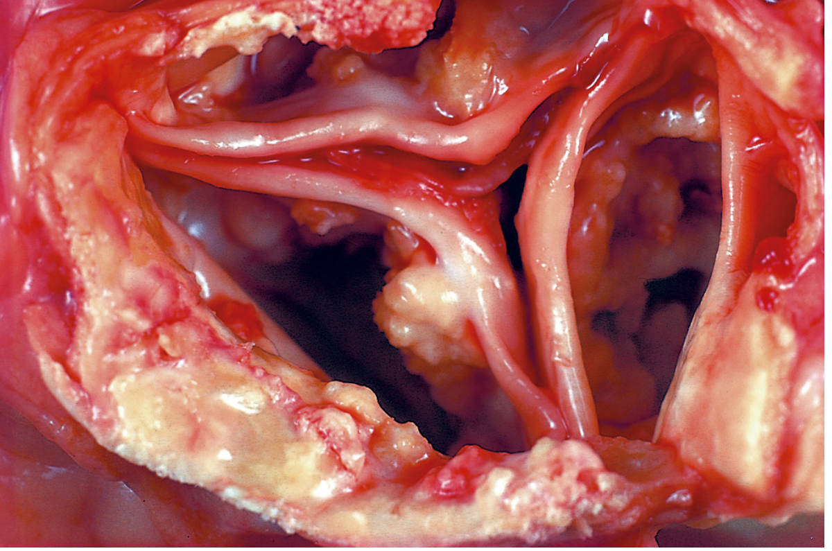
- Aging or damaged heart valves - especially calcific aortic stenosis
- Tuberculous lymph nodes - may become virtually converted to stone
- Tumors - papillary cancers (thyroid) form psammoma bodies
- Parasitic infections - calcified cysts around Taenia solium, Onchocerca volvulus
- Scars, burns, keloids - any site of local tissue injury
Mechanism
- Dying cells lose membrane integrity, allowing intracellular calcium influx
- The acidity associated with cell damage may disrupt normal calcium handling
- Necrotic cells or their debris serve as seed crystals (nidi)
- Progressive mineral encrustation around the nidus
Morphology
- Macroscopic: fine white granules or clumps; gritty texture on cut section
- Microscopic (H&E): basophilic, amorphous granular to clumped deposits; can be intracellular or extracellular
- Special forms:
- Psammoma bodies - lamellated (concentric ring) calcifications; seen in papillary thyroid carcinoma, meningioma, serous ovarian tumors
- Asbestos bodies - calcium and iron deposited around asbestos spicules creating beaded dumbbell shapes
- Heterotopic bone - may eventually form within old calcified foci
Clinical Significance
- May be an incidental marker of past cell injury (e.g., old TB)
- Can cause significant organ dysfunction: calcific aortic stenosis, acceleration of atherosclerosis
Gross pathology - dystrophic calcification of the aortic valve (calcific aortic stenosis):
2. Metastatic Calcification
Definition
Calcium deposition in otherwise normal tissues, almost always caused by hypercalcemia from a systemic disturbance in calcium metabolism.
Key Features
| Feature | Detail |
|---|---|
| Serum calcium | Elevated (hypercalcemia) |
| Tissue status | Normal (viable) |
| Distribution | Widespread / systemic |
Causes of Hypercalcemia
- Increased PTH secretion - primary hyperparathyroidism (parathyroid adenoma); ectopic PTH-related protein (PTHrP) from malignant tumors
- Bone resorption - multiple myeloma, diffuse skeletal metastases (breast cancer), Paget disease, immobilization
- Vitamin D-related disorders - vitamin D intoxication; sarcoidosis (macrophage-activated vitamin D precursor); Williams syndrome (idiopathic hypercalcemia of infancy)
- Renal failure - phosphate retention triggers secondary hyperparathyroidism
- Other - milk-alkali syndrome (excess calcium + absorbable antacids); aluminum intoxication (in dialysis patients)
In renal disease, metastatic calcification occurs when the calcium-phosphate product [Ca²⁺ (mg/dL) × PO₄ (mg/dL)] exceeds 70-80. - Tintinalli's Emergency Medicine
Preferred Sites
Tissues that secrete acid have an internal alkaline compartment that predisposes them to calcium deposition:
- Lungs (alveolar septa, bronchiolar and vessel walls - especially elastic fibers)
- Kidneys (nephrocalcinosis)
- Gastric mucosa
- Systemic arteries and pulmonary veins
Morphology
- Calcium salts appear identical to those in dystrophic calcification: basophilic, amorphous, granular deposits on H&E
- May be non-crystalline amorphous deposits or hydroxyapatite crystals
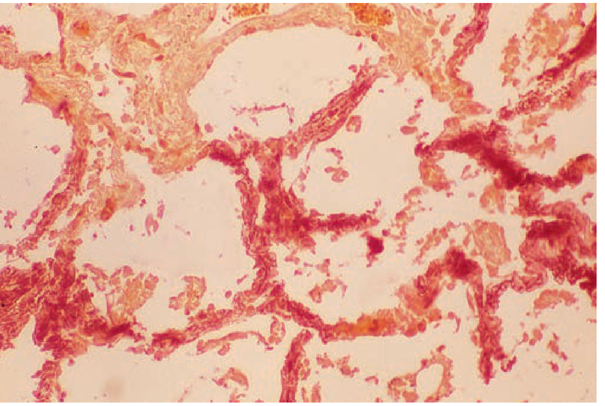
- In the lung: selective early deposition on elastic fibers (ultrastructurally)
Histology - metastatic calcification in alveolar septa (renal dialysis patient, Alizarin red stain):
Clinical Significance
- Usually clinically silent
- Massive pulmonary involvement can cause radiographic opacities mimicking pneumonia and respiratory compromise
- Massive renal deposits (nephrocalcinosis) can lead to renal failure
- Chest CT is more sensitive than plain film for detection; bone scan agents (⁹⁹ᵐTc-MDP, ¹⁸F-NaF) show increased uptake
Side-by-Side Comparison
| Feature | Dystrophic | Metastatic |
|---|---|---|
| Serum calcium | Normal | Elevated |
| Tissue affected | Dead / necrotic | Viable / normal |
| Calcium metabolism | Normal | Disturbed |
| Distribution | Local | Widespread / systemic |
| Classic sites | Atheromas, heart valves, tuberculous nodes, necrotic tumors | Lungs, kidneys, gastric mucosa, arteries |
| Psammoma bodies | Yes | No |
| H&E appearance | Basophilic granular deposits | Identical - basophilic granular deposits |
| Organ dysfunction | Yes (valvular disease, atherosclerosis) | Usually minimal; can cause nephrocalcinosis, respiratory failure |
Mixed Calcification
A combination of both types can occur - metastatic calcification (from calcium dysregulation) can be initiated systemically, and then local trauma or tissue injury propagates dystrophic calcification at the same site. - Dermatology 2-Volume Set 5e
Sources: Robbins, Cotran & Kumar Pathologic Basis of Disease (pp. 59-60, 76); Fishman's Pulmonary Diseases and Disorders (p. 1100); Tintinalli's Emergency Medicine; Dermatology 2-Volume Set 5e
This is a shared conversation. Sign in to Orris to start your own chat.