
Make a list of important vacutaines compound used in them and how they are used and for which test they can be used
- Red (no additive/clot activator) - serum chemistry
- Lavender/Purple (EDTA) - CBC, blood typing
- Blue (sodium citrate) - coagulation tests (PT, aPTT)
- Green (heparin) - plasma chemistry, ABGs
- Grey (sodium fluoride/potassium oxalate) - glucose, lactate
- Yellow (SPS or ACD) - blood cultures, HLA typing
- Gold/SST (gel + clot activator) - most serum chemistry panels
vacutainer tube colors additives tests complete guide 2024
vacutainer tube colors additives chart blood collection
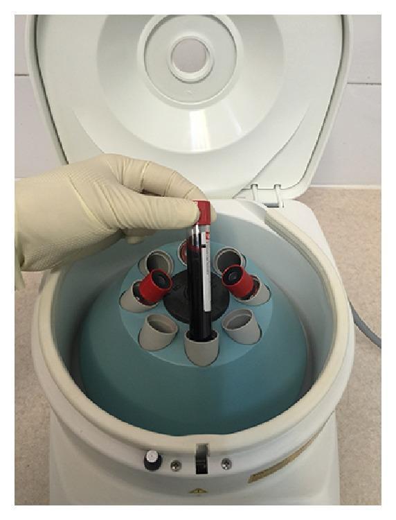
This clinical photograph displays the initial stage of blood processing for autologous platelet concentrates, specifically Platelet-Rich Fibrin (PRF) or Plasma Rich in Growth Factors (PRGF). A gloved hand is shown inserting a vacuum-sealed blood collection tube (Vacutainer style) into a medical centrifuge. The tube contains whole human blood and features a red stopper, typically indicating a serum tube or one without anticoagulants depending on the specific protocol (e.g., L-PRF). The centrifuge is a benchtop model with an open lid, revealing a blue fixed-angle rotor with eight bucket positions designed for standard 5ml or 10ml tubes. The image illustrates a critical laboratory step in regenerative medicine where controlled centrifugation parameters (e.g., 400g) are used to separate blood components—red blood cells, white blood cells, and plasma—to create a fibrin clot or plasma rich in growth factors for clinical applications such as wound healing or tissue engineering.
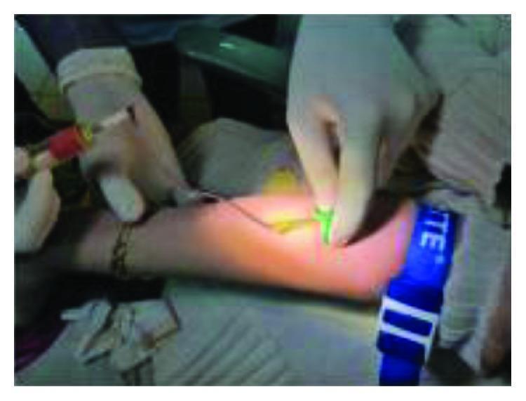
This clinical photograph illustrates a venipuncture procedure for blood collection from the antecubital fossa of a patient's arm. A blue reusable tourniquet is applied to the mid-humerus region to facilitate venous distension. A healthcare professional, wearing white sterile gloves, is performing the procedure using a winged infusion set (butterfly needle), indicated by the green plastic 'wings' held at the puncture site. The needle is connected via flexible tubing to a vacuum-sealed blood collection tube (vacutainer) with a red-stoppered cap. The setup demonstrates standard aseptic technique and equipment used for obtaining venous blood samples, which in this clinical context is being utilized for the subsequent preparation of Leukocyte-Platelet-Rich Fibrin (L-PRF) for surgical use.
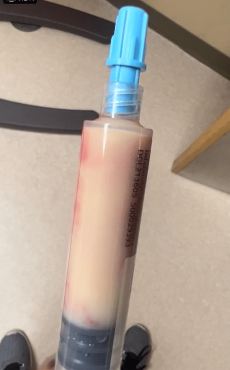
A clinical photograph of a lipemic blood sample contained within a clear plastic blood collection tube with a light blue stopper. The specimen exhibits extreme turbidity, appearing thick, opaque, and milky-white to creamy in color, characteristic of severe hypertriglyceridemia (triglycerides >4000 mg/dl in this clinical context). There is a visible separation of components: a dense, darker reddish-brown clotted layer at the bottom, and a predominant supernatant layer with a 'tomato soup' or 'creamy' appearance containing faint reddish streaks. This visual finding is a classic diagnostic sign of lactescent serum/plasma, often associated with acute pancreatitis and metabolic disorders. The tube is a standard Vacutainer labeled with alphanumeric manufacturing codes.
Vacutainer Tubes: Complete Reference Guide
Order of Draw (CLSI Standard)
- Blood culture bottles (Yellow/SPS)
- Sodium citrate (Light Blue)
- Serum tubes - plain Red, then Gold/SST
- Heparin (Green)
- EDTA (Lavender/Purple/Pink)
- Fluoride/oxalate (Gray)
Tube-by-Tube Breakdown
🔵 Light Blue Top
| Feature | Detail |
|---|---|
| Additive | Sodium citrate 0.109 M (3.2%) |
| Mechanism | Chelates calcium ions, reversibly inhibiting coagulation cascade |
| Sample type | Plasma |
| Inversions | 3-4 gentle inversions |
| Critical rule | Must be filled EXACTLY to the line (1:9 citrate-to-blood ratio) |
- Prothrombin Time (PT) / INR
- Activated Partial Thromboplastin Time (aPTT)
- Fibrinogen level
- D-dimer
- Thrombin time
- Factor assays (II, V, VII, VIII, IX, X, XI)
- Platelet function assay (PFA-100)
- Mixing studies
- Lupus anticoagulant / Antiphospholipid antibody tests
- TEG (Thromboelastography)
🔴 Red Top (Plain)
| Feature | Detail |
|---|---|
| Additive | None (glass tube) OR clot activator - silicone coating (plastic) |
| Mechanism | Allows natural clotting; serum is separated after centrifugation |
| Sample type | Serum |
| Inversions | 0 (glass) / 5 (plastic with clot activator) |
- Drug levels (digoxin, phenytoin, lithium, vancomycin)
- Blood bank / compatibility testing
- Serology (syphilis RPR, rubella, CMV)
- Viral antibodies (HIV, Hepatitis B & C serology)
- Routine chemistry where gel interference is unwanted
🟡 Gold Top / Tiger Top (SST - Serum Separator Tube)
| Feature | Detail |
|---|---|
| Additive | Clot activator (silica particles) + polymer gel barrier |
| Mechanism | Silica accelerates clotting; gel migrates during centrifugation to form a physical barrier between serum and cells |
| Sample type | Serum |
| Inversions | 5 inversions; allow 30 min clotting before centrifuge |
- Comprehensive Metabolic Panel (CMP) - glucose, BUN, creatinine, electrolytes, LFTs
- Lipid panel (cholesterol, triglycerides, HDL, LDL)
- Thyroid panel (TSH, T3, T4)
- Hormones (testosterone, estrogen, FSH, LH, prolactin)
- Vitamins (B12, folate, Vitamin D, Vitamin B1)
- Tumor markers (PSA, CEA, CA-125, CA 19-9, AFP)
- Cardiac markers (Troponin I/T, CK-MB, BNP)
- Hepatitis B surface antigen, HIV Ag/Ab combo
- Therapeutic drug monitoring
- ANA, anti-dsDNA, RF, CRP, ESR (some labs)
🟢 Green Top
| Feature | Detail |
|---|---|
| Additive | Lithium heparin OR sodium heparin |
| Mechanism | Activates antithrombin III, inhibits thrombin and factor Xa - prevents clotting without chelating calcium |
| Sample type | Plasma (fast availability - no need to wait for clotting) |
| Inversions | 8 inversions |
| Note | Lithium heparin used for most chemistries; sodium heparin for some special tests |
- STAT/urgent chemistry panels
- Ammonia level (must be placed on ice immediately)
- Arterial Blood Gas (ABG) - special heparinized syringe version
- Chromosomal analysis / cytogenetics (sodium heparin)
- HLA typing (sodium heparin)
- Lead levels
- Carboxyhemoglobin
- Plasma therapeutic drug monitoring
🟣 Lavender / Purple Top
| Feature | Detail |
|---|---|
| Additive | K2-EDTA or K3-EDTA (ethylenediaminetetraacetic acid) |
| Mechanism | Chelates calcium irreversibly - prevents ALL coagulation; preserves cell morphology |
| Sample type | Whole blood |
| Inversions | 8 inversions |
- Complete Blood Count (CBC) with differential
- Hemoglobin A1c (HbA1c)
- ESR (Erythrocyte Sedimentation Rate)
- Peripheral blood smear / morphology
- Reticulocyte count
- Blood typing (ABO/Rh) in some labs
- Sickle cell screening
- Malaria smear
- Direct Coombs test
- Immunosuppressive drug levels (tacrolimus, cyclosporine)
- Whole blood lead levels
- EBV / CMV by PCR
🩷 Pink Top
| Feature | Detail |
|---|---|
| Additive | K2-EDTA (blood bank grade, strictly labeled) |
| Mechanism | Same as lavender EDTA |
| Sample type | Whole blood |
| Inversions | 8 inversions |
| Key distinction | Dedicated exclusively to blood bank; strict labeling requirements to ensure positive patient ID |
- Type and Screen
- Type and Crossmatch
- Antibody identification
- Direct antiglobulin test (DAT)
- Transfusion compatibility testing
⚫ Gray Top
| Feature | Detail |
|---|---|
| Additive | Potassium oxalate + sodium fluoride (most common); OR sodium fluoride/Na2-EDTA |
| Mechanism | NaF inhibits enolase enzyme (stops glycolysis), preventing glucose from being metabolized by RBCs. Oxalate/EDTA act as anticoagulants |
| Sample type | Plasma |
| Inversions | 8 inversions |
- Fasting glucose and GTT (Glucose Tolerance Test)
- Lactate / lactic acid (on ice)
- Blood alcohol level (ethanol)
- Fluoride-sensitive pesticide levels
🟡 Yellow Top
| Feature | Detail |
|---|---|
| Additive | SPS (Sodium Polyanethol Sulfonate) OR ACD (Acid Citrate Dextrose - Solution A or B) |
| Mechanism | SPS: anticoagulant + inhibits complement, phagocytosis, and certain antibiotics, improving bacterial recovery. ACD: citrate chelates calcium + dextrose preserves RBC metabolism |
| Sample type | Whole blood |
| Inversions | 8 inversions |
- SPS: Blood culture specimen collection (microbiology)
- ACD: Blood bank studies, HLA antigen typing
- ACD: Paternity testing / DNA studies
- ACD: Adoptee workups, forensic identity testing
🔵 Royal Blue Top
| Feature | Detail |
|---|---|
| Additive | K2-EDTA (plastic) OR clot activator (plastic) OR no additive |
| Mechanism | Tube is manufactured with minimal trace metal contamination |
| Sample type | Serum or plasma depending on version |
| Inversions | 8 (EDTA version) |
- Trace metal analysis (zinc, copper, selenium, manganese)
- Heavy metal screening
- Toxicology panels
- Nutritional element testing
⬜ White Top
| Feature | Detail |
|---|---|
| Additive | K2-EDTA + gel separator |
| Mechanism | EDTA prevents clotting; gel separates plasma from cells |
| Sample type | Plasma |
| Inversions | 8 inversions |
- Molecular diagnostic testing (PCR-based assays)
- Branched DNA (bDNA) amplification
- Viral load testing (HIV RNA, HCV RNA, CMV viral load)
🔴 Clear Red / Light Gray (Discard Tube)
| Feature | Detail |
|---|---|
| Additive | None |
| Use | Discard tube (drawn before light blue to remove tissue thromboplastin from needle) or secondary specimen tube |
Quick-Reference Color Summary Table
| Tube Color | Additive | Sample | Key Tests |
|---|---|---|---|
| Light Blue | Sodium citrate | Plasma | PT/INR, aPTT, D-dimer, fibrinogen |
| Red (plain) | None / clot activator | Serum | Drug levels, serology, blood bank |
| Gold / SST | Clot activator + gel | Serum | CMP, lipids, hormones, TSH, troponin |
| Green | Lithium/sodium heparin | Plasma | Ammonia, STAT chemistry, cytogenetics |
| Lavender / Purple | K2-EDTA | Whole blood | CBC, HbA1c, blood smear, tacrolimus |
| Pink | K2-EDTA (blood bank) | Whole blood | Crossmatch, type & screen |
| Gray | NaF + potassium oxalate | Plasma | Glucose, lactate, blood alcohol |
| Yellow (SPS) | SPS | Whole blood | Blood cultures |
| Yellow (ACD) | ACD solution A/B | Whole blood | HLA typing, DNA/paternity |
| Royal Blue | EDTA or none | Serum/Plasma | Trace metals, heavy metals |
| White | K2-EDTA + gel | Plasma | PCR, viral loads |
Key Tips for Phlebotomists
- Never shake tubes - invert gently to mix. Shaking causes hemolysis and will invalidate the specimen.
- Fill citrate tubes exactly - under-filling the blue top dilutes the citrate ratio and produces false coagulation results.
- SST tubes need 30 minutes before centrifugation for proper clot formation; do not centrifuge too early.
- Lavender ≠ Pink - both contain EDTA but pink tubes are strictly for blood bank use and require a second positive patient ID per most transfusion safety protocols.
- Gray top glucose is stable for 24 hours because NaF halts glycolysis; without it, serum glucose drops ~10 mg/dL per hour at room temperature.
- Green top for ammonia must go on ice immediately and be processed within 15 minutes to prevent falsely elevated results.
- Royal blue tubes are low-trace-metal certified; using a regular tube for heavy metal testing will give falsely elevated results from tube contamination.
Generate a printable quick-reference PDF chart of tube colors & tests
mkdir -p /tmp/workspace/vacutainer-chart
~/vacutainer-chart/generate_chart.py
from reportlab.lib.pagesizes import A4
from reportlab.lib import colors
from reportlab.lib.units import mm
from reportlab.platypus import (
    SimpleDocTemplate, Table, TableStyle, Paragraph, Spacer, HRFlowable
)
from reportlab.lib.styles import getSampleStyleSheet, ParagraphStyle
from reportlab.lib.enums import TA_CENTER, TA_LEFT
from reportlab.platypus import KeepTogether

OUTPUT = "/tmp/workspace/vacutainer-chart/Vacutainer_Quick_Reference.pdf"

# ── Colour palette (RGB 0-1) ────────────────────────────────────────────────
LIGHT_BLUE_CAP  = colors.HexColor("#AED6F1")
RED_CAP         = colors.HexColor("#E74C3C")
GOLD_CAP        = colors.HexColor("#F0B327")
GREEN_CAP       = colors.HexColor("#2ECC71")
LAVENDER_CAP    = colors.HexColor("#9B59B6")
PINK_CAP        = colors.HexColor("#F48FB1")
GRAY_CAP        = colors.HexColor("#95A5A6")
YELLOW_CAP      = colors.HexColor("#F9E231")
ROYAL_BLUE_CAP  = colors.HexColor("#1A5276")
WHITE_CAP       = colors.HexColor("#D5D8DC")
HEADER_BG       = colors.HexColor("#1B2631")
PAGE_BG         = colors.HexColor("#FDFEFE")
STRIPE          = colors.HexColor("#EBF5FB")

doc = SimpleDocTemplate(
    OUTPUT,
    pagesize=A4,
    leftMargin=12*mm, rightMargin=12*mm,
    topMargin=14*mm, bottomMargin=14*mm,
    title="Vacutainer Quick Reference Chart",
    author="Orris Medical Reference"
)

W, H = A4
styles = getSampleStyleSheet()

title_style = ParagraphStyle(
    "title", fontName="Helvetica-Bold", fontSize=17,
    textColor=colors.white, alignment=TA_CENTER, spaceAfter=2
)
sub_style = ParagraphStyle(
    "sub", fontName="Helvetica", fontSize=9,
    textColor=colors.HexColor("#AEB6BF"), alignment=TA_CENTER, spaceAfter=0
)
col_header_style = ParagraphStyle(
    "col_hdr", fontName="Helvetica-Bold", fontSize=8.5,
    textColor=colors.white, alignment=TA_CENTER
)
cell_style = ParagraphStyle(
    "cell", fontName="Helvetica", fontSize=7.5,
    textColor=colors.HexColor("#1B2631"), alignment=TA_LEFT, leading=10
)
cell_bold = ParagraphStyle(
    "cell_bold", fontName="Helvetica-Bold", fontSize=8,
    textColor=colors.HexColor("#1B2631"), alignment=TA_LEFT, leading=11
)
small_style = ParagraphStyle(
    "small", fontName="Helvetica-Oblique", fontSize=6.5,
    textColor=colors.HexColor("#717D7E"), alignment=TA_CENTER
)
note_style = ParagraphStyle(
    "note", fontName="Helvetica", fontSize=7,
    textColor=colors.HexColor("#1B2631"), alignment=TA_LEFT, leading=9
)

# ── Tube data ────────────────────────────────────────────────────────────────
# (cap_color, text_color_on_cap, tube_label, additive, mechanism_short, sample, tests)
TUBES = [
    (
        LIGHT_BLUE_CAP, colors.HexColor("#1A5276"),
        "LIGHT BLUE",
        "Sodium Citrate\n0.109 M (3.2%)",
        "Chelates Ca²⁺ (reversible)\n→ 3–4 inversions\n⚠ Fill EXACTLY to line",
        "Plasma",
        "PT / INR\naPTT\nFibrinogen\nD-Dimer\nThrombin Time\nFactor Assays (II,V,VII–XI)\nLupus Anticoagulant\nTEG / PFA-100",
    ),
    (
        RED_CAP, colors.white,
        "RED (Plain)",
        "None (glass) or\nSilicone clot activator (plastic)",
        "Natural clotting → serum\n0 inversions (glass)\n5 inversions (plastic)",
        "Serum",
        "Drug levels (digoxin, phenytoin,\n  lithium, vancomycin)\nSerology (RPR, rubella, CMV)\nViral antibodies (HIV, HBV, HCV)\nBlood bank (where gel avoided)",
    ),
    (
        GOLD_CAP, colors.HexColor("#1B2631"),
        "GOLD / SST",
        "Clot activator (silica)\n+ Polymer gel barrier",
        "Silica speeds clotting;\ngel forms barrier after spin\n5 inversions, wait 30 min",
        "Serum",
        "CMP (glucose, BUN, Cr, LFTs,\n  electrolytes)\nLipid panel\nTSH, T3, T4\nHormones (FSH, LH, testosterone)\nVitamin D / B12 / folate\nTumor markers (PSA, CEA, AFP,\n  CA-125, CA 19-9)\nTroponin, CK-MB, BNP\nHBsAg, HIV Ag/Ab combo",
    ),
    (
        GREEN_CAP, colors.white,
        "GREEN",
        "Lithium heparin or\nSodium heparin",
        "Activates antithrombin III\n→ inhibits thrombin & Xa\n8 inversions",
        "Plasma",
        "STAT / urgent chemistry\nAmmonia ⚠ (on ice, <15 min)\nABG (heparinised syringe)\nChromosomal / cytogenetics\nHLA typing\nLead level\nCarboxyhemoglobin",
    ),
    (
        LAVENDER_CAP, colors.white,
        "LAVENDER / PURPLE",
        "K₂-EDTA or K₃-EDTA",
        "Chelates Ca²⁺ (irreversible)\nPreserves cell morphology\n8 inversions",
        "Whole Blood",
        "CBC with differential\nHbA1c\nESR\nPeripheral blood smear\nReticulocyte count\nSickle cell screen / malaria smear\nDirect Coombs\nImmunosuppressants (tacrolimus,\n  cyclosporine)\nWhole blood lead\nEBV / CMV PCR",
    ),
    (
        PINK_CAP, colors.HexColor("#1B2631"),
        "PINK",
        "K₂-EDTA\n(blood bank grade)",
        "Same as lavender EDTA\nStrict 2-ID labelling required\n8 inversions",
        "Whole Blood",
        "Type & Screen\nType & Crossmatch\nAntibody identification\nDirect Antiglobulin Test (DAT)\nTransfusion compatibility",
    ),
    (
        GRAY_CAP, colors.white,
        "GRAY",
        "NaF + Potassium oxalate\nor NaF + Na₂-EDTA",
        "NaF inhibits enolase\n→ stops glycolysis\nOxalate/EDTA anticoagulate\n8 inversions",
        "Plasma",
        "Fasting glucose / GTT\nLactate ⚠ (on ice)\nBlood alcohol (ethanol)\nFluoride-sensitive pesticides",
    ),
    (
        YELLOW_CAP, colors.HexColor("#1B2631"),
        "YELLOW",
        "SPS (Sodium Polyanethol\n  Sulfonate)\nor ACD Solution A or B",
        "SPS: anticoagulant +\n  inhibits complement/phagocytosis\nACD: citrate + dextrose\n  preserves RBC metabolism",
        "Whole Blood",
        "SPS → Blood cultures\n  (microbiology)\nACD → HLA antigen typing\nACD → Paternity / DNA testing\nACD → Blood bank studies",
    ),
    (
        ROYAL_BLUE_CAP, colors.white,
        "ROYAL BLUE",
        "K₂-EDTA (plastic) or\nClot activator or\nNo additive",
        "Low trace-metal certified tube\nUse ONLY for metals\n8 inversions (EDTA version)",
        "Serum or Plasma",
        "Trace metals (zinc, copper,\n  selenium, manganese)\nHeavy metal screen\nToxicology panels\nNutritional element testing",
    ),
    (
        WHITE_CAP, colors.HexColor("#1B2631"),
        "WHITE",
        "K₂-EDTA + gel separator",
        "EDTA prevents clotting;\ngel separates plasma from cells\n8 inversions",
        "Plasma",
        "Molecular PCR assays\nViral loads (HIV RNA, HCV RNA,\n  CMV viral load)\nbDNA amplification",
    ),
]

# ── Build table rows ──────────────────────────────────────────────────────────
col_widths = [26*mm, 38*mm, 38*mm, 18*mm, 56*mm]

def cap_cell(cap_color, text_color, label):
    """Rounded coloured cell simulated via a 1-cell mini-table."""
    p = Paragraph(f"<b>{label}</b>", ParagraphStyle(
        "cap", fontName="Helvetica-Bold", fontSize=7.5,
        textColor=text_color, alignment=TA_CENTER, leading=10
    ))
    return p

header_row = [
    Paragraph("TUBE COLOR", col_header_style),
    Paragraph("ADDITIVE", col_header_style),
    Paragraph("MECHANISM / NOTES", col_header_style),
    Paragraph("SAMPLE", col_header_style),
    Paragraph("TESTS / USES", col_header_style),
]

table_data = [header_row]
row_bg_colors = []  # (row_index, color)

for i, (cap_color, text_color, label, additive, mechanism, sample, tests) in enumerate(TUBES):
    row_idx = i + 1
    row = [
        cap_cell(cap_color, text_color, label),
        Paragraph(additive, cell_style),
        Paragraph(mechanism, cell_style),
        Paragraph(sample, cell_style),
        Paragraph(tests, cell_style),
    ]
    table_data.append(row)
    row_bg_colors.append((row_idx, cap_color))

table = Table(table_data, colWidths=col_widths, repeatRows=1)

# ── TableStyle ────────────────────────────────────────────────────────────────
ts = TableStyle([
    # Header
    ("BACKGROUND",  (0,0), (-1,0), HEADER_BG),
    ("TEXTCOLOR",   (0,0), (-1,0), colors.white),
    ("FONTNAME",    (0,0), (-1,0), "Helvetica-Bold"),
    ("FONTSIZE",    (0,0), (-1,0), 8.5),
    ("ALIGN",       (0,0), (-1,0), "CENTER"),
    ("VALIGN",      (0,0), (-1,-1), "MIDDLE"),
    ("TOPPADDING",  (0,0), (-1,0), 5),
    ("BOTTOMPADDING",(0,0),(-1,0), 5),
    # All cells
    ("FONTSIZE",    (0,1), (-1,-1), 7.5),
    ("TOPPADDING",  (0,1), (-1,-1), 4),
    ("BOTTOMPADDING",(0,1),(-1,-1), 4),
    ("LEFTPADDING", (0,0), (-1,-1), 4),
    ("RIGHTPADDING",(0,0), (-1,-1), 4),
    # Grid
    ("GRID",        (0,0), (-1,-1), 0.4, colors.HexColor("#BDC3C7")),
    ("LINEBELOW",   (0,0), (-1,0), 1.2, colors.HexColor("#BDC3C7")),
    # Alternate stripe
    *[("BACKGROUND", (0, r), (-1, r), STRIPE) for r in range(2, len(TUBES)+1, 2)],
])

# Apply per-row cap-column background colours
for row_idx, cap_color in row_bg_colors:
    ts.add("BACKGROUND", (0, row_idx), (0, row_idx), cap_color)

table.setStyle(ts)

# ── Order of draw box ─────────────────────────────────────────────────────────
draw_order_data = [
    [Paragraph("<b>ORDER OF DRAW  (CLSI Standard)</b>", ParagraphStyle(
        "od_title", fontName="Helvetica-Bold", fontSize=8,
        textColor=colors.white, alignment=TA_CENTER))],
    [Paragraph(
        "1. Blood Cultures (Yellow/SPS)  →  2. Sodium Citrate (Light Blue)  →  "
        "3. Serum tubes (Red, then Gold/SST)  →  4. Heparin (Green)  →  "
        "5. EDTA (Lavender / Pink)  →  6. Fluoride/Oxalate (Gray)",
        ParagraphStyle("od_body", fontName="Helvetica", fontSize=7.5,
                       textColor=colors.HexColor("#1B2631"), alignment=TA_CENTER, leading=11)
    )],
]
draw_order_table = Table(draw_order_data, colWidths=[176*mm])
draw_order_table.setStyle(TableStyle([
    ("BACKGROUND",   (0,0), (-1,0), HEADER_BG),
    ("BACKGROUND",   (0,1), (-1,1), colors.HexColor("#D6EAF8")),
    ("TOPPADDING",   (0,0), (-1,-1), 4),
    ("BOTTOMPADDING",(0,0), (-1,-1), 4),
    ("LEFTPADDING",  (0,0), (-1,-1), 6),
    ("RIGHTPADDING", (0,0), (-1,-1), 6),
    ("BOX",          (0,0), (-1,-1), 0.8, colors.HexColor("#85929E")),
]))

# ── Key tips ──────────────────────────────────────────────────────────────────
tips = [
    "• <b>Never shake</b> – invert gently; shaking causes haemolysis.",
    "• <b>Light Blue must be filled exactly</b> to the line (1:9 citrate:blood ratio); under-fill invalidates coagulation results.",
    "• <b>SST (Gold)</b> – allow 30 min clotting before centrifugation.",
    "• <b>Lavender ≠ Pink</b> – both are EDTA, but Pink requires a second positive patient ID for transfusion safety.",
    "• <b>Gray tube glucose</b> is stable for 24 h (NaF halts glycolysis); without NaF, glucose drops ~10 mg/dL per hour at room temp.",
    "• <b>Ammonia (Green)</b> – place on ice immediately; process within 15 min to avoid falsely elevated values.",
    "• <b>Royal Blue</b> – use only low-trace-metal certified tubes for heavy-metal testing; a standard tube will contaminate the sample.",
]
tips_data = [[Paragraph("<b>KEY TIPS FOR PHLEBOTOMISTS</b>", ParagraphStyle(
    "tip_title", fontName="Helvetica-Bold", fontSize=8,
    textColor=colors.white, alignment=TA_CENTER))]]
for t in tips:
    tips_data.append([Paragraph(t, note_style)])

tips_table = Table(tips_data, colWidths=[176*mm])
tips_style = TableStyle([
    ("BACKGROUND",   (0,0), (-1,0), HEADER_BG),
    ("BACKGROUND",   (0,1), (-1,-1), colors.HexColor("#FDFEFE")),
    ("TOPPADDING",   (0,0), (-1,-1), 3),
    ("BOTTOMPADDING",(0,0), (-1,-1), 3),
    ("LEFTPADDING",  (0,0), (-1,-1), 6),
    ("RIGHTPADDING", (0,0), (-1,-1), 6),
    ("BOX",          (0,0), (-1,-1), 0.8, colors.HexColor("#85929E")),
    ("LINEBELOW",    (0,0), (-1,0), 0.8, colors.HexColor("#85929E")),
    *[("BACKGROUND", (0, r), (-1, r), STRIPE) for r in range(2, len(tips)+1, 2)],
])
tips_table.setStyle(tips_style)

# ── Page background colour ───────────────────────────────────────────────────
def add_bg(canvas, doc):
    canvas.saveState()
    canvas.setFillColor(PAGE_BG)
    canvas.rect(0, 0, A4[0], A4[1], fill=1, stroke=0)
    canvas.restoreState()

# ── Assemble document ─────────────────────────────────────────────────────────
title_table_data = [[
    Paragraph("💉 VACUTAINER QUICK-REFERENCE CHART", title_style),
    Paragraph("Orris Medical Reference  •  BD CLSI Standard  •  July 2026", sub_style),
]]
title_block = Table(title_table_data, colWidths=[176*mm])
title_block.setStyle(TableStyle([
    ("BACKGROUND",   (0,0), (-1,-1), HEADER_BG),
    ("TOPPADDING",   (0,0), (-1,-1), 7),
    ("BOTTOMPADDING",(0,0), (-1,-1), 7),
    ("LEFTPADDING",  (0,0), (-1,-1), 8),
    ("RIGHTPADDING", (0,0), (-1,-1), 8),
    ("BOX",          (0,0), (-1,-1), 1, HEADER_BG),
]))

story = [
    title_block,
    Spacer(1, 4*mm),
    table,
    Spacer(1, 4*mm),
    draw_order_table,
    Spacer(1, 3*mm),
    tips_table,
    Spacer(1, 3*mm),
    Paragraph(
        "Reference: BD Vacutainer® Venous Blood Collection Tube Guide (BD-83178) | CLSI GP41-A6 | myOnsite Healthcare",
        small_style
    ),
]

doc.build(story, onFirstPage=add_bg, onLaterPages=add_bg)
print("PDF generated:", OUTPUT)
cd /tmp/workspace/vacutainer-chart && python generate_chart.py 2>&1
ls -lh /tmp/workspace/vacutainer-chart/Vacutainer_Quick_Reference.pdf
cd /tmp/workspace/vacutainer-chart && python -c " from pypdf import PdfReader r = PdfReader('Vacutainer_Quick_Reference.pdf') print('Pages:', len(r.pages)) print('Text sample:', r.pages[0].extract_text()[:300]) "
cd /tmp/workspace/vacutainer-chart && python -c " from pypdf import PdfReader r = PdfReader('Vacutainer_Quick_Reference.pdf') print('Pages:', len(r.pages)) # Check page size page = r.pages[0] print('Page size (pts):', float(page.mediabox.width), 'x', float(page.mediabox.height)) print('A4 should be ~595 x 842') # Print full text to check all 10 tubes are there text = page.extract_text() print('Total chars:', len(text)) # Check tubes present for tube in ['LIGHT BLUE', 'RED', 'GOLD', 'GREEN', 'LAVENDER', 'PINK', 'GRAY', 'YELLOW', 'ROYAL BLUE', 'WHITE']: print(f' {tube}: {tube in text}') "
Vacutainer Quick Reference
PDF Document · PDF
- Dark header banner with chart title and reference date
- Color-coded table with all 10 major tube types:
- Each row's first column is highlighted in the actual tube cap color (Light Blue, Red, Gold, Green, Lavender, Pink, Gray, Yellow, Royal Blue, White)
- Columns: Tube Color | Additive | Mechanism & Notes | Sample Type | Tests/Uses
- Order of Draw box (CLSI standard) - Blue → Red → Gold → Green → Lavender → Gray
- Key Tips section - 7 phlebotomy practice points (no shaking, fill citrate to line, SST wait time, Lavender vs Pink, gray tube glycolysis, ammonia on ice, royal blue contamination warning)
- Reference footer citing BD Vacutainer guide and CLSI GP41-A6
Erythrocyte normal value for adult male and adult female give accurate answer
Erythrocyte (RBC) Normal Reference Values - Adult
RBC Count (Erythrocyte Count)
| Parameter | Adult Male | Adult Female |
|---|---|---|
| RBC Count | 4.5 – 5.5 × 10⁶/µL (4.5–5.5 million/mm³) | 4.0 – 5.0 × 10⁶/µL (4.0–5.0 million/mm³) |
| SI Units | 4.5–5.5 × 10¹²/L | 4.0–5.0 × 10¹²/L |
Full Erythrocyte Panel (CBC Red Cell Parameters)
| Parameter | Adult Male | Adult Female | Units |
|---|---|---|---|
| RBC Count | 4.5 – 5.5 | 4.0 – 5.0 | × 10⁶/µL |
| Hemoglobin (Hb) | 13.5 – 17.5 | 12.0 – 15.5 | g/dL |
| Hematocrit (Hct / PCV) | 41.5 – 50.4% | 35.9 – 44.6% | % |
| MCV (Mean Corpuscular Volume) | 80 – 100 | 80 – 100 | fL |
| MCH (Mean Corpuscular Hgb) | 26 – 34 | 26 – 34 | pg |
| MCHC (Mean Corpuscular Hgb Conc.) | 33 – 36 | 33 – 36 | g/dL |
| RDW (Red Cell Distribution Width) | 11.5 – 14.5 | 11.5 – 14.5 | % |
| Red Cell Volume | 20 – 36.3 | 19 – 32.3 | mL/kg body weight |
| Reticulocytes | 0.5 – 1.5 | 0.5 – 1.5 | % of RBCs |
Why Are Male Values Higher?
Important Notes
- Altitude effect - People living at high altitude have higher RBC counts due to lower ambient oxygen driving up erythropoietin. Labs at altitude use adjusted reference ranges.
- Anemia threshold - An RBC count below the lower limit of the reference range suggests anemia. Hemoglobin is the most practical indicator clinically.
- The "rule of 3" (Wintrobe's rule) - In a normal sample: Hb × 3 ≈ Hct; RBC × 3 ≈ Hb. This is a quick bedside check for internal consistency.
- Lab variation - Exact reference ranges vary slightly between laboratories based on the automated analyzer used and the reference population.
- Henry's Clinical Diagnosis and Management by Laboratory Methods, Table A5.9 (Hematology Reference Intervals) - Hematocrit Male: 41.5–50.4%, Female: 35.9–44.6%
- Goldman-Cecil Medicine, Ch. 144 - RBC differences between sexes attributed to testosterone action on erythroid progenitors
- Basic Medical Biochemistry (6e) - Female RBC reference range stated as 4.0–5.2 × 10⁶/mm³
Erythrocyte Sedimentation Rate normal value of adult male and adult female
- Adult Male: 0–15 mm/hr (Westergren method)
- Adult Female: 0–20 mm/hr (Westergren method)
- Values increase with age (Westergren age-adjusted formula commonly used)
ESR (Erythrocyte Sedimentation Rate) - Normal Values
Standard Reference Values (Westergren Method)
| Age Group | Adult Male | Adult Female |
|---|---|---|
| Below 50 years | 0 – 15 mm/hr | 0 – 20 mm/hr |
| Above 50 years | 0 – 20 mm/hr | 0 – 30 mm/hr |
| Above 85 years | 0 – 30 mm/hr | 0 – 42 mm/hr |
Source: Böttiger & Svedberg (1967) and Zauber & Zauber (1987), as cited in Henry's Clinical Diagnosis and Management by Laboratory Methods
Age-Adjusted Formula (Miller's Formula)
| Sex | Formula |
|---|---|
| Men | Age (years) ÷ 2 |
| Women | (Age (years) + 10) ÷ 2 |
- A 40-year-old man: upper limit = 40 ÷ 2 = 20 mm/hr
- A 40-year-old woman: upper limit = (40 + 10) ÷ 2 = 25 mm/hr
- A 70-year-old man: upper limit = 70 ÷ 2 = 35 mm/hr
- A 70-year-old woman: upper limit = (70 + 10) ÷ 2 = 40 mm/hr
Which Method? Westergren vs. Wintrobe
| Feature | Westergren | Wintrobe |
|---|---|---|
| Tube length | 200 mm | 100 mm |
| Anticoagulant | Sodium citrate (1:4 dilution) or EDTA | EDTA (undiluted) |
| ICSH recommended | ✅ Yes | No |
| Sensitivity | Higher (better for elevated ESR) | Lower range |
| Standard normal (male) | 0–15 mm/hr | 0–9 mm/hr |
| Standard normal (female) | 0–20 mm/hr | 0–20 mm/hr |
Key Facts About ESR
- Why females are higher - Women naturally have lower hematocrit and higher fibrinogen levels, both of which increase rouleaux formation (RBC stacking) and accelerate sedimentation.
- Age effect - ESR increases progressively with age. Westergren's original limits (10 mm/hr for men, 20 mm/hr for women) are now considered too strict based on population studies.
- Not a specific test - ESR is a non-specific marker of inflammation, infection, malignancy, or tissue injury. CRP is more sensitive and specific for acute inflammation.
- Factors that raise ESR - Pregnancy, anemia, infections, autoimmune disease (especially SLE, RA, temporal arteritis), malignancy, hyperfibrinogenemia.
- Factors that falsely lower ESR - Polycythemia, sickle cell disease, extreme leukocytosis, low fibrinogen, heparin (cannot be used as anticoagulant for ESR).
- Technical note - Tilting the tube even 3° from vertical can accelerate ESR by up to 30%; temperature must be 20–25°C; test must be set up within 2 hours of collection.
- Henry's Clinical Diagnosis and Management by Laboratory Methods - Modified Westergren Method section (Böttiger/Svedberg/Zauber age-stratified upper limits)
- Firestein & Kelley's Textbook of Rheumatology - "the usual accepted upper limits of normal are 15 mm/hr for males and 20 mm/hr for females... A simple formula: men = age ÷ 2; women = (age + 10) ÷ 2"
Causes of splenomegaly and divide splenomegaly into mild moderate and massive and give examples also
Splenomegaly - Causes & Classification
Normal Spleen Reference Points
- Normal weight: < 250 g
- Normal size (ultrasound): max cephalocaudad diameter ≤ 13 cm
- Not normally palpable on examination
- The spleen must enlarge threefold before it becomes palpable
Classification by Size
🟡 Mild Splenomegaly
| Category | Examples |
|---|---|
| Infections (viral) | Infectious mononucleosis (EBV), CMV, HIV |
| Bacterial infections | Infective endocarditis, typhoid fever, brucellosis |
| Autoimmune | SLE, rheumatoid arthritis (Felty's syndrome), autoimmune haemolytic anaemia |
| Congestive | Early congestive heart failure, early cirrhosis |
| Haematological | Immune thrombocytopenic purpura (ITP) |
| Miscellaneous | Sarcoidosis, drug reactions (phenytoin) |
🟠 Moderate Splenomegaly
| Category | Examples |
|---|---|
| Haemolytic anaemias | Hereditary spherocytosis, thalassaemia major, sickle cell disease |
| Portal hypertension | Cirrhosis (established), portal vein thrombosis, Budd-Chiari syndrome |
| Lymphomas | Hodgkin's lymphoma, Non-Hodgkin's lymphoma (moderate) |
| Chronic leukaemias | Chronic lymphocytic leukaemia (CLL) - early/moderate stage |
| Infections (parasitic) | Malaria (subacute), visceral leishmaniasis (kala-azar) - early stages |
| Storage diseases | Gaucher's disease, Niemann-Pick disease |
| Myeloproliferative | Polycythaemia vera, essential thrombocythaemia |
🔴 Massive Splenomegaly
| Condition | Notes |
|---|---|
| Chronic Myeloid Leukaemia (CML) | Classic cause; BCR-ABL driven massive splenomegaly |
| Myelofibrosis with myeloid metaplasia | Extramedullary haematopoiesis drives massive enlargement |
| Non-Hodgkin's Lymphoma | Especially splenic marginal zone lymphoma |
| Chronic Lymphocytic Leukaemia (CLL) | Advanced disease |
| Hairy Cell Leukaemia | Often presents with massive splenomegaly |
| Polycythaemia Vera | Myeloproliferative disorder |
| Visceral Leishmaniasis (Kala-azar) | Leishmania donovani - hallmark massive splenomegaly in tropics |
| Hyperreactive Malarial Splenomegaly | Tropical splenomegaly syndrome; chronic Plasmodium infection |
| Thalassaemia Major | Untreated; due to extramedullary haematopoiesis + haemolysis |
| Gaucher's Disease (Type 1) | Glucocerebrosidase deficiency; massive spleen + liver |
Causes by Mechanism (Harrison's Classification)
| Mechanism | Examples |
|---|---|
| Reticuloendothelial hyperplasia (work hypertrophy) | Hereditary spherocytosis, thalassaemia - removal of defective RBCs |
| Immune hyperplasia | Infectious mononucleosis, SBE, immune thrombocytopenia, SLE, Felty's syndrome |
| Passive congestion | Cirrhosis, portal vein thrombosis, Budd-Chiari syndrome, congestive heart failure |
| Infiltrative disease | Lymphomas, metastatic cancer, amyloidosis, Gaucher's disease, myeloproliferative disorders with extramedullary haematopoiesis |
Full Causes Table (Goldman-Cecil Medicine, Table 154-5)
| Category | Specific Causes |
|---|---|
| Infectious - Bacterial | Endocarditis, brucellosis, syphilis, tuberculosis, Rocky Mountain spotted fever |
| Infectious - Fungal | Histoplasmosis, toxoplasmosis |
| Infectious - Parasitic | Malaria, leishmaniasis |
| Infectious - Viral | EBV (infectious mononucleosis), HIV, CMV |
| Immune-mediated | Rheumatoid arthritis, SLE, autoimmune haemolytic anaemia, phenytoin (drug) |
| Malignant - Haematological | Lymphoma, leukaemia, myeloproliferative neoplasms |
| Malignant - Solid tumour | Metastatic disease to spleen |
| Congestive | Liver disease, portal vein thrombosis, congestive heart failure |
| Non-malignant haematological | Hereditary spherocytosis, haemolytic anaemias, haemoglobinopathies, extramedullary haematopoiesis |
| Storage diseases | Gaucher's disease, Niemann-Pick disease |
| Other | Sarcoidosis, splenic cysts/pseudocysts, amyloidosis |
Clinical Tips
- Hypersplenism occurs when an enlarged spleen causes anaemia, leucopenia, or thrombocytopenia (or a combination) due to increased sequestration and destruction of blood cells. It improves after splenectomy.
- Symptoms of massive splenomegaly - early satiety (stomach compressed), LUQ pain, referred left shoulder pain (Kehr's sign from diaphragmatic irritation), risk of spontaneous rupture.
- Most common overall causes - In developed countries: infections, autoimmune disorders, and malignancies. In the tropics: malaria and kala-azar dominate.
- Imaging of choice - Ultrasound (safe, fast, bedside-capable). CT/MRI for structural lesions. Radionuclide scan best for accessory splenic tissue.
- Spleen is never palpably enlarged in ITP (despite sometimes being involved) - spleen is rarely palpable even when affected.
- Harrison's Principles of Internal Medicine 22E (2025), Ch. 70 - Splenomegaly, Table 70-2 & 70-3, mechanism classification
- Goldman-Cecil Medicine, Ch. 154 - Table 154-5: Differential Diagnosis of Splenomegaly
- Bailey and Love's Short Practice of Surgery 28th Ed., Ch. 70 - Splenomegaly and Hypersplenism