
Rheumatic feverl in detail based on USMLE
"rheumatic fever" AND diagnosis
rheumatic fever USMLE Jones criteria pathophysiology
erythema marginatum rheumatic fever rash
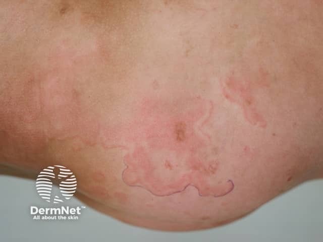
Clinical photograph (dermatology) showing erythema marginatum, an annular, non-itchy rash characteristic of acute rheumatic fever. The image depicts multiple pink to red circular to oval patches on the skin of the lower extremity with sharply demarcated, serpiginous margins and centers that appear pale or clearing. Lesions are non-vesicular, smooth-surfaced, and vary in size, with a tendency to migrate or transiently flare. The distribution favors the trunk and proximal limbs and typically spares the face. On close inspection the patches lack scaling and crusting, consistent with erythema marginatum rather than dermatitis or fungal infection. This cutaneous manifestation is part of the Jones criteria and may correlate with concurrent migratory polyarthritis, fever, and evidence of preceding group A Streptococcus infection. Histologic features (if biopsied) would show superficial perivascular lymphocytic infiltrate and dermal edema with minimal epidermal changes; clinically, the rash is a marker for systemic inflammatory involvement. Differential considerations include urticaria, erythema multiforme, tinea corporis, pityriasis rosea, and other annular dermatoses. Clinically, recognizing erythema marginatum prompts evaluation for acute rheumatic fever and guides management including antibiotic therapy and anti-inflammatory treatment. The image serves educational and diagnostic-reference purposes for dermatology, pediatrics, infectious diseases, and rheumatology training.
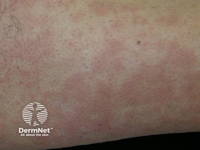
This is a clinical dermatology photograph capturing erythema marginatum on the skin of the lower limb. The modality is Clinical photography; technique: naked-eye, color-balanced image. The observed lesions are erythematous, annular plaques with central clearing and pale, sometimes hypopigmented centers. Lesions appear as rings with slightly raised margins and a uniform pink-to red hue; they range from 0.5 to 2 cm in diameter and may coalesce into larger plaques. The rash is typically non-pruritic and evanescent, with temporary eruption that may migrate or fade over hours to days; distribution commonly involves the trunk and proximal limbs, with sparing of the face. The image may reflect erythema marginatum, a cutaneous manifestation associated with acute rheumatic fever and autoimmune inflammatory processes. In longer-standing presentations, the rash signals preceding or concurrent carditis and migratory arthritis; therefore clinical correlation with recent group A Streptococcus infection, elevated ASO titers, and echocardiography is essential. Differential diagnoses include urticaria, erythema multiforme, pityriasis rosea, and drug-induced eruptions. Clinically, this finding has diagnostic significance as a major criterion in rheumatic fever per Jones criteria; management focuses on antibiotic treatment of streptococcal infection and secondary prophylaxis. This image serves educational purposes for medical students, residents, and researchers studying connective tissue disease and pediatric rheumatology.
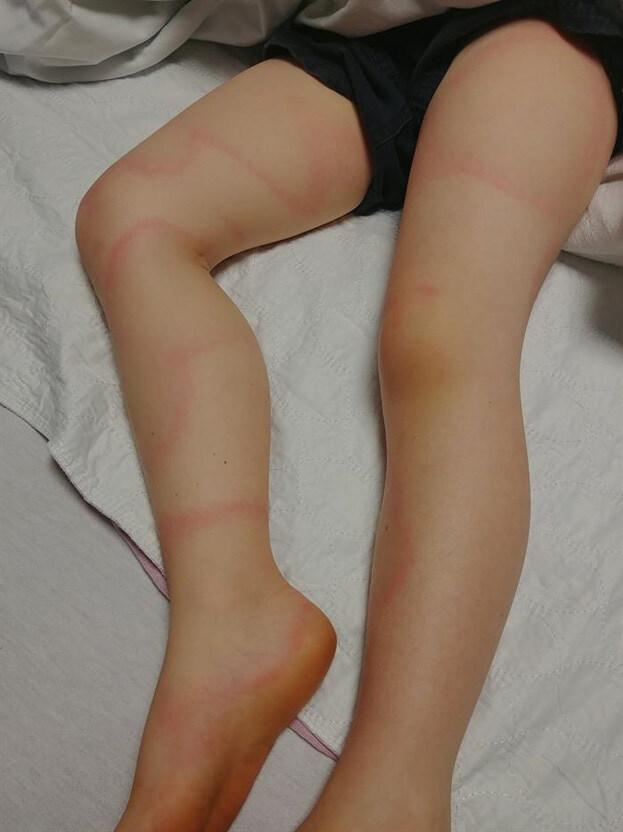
A clinical photograph of the lower extremities of a pediatric patient, demonstrating a distinctive cutaneous manifestation. The image shows multiple erythematous, annular (ring-shaped) lesions distributed across the thighs, knees, shins, and ankles. The rash is characterized by raised, pinkish-red borders that form large, asymmetric, and serpiginous patterns, some partially encircling the limbs. These lesions appear papular and lack visible scaling or central crusting, suggesting an inflammatory or migratory process such as Erythema Marginatum or Erythema Migrans. A localized area of yellowish discoloration (bruising or ecchymosis) is visible on the right knee. The morphology of these blanchable, annular lesions is a key diagnostic feature often associated with systemic conditions such as acute rheumatic fever or early-stage Lyme disease. This visual is intended for medical education in dermatology, pediatrics, and infectious diseases to assist in the identification of classic annular rash patterns.
Aschoff body rheumatic heart histology
Rheumatic Fever (Acute Rheumatic Fever) - USMLE Review
1. Definition and Etiology
2. Pathophysiology (frequently tested mechanism)
- Certain "rheumatogenic" GAS M protein serotypes (e.g., types 1, 3, 5, 6, 18) share antigenic epitopes with human tissue proteins - this is molecular mimicry.
- Antibodies raised against the bacterial M protein cross-react with cardiac myosin, tropomyosin, laminin (valve endothelium), synovium, and neuronal tissue (basal ganglia).
- This is classically taught as a type II hypersensitivity reaction (antibody-mediated), although T-cell mediated mechanisms also contribute.
- Result: valvulitis, pancarditis (endocarditis + myocarditis + pericarditis), migratory synovitis, and basal ganglia dysfunction (chorea) (Medical Microbiology 9e, p. 229; Firestein & Kelley's Textbook of Rheumatology).
- Pathology hallmark: Aschoff bodies - foci of fibrinoid necrosis surrounded by lymphocytes, plasma cells, and activated macrophages ("Anitschkow cells" / "caterpillar cells" with slender, wavy nuclear chromatin) found in the myocardium.
- Mnemonic often used: "Rheumatic fever licks the joints but bites the heart" - arthritis is more common but self-limited; carditis causes the lasting damage.
3. Epidemiology
- Peaks in children ages 4-9; rare before age 3 and uncommon after 15.
- Only about 0.3-3% of untreated GAS pharyngitis cases progress to ARF, but risk is higher with rheumatogenic strains and genetic susceptibility (certain HLA class II alleles).
- Now rare in high-income countries (largely eradicated) but remains a major cause of morbidity/mortality (rheumatic heart disease, RHD) in low- and middle-income countries, causing an estimated 233,000-500,000 deaths/year globally (Firestein & Kelley's Textbook of Rheumatology).
4. Diagnosis - Revised/Modified Jones Criteria
- 2 major criteria, OR
- 1 major + 2 minor criteria
| Major Criteria (JONES mnemonic) | Minor Criteria |
|---|---|
| J – Joints: migratory polyarthritis (large joints, 60-75% of cases) | Fever |
| O – (o-shaped) Carditis (pancarditis; new/changing murmur, cardiomegaly, CHF, pericarditis) | Arthralgia (if arthritis already counted as major) |
| N – Nodules (subcutaneous, painless, over extensor surfaces/bony prominences) | Elevated ESR/CRP |
| E – Erythema marginatum (evanescent, serpiginous, non-pruritic annular rash, trunk/proximal limbs) | Prolonged PR interval on ECG |
| S – Sydenham chorea ("St. Vitus dance" - involuntary, purposeless movements; can appear in isolation and alone satisfies diagnostic criteria) | History of prior ARF |
5. Clinical Features in Detail
- Carditis (~50-70%): the only manifestation that causes chronic disease. Order of frequency of valve involvement: mitral valve alone most common, then mitral + aortic combined, aortic alone least common. Acute phase causes mitral regurgitation (valvulitis); chronic recurrent disease over years causes mitral stenosis (fibrosis, fusion of chordae, "fish-mouth" or "buttonhole" valve) - one of the few causes of pure mitral stenosis without prior regurgitation history.
- Arthritis: migratory, asymmetric, affects large joints (knees, ankles, elbows, wrists); dramatically responsive to aspirin/NSAIDs (a diagnostic clue - if migratory arthritis does NOT respond to salicylates, reconsider the diagnosis, e.g., post-streptococcal reactive arthritis instead).
- Subcutaneous nodules: firm, painless, over extensor tendons/bony prominences; associated with more severe carditis.
- Erythema marginatum: rare (<6%) but highly specific; pink, serpiginous, blanching rings with central clearing, spares the face, worsens with heat.
- Sydenham chorea: rare, delayed onset, more common in girls; resolves over weeks to months. Distinguish from PANDAS (pediatric autoimmune neuropsychiatric disorder associated with streptococcus), a related but distinct clinical entity.
6. Treatment
- Eradicate GAS: Penicillin (benzathine penicillin G IM single dose, or oral penicillin V/amoxicillin for 10 days; erythromycin/azithromycin if penicillin-allergic) - given even if throat culture is negative at presentation, since the triggering infection has usually cleared by the time ARF manifests.
- Anti-inflammatory therapy: High-dose aspirin (or NSAIDs) for arthritis/arthralgia and mild carditis; corticosteroids for moderate-to-severe carditis or pancarditis.
- Heart failure management if carditis causes decompensation (diuretics, ACE inhibitors as needed).
- Chorea: supportive care; can use haloperidol or valproic acid if severe.
- Secondary prophylaxis (the single most important long-term intervention to prevent recurrence and progression to rheumatic heart disease): monthly (every 3-4 weeks) IM benzathine penicillin G, continued for:
- No carditis: 5 years or until age 21 (whichever is longer)
- Carditis without residual valve disease: 10 years or until age 21
- Carditis with residual valvular disease: 10 years or until age 40, sometimes lifelong
7. High-Yield USMLE Pearls
- Rheumatic fever follows strep throat, not strep skin infection.
- Diagnosis is clinical (Jones criteria) - there is no single confirmatory lab test.
- Chorea or indolent carditis alone can establish the diagnosis without meeting full Jones criteria.
- The valve most commonly damaged is the mitral valve; chronic disease causes mitral stenosis.
- Aschoff bodies and Anitschkow cells are the pathognomonic histologic findings in the myocardium.
- Mechanism = molecular mimicry / type II hypersensitivity against M protein-related epitopes.
- Secondary penicillin prophylaxis is the key preventive measure and a common answer for "next best step" recurrence-prevention questions.