
non variceal upper gi bleeding
"nonvariceal upper gastrointestinal bleeding" AND management
Forrest classification peptic ulcer bleeding endoscopy
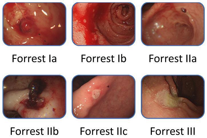
This composite educational clinical image demonstrates the Forrest classification for peptic ulcer bleeding (PUB) via six gastroscopic panels. Top row (Active Bleeding): Forrest Ia shows 'spurting hemorrhage' with an arterial jet; Forrest Ib shows 'oozing hemorrhage' without a visible vessel. Middle and bottom rows (Recent Hemorrhage Signs): Forrest IIa depicts a 'non-bleeding visible vessel' as a protuberant lesion; Forrest IIb shows an 'adherent clot' covering the ulcer base; Forrest IIc presents 'flat pigmented spots' (hematin) indicating recent stasis; and Forrest III displays a 'clean-based ulcer' with a white fibrin base and no stigmata of hemorrhage. This classification system is essential for gastroenterologists to assess rebleeding risk and determine the necessity of endoscopic hemostasis therapy during emergency esophagogastroduodenoscopy (EGD).
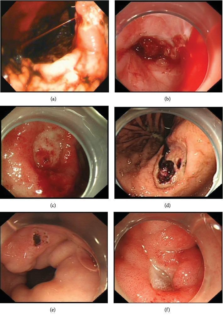
A composite of six endoscopic images illustrating the Forrest classification for upper gastrointestinal bleeding risk assessment in peptic ulcer disease. (a) Forrest Ia: demonstrates active, high-pressure spurting bleeding from an arterial source. (b) Forrest Ib: shows active oozing bleeding without pulsatility. (c) Forrest IIa: displays a non-bleeding visible vessel appearing as a raised, red protuberance within the ulcer bed. (d) Forrest IIb: shows an adherent clot, a dark, thick mass of coagulated blood firmly attached to the ulcer. (e) Forrest IIc: depicts a flat, pigmented spot (haematin) indicating recent hemorrhage. (f) Forrest III: shows a clean-based ulcer with a white or yellow fibrin base and no signs of recent bleeding. This series is a critical educational tool for gastroenterology to differentiate between high-risk (Ia-IIb) and low-risk (IIc-III) lesions, guiding endoscopic intervention and pharmacological management.
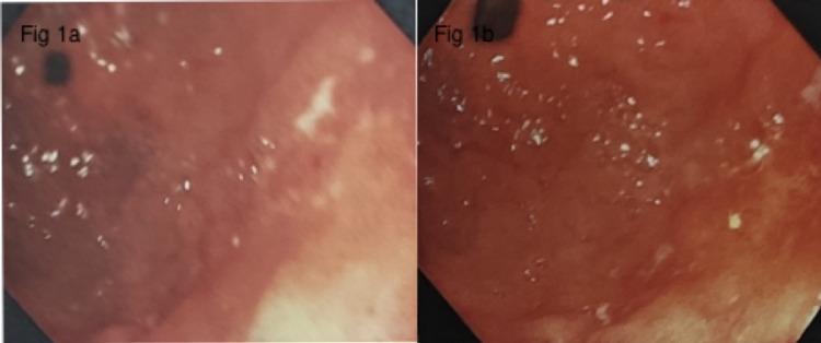
Comparison of two upper gastrointestinal endoscopy (UGIE) frames of the gastric antrum, labeled Figure 1a and Figure 1b, documenting the progression of a gastric ulcer. Figure 1a shows the initial presentation of a gastric ulcer located along the lesser curvature, characterized by the presence of an adherent clot, consistent with a Forrest class IIb bleeding classification. The surrounding mucosa displays patchy erythema. Figure 1b, captured two days post-intervention, shows the same ulcer at the incisura now featuring a clean base without an adherent clot, corresponding to Forrest class III. The surrounding gastric tissue exhibits signs of induration and persistent erythema. These images illustrate the diagnostic and monitoring utility of endoscopy in assessing upper gastrointestinal bleeding and the evolution of peptic ulcer disease in the context of H. pylori-positive gastritis.
Rockall score Blatchford score risk stratification upper GI bleeding table
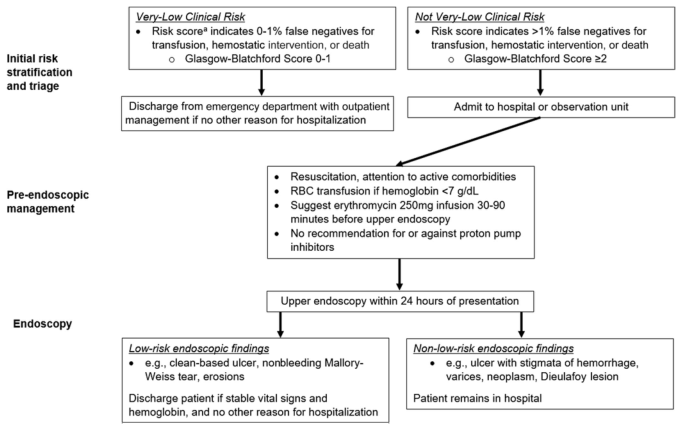
Summary : This flowchart outlines the recommended clinical management pathway for patients presenting with upper gastrointestinal bleeding, based on initial risk stratification using the Glasgow-Blatchford Score, pre-endoscopic management, and endoscopic findings. flowchart: # Nodes : • Initial risk stratification and triage (rectangle) – Very-Low Clinical Risk (rectangle) • Risk score indicates 0–1% false negatives for transfusion, hemostatic intervention, or death • Glasgow-Blatchford Score 0–1 – Not Very-Low Clinical Risk (rectangle) • Risk score indicates >1% false negatives for transfusion, hemostatic intervention, or death • Glasgow-Blatchford Score ≥2 – Discharge from emergency department with outpatient management if no other reason for hospitalization (rectangle) – Admit to hospital or observation unit (rectangle) • Pre-endoscopic management (rectangle) – Resuscitation, attention to active comorbidities – RBC transfusion if hemoglobin <7 g/dL – Suggest erythromycin 250mg infusion 30–90 minutes before upper endoscopy – No recommendation for or against proton pump inhibitors • Upper endoscopy within 24 hours of presentation (rectangle) • Endoscopy (rectangle) – Low-risk endoscopic findings (rectangle) • e.g., clean-based ulcer, nonbleeding Mallory-Weiss tear, erosions • Discharge patient if stable vital signs and hemoglobin, and no other reason for hospitalization – Non-low-risk endoscopic findings (rectangle) • e.g., ulcer with stigmata of hemorrhage, varices, neoplasm, Dieulafoy lesion • Patient remains in hospital # Connectors : • From "Initial risk stratification and triage" split into two branches: – Very-Low Clinical Risk → Discharge from emergency department – Not Very-Low Clinical Risk → Admit to hospital or observation unit • Both branches converge to "Pre-endoscopic management" • "Pre-endoscopic management" leads to "Upper endoscopy within 24 hours of presentation" • "Endoscopy" splits into: – Low-risk endoscopic findings → Discharge patient if stable – Non-low-risk endoscopic findings → Patient remains in hospital # Layout : • Vertical flow from top (Initial risk stratification) to bottom (Endoscopy outcomes) • Two main branches at the top (Very-Low vs Not Very-Low Clinical Risk) merge before pre-endoscopic management • Endoscopy results split into two outcome branches (discharge vs remain in hospital) # Analysis : • The flowchart provides a clear, stepwise approach for triaging and managing upper GI bleeding based on risk assessment and endoscopic findings. • Patients with very-low clinical risk (Glasgow-Blatchford Score 0–1) are discharged early, while higher-risk patients are admitted. • Pre-endoscopic management is standardized for all admitted patients, focusing on resuscitation and transfusion thresholds. • Endoscopic findings further stratify patients into those suitable for discharge versus those requiring continued hospitalization. • The process emphasizes early risk stratification and timely endoscopy (within 24 hours) to optimize patient outcomes.
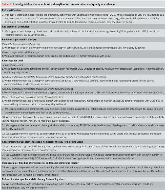
<table> <tr> <td colspan="2"><b>Table 1. </b>List of guideline statements with strength of recommendation and quality of evidence</td> </tr> <tr> <td colspan="2"><b>Risk stratification</b></td> </tr> <tr> <td>1.</td> <td>We suggest that patients presenting to the emergency department with upper gastrointestinal bleeding (UGIB) who are classified as very low risk, defined as a risk assessment score with ≤1% false negative rate for the outcome of hospital-based intervention or death (e.g., Glasgow-Blatchford score = 0–1), be discharged with outpatient follow-up rather than admitted to hospital (conditional recommendation, very-low-quality evidence).</td> </tr> <tr> <td colspan="2"><b>Red blood cell transfusion</b></td> </tr> <tr> <td>2.</td> <td>We suggest a restrictive policy of red blood cell transfusion with a threshold for transfusion at a hemoglobin of 7 g/dL for patients with UGIB (conditional recommendation, low-quality evidence).</td> </tr> <tr> <td colspan="2"><b>Pre-endoscopic medical therapy</b></td> </tr> <tr> <td colspan="2"><i>Prokinetic therapy with erythromycin</i></td> </tr> <tr> <td>3.</td> <td>We suggest an infusion of erythromycin before endoscopy in patients with UGIB (conditional recommendation, very-low-quality evidence).</td> </tr> <tr> <td colspan="2"><i>Proton pump inhibitor (PPI) therapy</i></td> </tr> <tr> <td>4.</td> <td>We could not reach a recommendation for or against pre-endoscopic PPI therapy for patients with UGIB.</td> </tr> <tr> <td colspan="2"><b>Endoscopy for UGIB</b></td> </tr> <tr> <td colspan="2"><i>Timing of endoscopy</i></td> </tr> <tr> <td>5.</td> <td>We suggest that patients admitted to or under observation in hospital for UGIB undergo endoscopy within 24 hr of presentation (conditional recommendation, very-low-quality evidence).</td> </tr> <tr> <td colspan="2"><i>Need for endoscopic hemostatic therapy for ulcers with active bleeding or nonbleeding visible vessels</i></td> </tr> <tr> <td>6.</td> <td>We recommend endoscopic therapy in patients with UGIB due to ulcers with active spurting, active oozing, and nonbleeding visible vessels (strong recommendation, moderate-quality evidence).</td> </tr> <tr> <td colspan="2"><i>Need for endoscopic hemostatic therapy for ulcers with adherent clot</i></td> </tr> <tr> <td>7.</td> <td>We could not reach a recommendation for or against endoscopic therapy in patients with UGIB due to ulcers with adherent clot resistant to vigorous irrigation.</td> </tr> <tr> <td colspan="2"><i>Choice of endoscopic hemostatic therapy for bleeding ulcers</i></td> </tr> <tr> <td>8.</td> <td>We recommend endoscopic hemostatic therapy with bipolar electrocoagulation, heater probe, or injection of absolute ethanol for patients with UGIB due to ulcers (strong recommendation, moderate-quality evidence).</td> </tr> <tr> <td>9.</td> <td>We suggest endoscopic hemostatic therapy with clips, argon plasma coagulation, or soft monopolar electrocoagulation for patients with UGIB due to ulcers (conditional recommendation, very-low- to low-quality evidence).</td> </tr> <tr> <td>10.</td> <td>We recommend that epinephrine injection not be used alone for patients with UGIB due to ulcers but rather in combination with another hemostatic modality (strong recommendation, very-low- to moderate-quality evidence).</td> </tr> <tr> <td>11.</td> <td>We suggest endoscopic hemostatic therapy with hemostatic powder spray TC-325 for patients with actively bleeding ulcers (conditional recommendation, very-low-quality evidence).</td> </tr> <tr> <td>12.</td> <td>We suggest over-the-scope clips as a hemostatic therapy for patients who develop recurrent bleeding due to ulcers after previous successful endoscopic hemostasis (conditional recommendation, low-quality evidence).</td> </tr> <tr> <td colspan="2"><i>Antisecretory therapy after endoscopic hemostatic therapy for bleeding ulcers</i></td> </tr> <tr> <td>13.</td> <td>We recommend high-dose PPI therapy given continuously or intermittently for 3 d after successful endoscopic hemostatic therapy of a bleeding ulcer (strong recommendation, moderate- to high-quality evidence).</td> </tr> <tr> <td>14.</td> <td>We suggest that high-risk patients with UGIB due to ulcers who received endoscopic hemostatic therapy followed by short-term high-dose PPI therapy in hospital continue on twice-daily PPI therapy until 2 wk after index endoscopy (conditional recommendation, low-quality evidence).</td> </tr> <tr> <td colspan="2"><i>Recurrent ulcer bleeding after successful endoscopic hemostatic therapy</i></td> </tr> <tr> <td>15.</td> <td>We suggest that patients with recurrent bleeding after endoscopic therapy for a bleeding ulcer undergo repeat endoscopy and endoscopic therapy rather than undergo surgery or transcatheter arterial embolization (conditional recommendation, low-quality evidence for comparison with surgery, very-low-quality evidence for comparison with transcatheter arterial embolization)</td> </tr> <tr> <td colspan="2"><i>Failure of endoscopic hemostatic therapy for bleeding ulcers</i></td> </tr> <tr> <td>16.</td> <td>We suggest patients with bleeding ulcers who have failed endoscopic therapy next be treated with transcatheter arterial embolization (conditional recommendation, very-low-quality evidence).</td> </tr> </table>
Non-Variceal Upper GI Bleeding (NVUGIB)
Definition & Scope
Causes (by frequency)
| Cause | Frequency |
|---|---|
| Peptic ulcer | 35.2% |
| Esophagitis | 4.6% |
| Angioectasia / telangiectasia | 4.0% |
| Mallory-Weiss tear | 4.0% |
| Dieulafoy lesion | 3.2% |
| UGI tract neoplasm | 3.1% |
| Epistaxis (swallowed) | 2.2% |
| Erosions | 1.2% |
| No cause found | 7.3% |
Clinical Presentation
- Hematemesis (vomiting bright red blood or "coffee grounds")
- Melena (black, tarry stools - digested blood)
- Hematochezia (only if bleeding is very brisk)
- Elevated BUN:creatinine ratio - digested blood raises BUN (a useful biochemical clue)
- Shock (hypotension, tachycardia) in severe cases
- Nasogastric lavage showing red blood or coffee-ground aspirate confirms upper GI source
Risk Stratification
Pre-endoscopy Scores
- Blood pressure
- BUN level
- Hemoglobin
- Heart rate
- Syncope, melena, liver disease, heart failure
- GBS 0-1 = very low risk; safe for outpatient management / early discharge
- Albumin < 3.0 g/dL
- INR > 1.5
- Altered mental status
- Systolic BP < 90 mmHg
- Age > 65 years
- Score ≥ 2 = higher risk of mortality, longer stay, higher cost
Post-endoscopy Score
- Pre-endoscopy: age, shock, comorbidities
- Endoscopic: diagnosis + stigmata of recent hemorrhage (SRH)
- Score 0-2 = low risk; may consider early discharge
- Correlates well with mortality but less so with rebleeding risk
Forrest Classification (Endoscopic SRH)
| Class | Finding | Rebleeding Risk |
|---|---|---|
| Ia | Active spurting arterial bleeding | ~55% |
| Ib | Active oozing | ~55% |
| IIa | Non-bleeding visible vessel | ~43% |
| IIb | Adherent clot | ~22% |
| IIc | Flat pigmented spot | ~10% |
| III | Clean-based ulcer | ~5% |
Forrest IIb (adherent clot) = controversial; vigorous irrigation recommended
Forrest IIc, III = low risk; no endoscopic therapy needed
Management
1. Initial Resuscitation
- Large-bore IV access, volume resuscitation
- RBC transfusion threshold: Hb < 7 g/dL (restrictive strategy - reduces mortality vs. liberal transfusion in trials)
- Correct coagulopathy (platelets, FFP as needed)
- Airway protection - consider prophylactic intubation if massive active bleeding before endoscopy
- Admit if GBS ≥ 2; GBS 0-1 can be discharged from ED with outpatient follow-up
2. Pre-endoscopic Medical Therapy
- Erythromycin 250 mg IV infusion 30-90 min before endoscopy (prokinetic - clears blood from stomach, improves mucosal visualization) - conditional recommendation
- Pre-endoscopy PPI: no consensus recommendation for or against; many centers give it empirically
- Octreotide: not routinely used in NVUGIB (main role is variceal bleeding)
3. Endoscopy
- Timing: within 24 hours of presentation after adequate resuscitation (strong recommendation)
- Upper endoscopy is both diagnostic AND therapeutic
- Therapeutic endoscope with large suction channel preferred
- A side-viewing duodenoscope should be used if duodenal ampullary cause is suspected
| Method | Notes |
|---|---|
| Injection therapy | Epinephrine - must NOT be used alone; always combine with a second modality |
| Thermal contact | Bipolar electrocoagulation, heater probe, soft monopolar - strong recommendation |
| Mechanical | Through-the-scope clips; over-the-scope clips (OTSC) for refractory/recurrent bleeding |
| Non-contact thermal | Argon plasma coagulation (APC) |
| Hemostatic powder | TC-325 (hemostatic spray) for actively bleeding ulcers |
| Absolute ethanol injection | Strong recommendation |
- Epinephrine alone is contraindicated as monotherapy - must combine with thermal or mechanical method
- OTSC (over-the-scope clip) is preferred for recurrent bleeding after prior successful hemostasis - supported by 2025 meta-analysis (PMID 39500365)
4. Post-endoscopy PPI Therapy
- High-dose PPI (e.g., IV omeprazole 80 mg bolus then 8 mg/hr infusion, or equivalent intermittent dosing) for 3 days after successful endoscopic hemostasis of a high-risk ulcer - strong recommendation (moderate-high quality evidence)
- Continue twice-daily oral PPI until 2 weeks post-index endoscopy in high-risk patients
5. H. pylori Management
- Test all bleeding peptic ulcer patients for H. pylori
- Eradicate if positive (reduces long-term rebleeding risk dramatically)
- Confirm eradication with urea breath test or stool antigen after treatment
6. NSAID/Aspirin Management
- Discontinue NSAIDs where possible
- If aspirin must be continued (cardiovascular indication), use lowest effective dose + PPI co-prescription
Salvage Therapies (Failed Endoscopy)
Transcatheter Arterial Embolization (TAE)
- Preferred over surgery when endoscopy fails (conditional recommendation)
- Celiac and SMA angiography performed; CO2 contrast useful for detecting slow bleeds
- Selective embolization of gastroduodenal artery (duodenal ulcers) or left gastric artery (gastric ulcers)
- "Prophylactic embolization" acceptable when massive bleeding has ceased at time of angiography
Surgery
- Considered when TAE fails or is unavailable
- Reserved as last resort given high operative mortality in this population
Management Flowchart
Cause-Specific Notes
Peptic Ulcer Disease
Mallory-Weiss Tear
- Caused by forceful retching - mucosal laceration at gastroesophageal junction
- Accounts for ~4-15% of UGIB
- Most stop spontaneously; rebleeding in ~10%
- Endoscopic options: bipolar electrocoagulation, epinephrine injection, clips, band ligation
Dieulafoy Lesion
- Aberrant submucosal artery that erodes through mucosa without an ulcer
- Often missed at first endoscopy; requires careful inspection
- Endoscopic hemostasis (clips, band ligation, thermal therapy) is effective
Esophagitis
- ~10% of UGIB, usually from GERD
- Less hemodynamic instability; excellent prognosis, low rebleeding risk
Angioectasia / AVMs
- Common in elderly, patients with renal failure, aortic stenosis (Heyde syndrome)
- Argon plasma coagulation is treatment of choice
Recent Evidence (2024-2025)
- [2024 Systematic Review, PMID 38446116]: Defined failure rates and predictors of failed endoscopic hemostasis in NVUGIB - identifies posterior duodenal ulcers and large ulcers as highest-risk for failure.
- [2025 Meta-analysis, PMID 39500365]: Over-the-scope clips (OTSC) significantly reduce rebleeding vs. standard endoscopic therapy in recurrent bleeding after initial hemostasis.
- [2025 Japanese Practice Guideline, PMID 40114631]: Second edition guidelines for endoscopic management, reinforcing role of combined hemostatic modalities and early high-dose PPI.
Key Points Summary
- Peptic ulcer = most common cause (~35%); driven by H. pylori and NSAIDs
- GBS 0-1 = safe for outpatient management; GBS ≥ 2 = admit
- Erythromycin 250 mg IV pre-endoscopy improves visualization
- Endoscopy within 24 hours for all admitted patients
- Forrest Ia/Ib/IIa = endoscopic therapy mandatory
- Never use epinephrine alone - combine with thermal or mechanical method
- High-dose PPI 72 hours post-hemostasis = strong recommendation
- Rebleed after endoscopy = repeat endoscopy first; if fails, TAE before surgery
- Test and eradicate H. pylori in all peptic ulcer bleeders