Tell me about acute pyelonephritis for step 1

Reading File
Finding Sources
Searching PubMed

"pyelonephritis"[MeSH Terms] AND "diagnosis"[MeSH Terms]

Reading File
Reading File
Reading File
Finding Sources
Finding Sources
Reading File
Reading File
Reading File
Loading Image
Reading File
Reading File
Loading Image
Reading File
I now have all the material needed for a comprehensive Step 1-oriented answer. Let me compile everything.

Acute Pyelonephritis - Step 1 Review

Definition

Acute pyelonephritis is suppurative (purulent) inflammation of the kidney and renal pelvis caused by bacterial infection. It is an upper urinary tract infection (UTI), in contrast to cystitis (lower UTI). One or both kidneys may be involved.
  • Robbins & Kumar Basic Pathology, p. 514
  • Robbins, Cotran & Kumar Pathologic Basis of Disease, p. 859

Pathogenesis - Routes of Infection
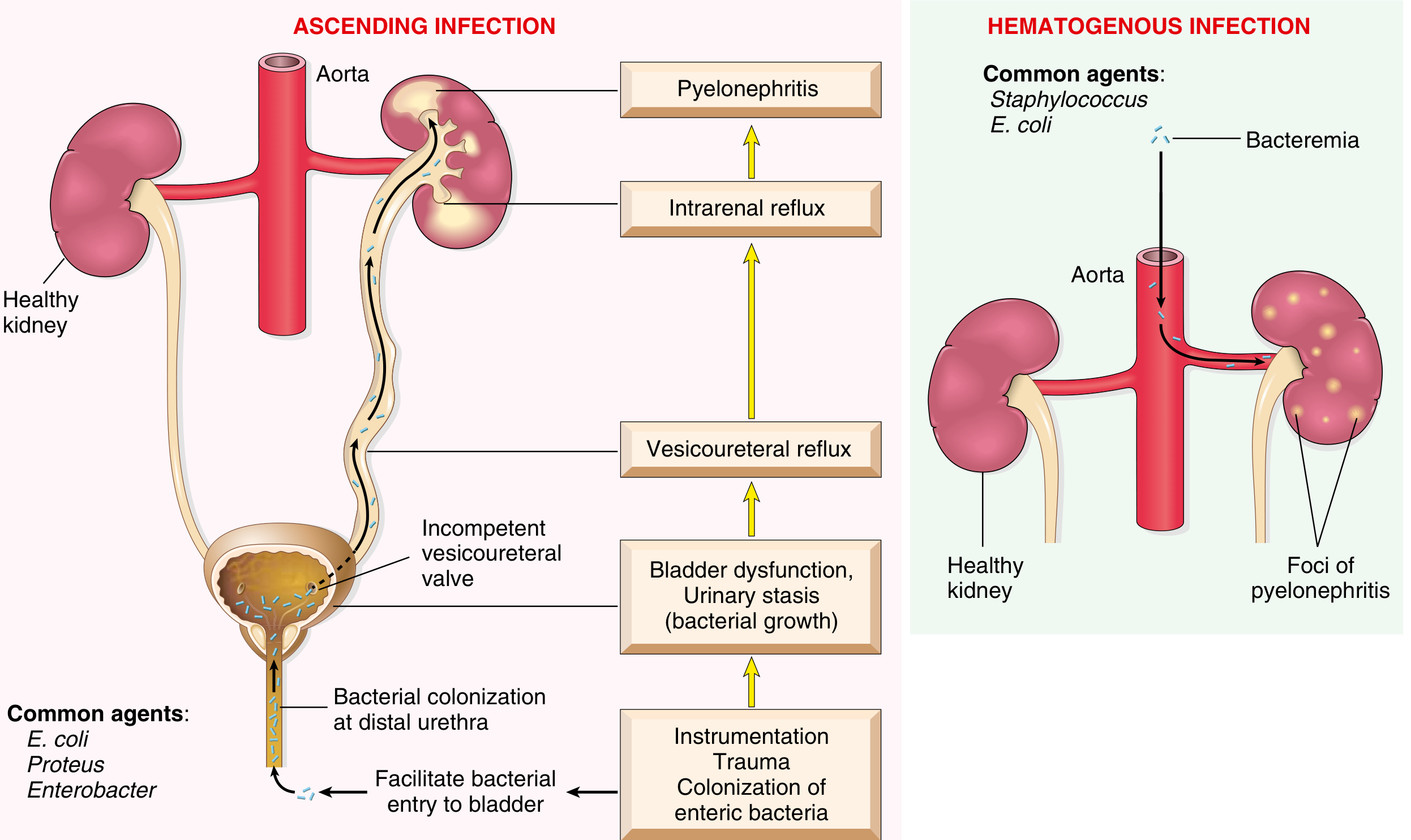
Two pathways exist; ascending is by far the more common:
Pathways of renal infection: ascending vs hematogenous
1. Ascending infection (most common)
  • Colonization of distal urethra/introitus by enteric bacteria (especially E. coli)
  • Instrumentation, trauma, or sexual activity facilitates bladder entry
  • Urinary stasis (obstruction, bladder dysfunction, neurogenic bladder, VUR) allows bacterial multiplication
  • Vesicoureteral reflux (VUR) - incompetent ureterovesical valve - propels infected urine up to the renal pelvis and parenchyma via intrarenal reflux
  • VUR present in 20-40% of children with UTI (usually congenital)
2. Hematogenous infection (less common)
  • Bacteremia (e.g., endocarditis, sepsis) seeds the kidney
  • Common agents here: Staphylococcus aureus, E. coli
  • Produces multifocal cortical abscesses rather than medullary/pelvic disease

Microbiology

OrganismNotes
E. coliMost common (80% of cases); possesses P pili that bind to P blood group antigen receptors on urothelium (virulence factor)
KlebsiellaCommon, especially in diabetics
ProteusUrease-producing - raises urinary pH, causes struvite stones
EnterobacterLess common gram-negative rod
PseudomonasAssociated with nosocomial/catheter infections
StaphylococcusHematogenous route; S. saprophyticus in young sexually active women
  • All gram-negative enteric bacilli are normal intestinal flora.
  • Campbell Walsh Wein Urology, p. 3308

Risk Factors / Predisposing Conditions

  • Female sex - short urethra close to rectum; most common in sexually active women
  • Urinary tract obstruction - BPH, calculi, uterine prolapse, posterior urethral valves
  • Vesicoureteral reflux (VUR)
  • Pregnancy - uterine compression causes urinary stasis; 4-6% develop bacteriuria; 20-40% of untreated bacteriuria progresses to pyelonephritis
  • Catheterization / urinary tract instrumentation
  • Diabetes mellitus - immunosuppression + neurogenic bladder dysfunction
  • Immunosuppression / immunodeficiency
  • Preexisting renal scarring

Clinical Presentation

Classic triad (classic Step 1 teaching):
Fever + Flank/CVA (costovertebral angle) pain/tenderness + Dysuria
More specifically:
  • Systemic: abrupt onset of chills, fever (≥100.4°F / 38°C), malaise, nausea/vomiting
  • Urinary: dysuria, frequency, urgency (lower tract symptoms may also be present or absent)
  • Physical exam: costovertebral angle (CVA) tenderness to deep palpation
  • Can simulate GI illness with abdominal pain, nausea, vomiting
  • Usually unilateral - does NOT typically cause renal failure in uncomplicated cases
  • Robbins & Kumar Basic Pathology, p. 516
  • Campbell Walsh Wein Urology, p. 3294

Lab Findings

TestFindingSignificance
UrinalysisPyuria (many WBCs), bacteriuriaCore finding
WBC castsNeutrophil-rich (pus) castsPathognomonic for kidney involvement (casts form only in tubules)
Urine culturePositive (usually >10^5 CFU/mL)Establishes diagnosis; ~20% have <10^5 CFU/mL
CBCLeukocytosis with left shift (neutrophilia)Systemic infection
Blood culturesMay be positiveBacteremia/urosepsis
CRP/ESRElevatedInflammatory markers
CreatinineMay be elevated in severe casesAKI (uncommon in uncomplicated cases)
Key Step 1 pearl: WBC casts = upper tract (renal parenchymal) involvement. You will NOT see WBC casts in uncomplicated cystitis.

Morphology / Pathology

Gross:
  • Discrete, raised, yellowish-white abscesses on the cortical surface
  • May form large wedge-shaped areas of necrosis
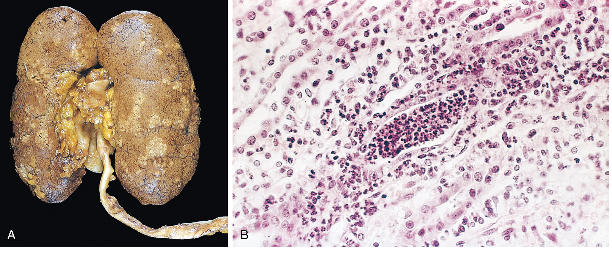
Acute pyelonephritis - gross specimen (A) and histology (B)
Fig. 20.23: (A) Cortical surface shows gray-white areas of inflammation and abscess formation. (B) Neutrophilic exudate within tubules and interstitial inflammation.
Histology:
  • Patchy interstitial suppurative inflammation
  • Intratubular aggregates of neutrophils (neutrophilic tubulitis)
  • Tubular injury with extension into the interstitium
  • Glomeruli are relatively SPARED (characteristically resistant)
  • Fungal infections (e.g., Candida) cause granulomatous interstitial inflammation instead

Complications

Three major complications can be superimposed:

1. Papillary Necrosis

  • Ischemic + suppurative necrosis of the tips/distal two-thirds of the renal papillae
  • Classic triad: Diabetes + Urinary obstruction + Sickle cell disease (also analgesic nephropathy)
  • Usually bilateral but can be unilateral
  • Microscopy: coagulative necrosis of papillary tips with preserved tubular outlines
  • May lead to acute renal failure if sloughed papillae cause obstruction

2. Pyonephrosis

  • Total or near-complete urinary obstruction, especially high in the urinary tract
  • Suppurative exudate fills the renal pelvis, calyces, and ureter with pus
  • Unable to drain - medical emergency

3. Perinephric Abscess

  • Extension of suppurative inflammation through the renal capsule into perinephric fat
  • May not respond to antibiotics alone; requires drainage

Treatment (Step 1 Overview)

SettingPreferred Agents
Mild-moderate, outpatientFluoroquinolones (ciprofloxacin 5-7 days; levofloxacin 5-7 days) or TMP-SMX x 14 days
Hospitalized / severeIV aminoglycoside (gentamicin) initial empiric therapy; transition to oral after 24-48 hrs of improvement
PregnancyIV cephalosporins or penicillins until afebrile, then 14 days total oral
Complicated/resistantMeropenem, piperacillin-tazobactam, plazomicin
  • Nitrofurantoin and fosfomycin are NOT appropriate for pyelonephritis (insufficient renal tissue levels)
  • Duration: 7-14 days total (5-7 days adequate for fluoroquinolones)
  • Goldman-Cecil Medicine, p. 2979

Key Distinctions for Step 1

FeatureCystitis (Lower UTI)Pyelonephritis (Upper UTI)
Fever/chillsUsually absentPresent
CVA tendernessAbsentPresent
WBC castsABSENTPresent (pathognomonic)
Systemic signsAbsentPresent
Renal impairmentNoPossible in severe/complicated cases
Treatment duration3-7 days7-14 days

Recent Evidence Note

A 2025 systematic review (PMID 40228579) comparing short vs. long antibiotic courses for pyelonephritis supports that shorter courses (5-7 days with fluoroquinolones) are non-inferior to traditional 10-14 day courses for uncomplicated disease - consistent with current guidelines. A 2025 network meta-analysis (PMID 39817442) confirms fluoroquinolones and beta-lactams as effective treatment options.

High-Yield Summary for Step 1

  • Most common organism: E. coli (gram-negative, enteric bacillus, P pili virulence factor)
  • Most common route: Ascending infection (lower UTI → VUR → upper tract)
  • Classic presentation: Fever + CVA tenderness + dysuria/pyuria
  • Key urinalysis finding: WBC casts (= renal tubular origin = upper tract)
  • Histology: Neutrophilic tubulitis + interstitial suppuration + spared glomeruli
  • Major complications: Papillary necrosis (diabetics/SCD/obstruction), pyonephrosis, perinephric abscess
  • First-line treatment: Fluoroquinolones (outpatient); aminoglycosides (empiric inpatient)
  • Do NOT use: Nitrofurantoin for upper tract infection

Acute interstatil nephritis

Finding Sources
Searching PubMed

"acute interstitial nephritis"[MeSH Terms]

Reading File
Reading File
Finding Sources
Reading File
Reading File
Reading File
Reading File
Finding Sources
Reading File
Reading File
Reading File
Finding Sources
Reading File
Loading Image
I now have comprehensive material. Let me compile the full Step 1-focused response.

Acute Interstitial Nephritis (AIN) - Step 1 Review

Definition

Acute interstitial nephritis (AIN) is an acute, often reversible form of AKI characterized by inflammatory cell infiltrates within the renal interstitium and tubules, with relative sparing of glomeruli and vessels. It accounts for 10-15% of all biopsy-proven AKI cases.
  • Comprehensive Clinical Nephrology, 7th Ed., p. 890
  • Robbins, Cotran & Kumar Pathologic Basis of Disease, p. 862

Causes / Etiology

AIN is broadly divided into three categories. Drugs cause 70-90% of all cases.

1. Drug-Induced AIN (Most Common ~70-90%)

Drug ClassKey Examples
Beta-lactam antibioticsMethicillin (classic/prototype), ampicillin, amoxicillin, cephalosporins
SulfonamidesFirst class ever reported to cause AIN
FluoroquinolonesCiprofloxacin, levofloxacin
Rifampin
NSAIDsSpecial: can co-present with minimal change disease + nephrotic syndrome
Proton pump inhibitors (PPIs)Omeprazole, pantoprazole - often diagnosed months later
DiureticsThiazides, furosemide
AllopurinolAssociated with granulomatous AIN
Immune checkpoint inhibitorsAnti-PD-1, anti-PD-L1, anti-CTLA-4 (rising incidence)
Cimetidine
Step 1 pearl: NSAIDs are unique - they can cause AIN plus minimal change disease simultaneously, giving nephrotic-range proteinuria.

2. Infection-Related AIN

  • Historically: Streptococcus (scarlet fever), diphtheria
  • Now: Legionella, Mycoplasma, EBV, CMV, leptospirosis, hantavirus
  • Key distinction from pyelonephritis: relative absence of neutrophils and failure to isolate organism from kidney parenchyma - suggests immune-mediated mechanism, not direct invasion

3. Systemic / Autoimmune

  • Sjogren syndrome - classic cause
  • SLE (with immune complex deposition)
  • Sarcoidosis - granulomatous AIN
  • TINU syndrome (Tubulointerstitial Nephritis and Uveitis) - young women + bilateral uveitis
  • IgG4-related disease

Pathogenesis

AIN is primarily a hypersensitivity (immune-mediated) reaction:
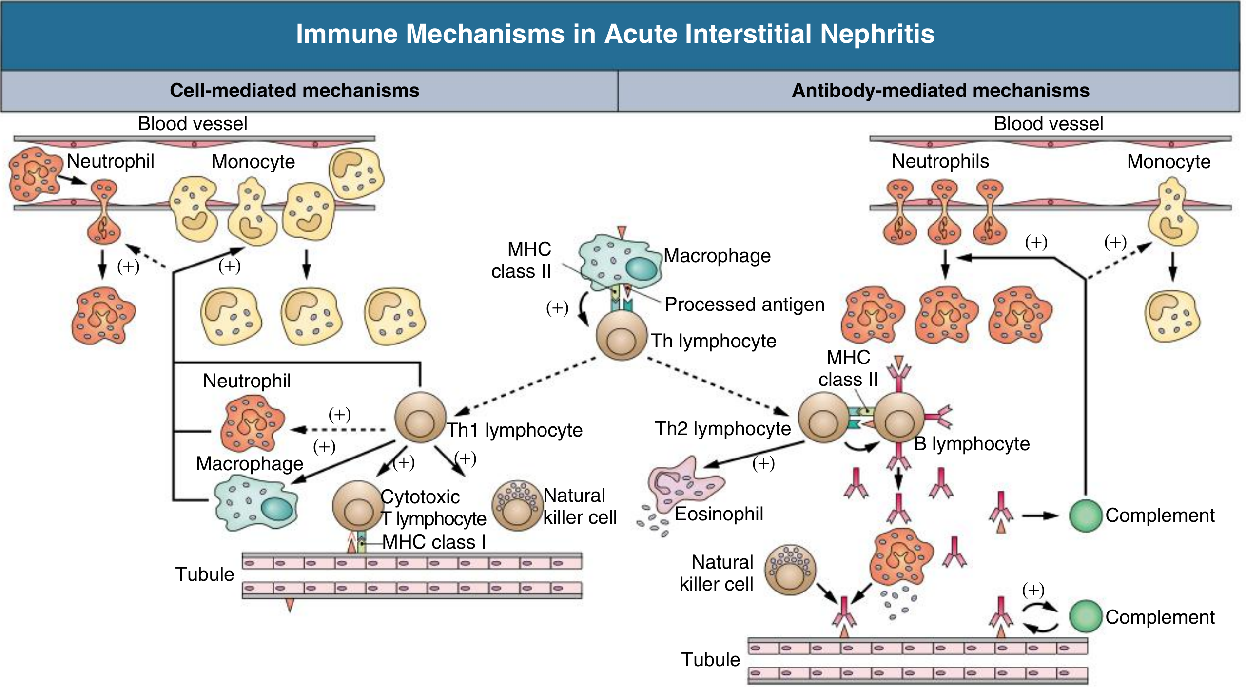
Immune mechanisms in AIN - cell-mediated and antibody-mediated pathways
Key evidence it is immune-mediated (NOT dose-dependent toxicity):
  • Occurs in only a small fraction of people taking the drug
  • Not dose-dependent
  • Associated with systemic hypersensitivity signs (rash, eosinophilia, fever)
  • Recurs on re-exposure to the same or related drug
  • Responds to steroids
Mechanism: Drug acts as a hapten → binds to tubular basement membrane (TBM) proteins → creates modified self-antigen → triggers:
  • Type IV (cell-mediated / delayed-type) hypersensitivity - T cells, macrophages (dominant in most cases)
  • Type I (IgE-mediated) - seen with eosinophilia, elevated IgE, in some cases
  • Cytokine release (TGF-β) → interstitial fibrosis if injury persists
  • Comprehensive Clinical Nephrology, 7th Ed., p. 890

Clinical Presentation

Classic Triad (Step 1 favorite)

Fever + Maculopapular rash + Eosinophilia
BUT - this full triad is present in only ~10-15% of cases overall (more common with methicillin, less common with NSAIDs or PPIs). Never use it to rule out AIN.

Full Clinical Picture

FeatureDetails
FeverPresent in <50% of cases
RashMaculopapular, ~25% of cases
Peripheral eosinophiliaVariable; may be transient
AKIMost common presentation - rising creatinine, often asymptomatic initially
Flank pain~33% - due to acute distension of kidney capsule
OliguriaPresent in severe cases; dialysis needed in ~1/3
Hypertension / edemaUsually absent (distinguishes from glomerulonephritis)
DRESS syndromeDrug Rash, Eosinophilia, Systemic Symptoms - occurs in up to 40% with certain drugs
Classic scenario: Patient on antibiotics for infection, defervesces appropriately, then develops recurrent fever + rising creatinine a few days later. Think AIN.
  • Onset: 2-40 days after drug exposure (NSAIDs/PPIs: often months later)
  • Goldman-Cecil Medicine, p. 1267

Laboratory Findings

TestFindingNotes
Serum creatinineElevated (first and most sensitive finding)May be significantly elevated before symptoms
UrinalysisPyuria, hematuria, mild proteinuria
WBC castsPresent (leukocyte casts)Key finding - indicates tubular origin
Eosinophiluria>5% eosinophils in urineSupportive but low sensitivity (31%) and specificity (68%) - NOT reliable to rule in or out
RBC castsABSENTIf present, suggests glomerulonephritis instead
Proteinuria<1-3 g/day (non-nephrotic range)Except NSAIDs (minimal change co-lesion → nephrotic range)
Peripheral eosinophiliaVariableMore with antibiotics; less with NSAIDs
FENaOften >1%Tubular dysfunction
Serum IgEMay be elevated

Morphology / Pathology

Gross:
  • Kidneys normal or slightly enlarged (edema)
  • Increased cortical echogenicity on ultrasound
Histology (renal biopsy - gold standard for diagnosis):
  • Interstitial edema (displaces tubules apart)
  • Mononuclear infiltrate: predominantly T lymphocytes (CD4+) and macrophages; also eosinophils, plasma cells, mast cells
  • Tubulitis - lymphocyte infiltration into tubular epithelium (tubules infiltrated by WBCs)
  • Inflammation often starts at corticomedullary junction where drug concentration is highest
  • Glomeruli SPARED (until late disease)
  • Granulomas - seen with methicillin, thiazides, allopurinol, sarcoidosis
  • No immune deposits by immunofluorescence (usually) - distinguishes from lupus nephritis, membranoproliferative GN
  • Robbins, Cotran & Kumar, p. 863

Comparison Table: AIN vs. Other AKI Causes

FeatureAINATNGlomerulonephritis
Fever/rashMay be presentAbsentAbsent
EosinophiliaYes (variable)NoNo
RBC castsABSENTAbsentPRESENT
WBC castsPRESENTGranular castsRare
Proteinuria<3 g/day (mild)MildOften heavy (nephrotic)
HematuriaMildAbsentProminent
BiopsyInterstitial infiltrateTubular necrosisGlomerular changes
FENa>1%>1%<1% (usually)

Diagnosis

  • Clinical suspicion + urinalysis + drug history
  • Eosinophiluria: supportive but not reliable (low PPV)
  • Renal biopsy: definitive diagnosis - indicated when:
    • AKI unexplained by prerenal causes or ATN
    • Diagnosis uncertain and tissue needed to guide therapy
    • No improvement after stopping offending drug
  • Goldman-Cecil Medicine, p. 1269

Treatment

  1. Remove the offending drug / treat underlying cause - most important step
  2. Corticosteroids - prednisone 0.75-1 mg/kg/day x ~1 week then taper (8-12 weeks total); benefit is modest but may accelerate recovery if started within 7-14 days of stopping drug and before significant fibrosis
  3. Dialysis if severe AKI (~1/3 of patients need it)
  4. Mycophenolate mofetil - alternative for steroid-refractory or steroid-intolerant cases
  5. Most cases are reversible with early recognition and intervention
  • Brenner and Rector's The Kidney, p. 3040

Special Associations for Step 1

Drug / ConditionSpecial Association
MethicillinPrototype drug; classic AIN description
NSAIDsAIN + minimal change disease simultaneously → nephrotic syndrome
PPIsDelayed onset (months); rising incidence; often missed
Allopurinol, thiazidesGranulomatous AIN
Checkpoint inhibitors(anti-PD1, anti-CTLA4) - AIN occurs ~3% of the time
Sjogren syndromeInterstitial nephritis + tubular dysfunction (RTA)
TINU syndromeAIN + bilateral anterior uveitis in young women
SarcoidosisGranulomatous AIN

High-Yield Step 1 Summary

  • Definition: Immune-mediated inflammatory infiltrate in renal interstitium → AKI
  • Most common cause: Drugs (antibiotics > NSAIDs > PPIs)
  • Classic triad: Fever + rash + eosinophilia (present in only ~10-15% - rarely complete!)
  • Key urine findings: WBC casts (leukocyte casts) + eosinophiluria; NO RBC casts
  • Proteinuria: Mild (<3 g/day), UNLESS NSAIDs → minimal change co-lesion → nephrotic
  • Biopsy: Interstitial lymphocytes/eosinophils + tubulitis + spared glomeruli + NO immune deposits (usually)
  • Treatment: Stop drug + corticosteroids
  • Prognosis: Usually reversible if caught early; chronic fibrosis if unrecognized

Interstatil renal inflammation

Asking for Preferences

Renal interstitium

Finding Sources
Finding Sources
Reading File
Reading File
Reading File
Reading File
Loading Image
I have rich content. Here is a thorough Step 1-relevant overview of the renal interstitium.

The Renal Interstitium

What Is the Renal Interstitium?

The renal interstitium is the connective tissue compartment that fills the space between the renal tubules, peritubular capillaries, vessels, and glomeruli. It is not just passive "filler" - it plays active roles in tubular function, urine concentration, erythropoietin production, and immune surveillance. Its expansion (by inflammation or fibrosis) is a hallmark of progressive kidney disease.
  • Tietz Textbook of Laboratory Medicine, p. 1850
  • Brenner and Rector's The Kidney, p. 117

Compartments of the Interstitium

The interstitium is divided into two major zones based on anatomy and function:

1. Cortical Interstitium

  • Very sparse in the normal kidney - only 7-9% of cortical volume (tubules are closely packed)
  • Divided into:
    • Peritubular interstitium: space between tubular basement membranes and peritubular capillaries
    • Periarterial interstitium: loose connective tissue sheath around interlobular and arcuate arteries; contains lymphatics and nerve fibers
  • The fenestrated portion of the peritubular capillary wall faces the narrow interstitial slit - this facilitates rapid reabsorption of fluid and solutes from the tubules into the bloodstream

2. Medullary Interstitium

  • Much larger volume fraction than the cortex (expands progressively from outer to inner medulla)
  • Contains lipid-laden interstitial cells arranged in a characteristic ladder-like pattern across the loops of Henle and vasa recta
  • The extracellular space is rich in glycosaminoglycans (GAGs), creating a gelatinous matrix
  • This gelatinous matrix contains osmolytes - osmotically active molecules that help stabilize the medullary osmotic gradient essential for the countercurrent concentration mechanism
  • The medullary interstitium is the functional core of urine concentration
  • Tietz Textbook of Laboratory Medicine, p. 1850

Cell Types of the Renal Interstitium

1. Fibroblasts (Most Abundant Cell)

  • Also called stellate or sustentacular cells in the cortex
  • Form a network spanning between capillaries and tubules via "attachment plaques" of actin fibers
  • Primary producers of extracellular matrix (collagen, fibronectin, laminin)
  • In the cortex, fibroblasts express ecto-5'-nucleotidase (5'NT) - a useful immunohistochemical marker
Special variant - Medullary fibroblasts ("lipid-laden cells"):
  • Located in the inner stripe of the outer medulla and inner medulla
  • Contain numerous lipid inclusions
  • Produce prostaglandins (especially PGE₂) - important in regulating blood pressure and medullary blood flow
  • Contain α-smooth muscle actin and vimentin filaments
In disease - Myofibroblasts:
  • In inflammatory disease, fibroblasts become activated → transform into myofibroblasts
  • Express α-smooth muscle actin (αSMA)
  • Drive interstitial fibrosis and tubular atrophy → progressive CKD
  • Pericytes (see below) are now recognized as the main progenitor of myofibroblasts

2. Pericytes

  • Contractile cells wrapped around peritubular capillaries
  • Most abundant in the inner stripe of the outer medulla around descending vasa recta
  • Regulate capillary tone and microvascular blood flow
  • In inflammation: pericytes detach from capillaries, proliferate, and differentiate into myofibroblasts - directly driving fibrosis and capillary rarefaction

3. Dendritic Cells

  • Most common immune cell in the healthy interstitium
  • Stellate shape with long cytoplasmic extensions
  • Sentinel cells - present antigens to T cells during immune responses
  • Distinguished from fibroblasts by: absence of actin bundles under the plasma membrane; organelles clustered near nucleus

4. Macrophages

  • Mostly found in the periarterial interstitium in healthy kidney
  • Not abundant in health; expand dramatically in injury
  • In inflammation, release cytokines that amplify fibrosis and tubular damage

5. Lymphocytes and Granulocytes

  • Rare in the healthy kidney
  • Prominent in acute interstitial nephritis (lymphocytes + eosinophils) and pyelonephritis (neutrophils)
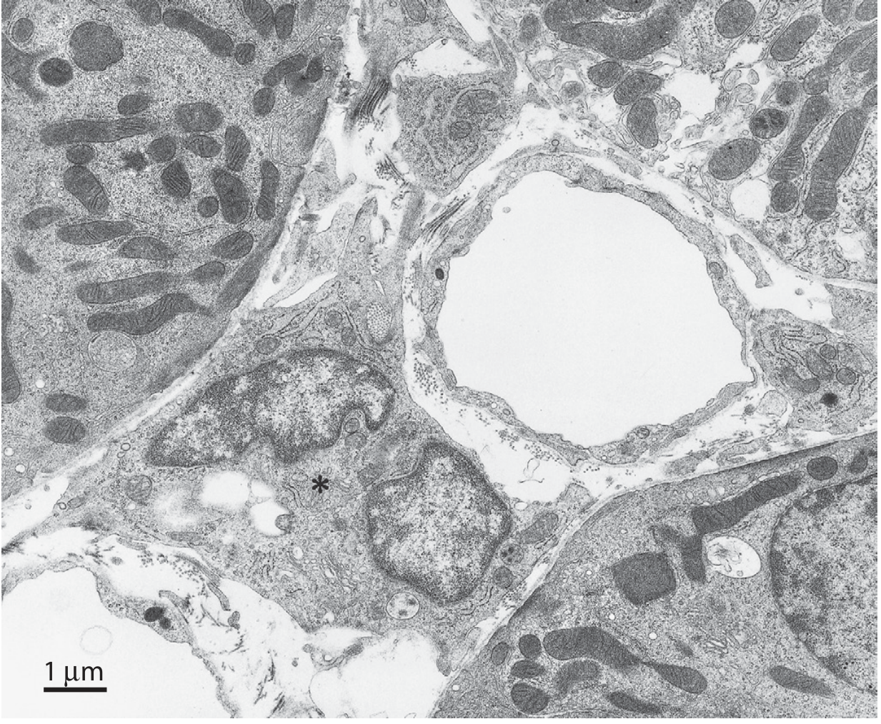
Transmission electron micrograph of cortical interstitial fibroblast (*) from a rat, with a peritubular capillary at right center
Fig. 2.53: TEM of cortical interstitial fibroblast (asterisk). Surrounding tubular cell mitochondria are visible; peritubular capillary lumen appears at right. - Brenner & Rector's The Kidney

Extracellular Matrix (ECM) of the Interstitium

ComponentDetails
Collagen fibrilsTypes I, III, V, VI, VII, XV
FibronectinCell adhesion, matrix organization
LamininBasement membrane component
Sulfated + nonsulfated GAGsGround substance; creates gelatinous matrix in medulla; stabilizes osmotic gradient
Interstitial fluidContinuous with the ground substance
  • Brenner and Rector's The Kidney, p. 117

Functions of the Renal Interstitium

FunctionMechanism
Structural supportFibroblasts scaffold tubules and capillaries; ECM provides framework
Urine concentrationMedullary interstitium maintains the corticomedullary osmotic gradient via GAG matrix and lipid-laden cells
Solute/fluid exchangePeritubular interstitium is the intermediate space for reabsorbed water and solutes moving from tubule lumen → capillary
Erythropoietin (EPO) productionCortical fibroblasts (specifically peritubular fibroblasts) are the primary source of EPO - this is why CKD with interstitial fibrosis → loss of EPO-producing cells → normocytic anemia
Immune surveillanceDendritic cells and macrophages monitor for pathogens and damaged cells
Prostaglandin synthesisMedullary lipid-laden fibroblasts produce PGE₂ - vasodilatory, regulates blood pressure and medullary perfusion
Vitamin D activationPeritubular cells contribute to 1α-hydroxylase activity

The Interstitium in Disease

Interstitial Expansion = Bad Prognostic Sign

Expansion of the interstitium is one of the best histological predictors of progression to CKD. It occurs by:
  1. Cellular infiltration: lymphocytes, macrophages, eosinophils (AIN, pyelonephritis)
  2. Edema: early AIN - kidneys enlarge due to interstitial fluid
  3. Interstitial fibrosis: myofibroblast activation → excess collagen deposition → tubular atrophy → nephron loss

Interstitial Fibrosis and Tubular Atrophy (IFTA)

  • Final common pathway of most kidney diseases
  • Driven by: TGF-β, PDGF, FGF-2 → fibroblast/myofibroblast proliferation
  • Characterized by: collagen accumulation (types I-V, VII), fibronectin, laminin deposition
  • Results in: capillary rarefaction, tubular dropout, glomerulosclerosis (eventually)

Specific Disease Correlates

DiseaseInterstitial Finding
Acute interstitial nephritis (AIN)Edema + lymphocyte/eosinophil infiltrate + tubulitis; NO immune deposits
PyelonephritisNeutrophilic infiltrate + tubular abscesses; begins in medulla → affects concentration first
Chronic pyelonephritisCoarse irregular scars, dilated calyces, patchy lymphocytic infiltrate
Sjogren syndromeLymphocytic interstitial nephritis + distal RTA
Analgesic nephropathyPapillary necrosis + chronic interstitial fibrosis
CKD (any cause)IFTA - correlates with GFR loss better than glomerular changes alone
  • Guyton and Hall Medical Physiology, p. 432

Key Step 1 Pearls

  • Normal cortical interstitium = 7-9% by volume - mostly empty space between packed tubules
  • EPO is made by cortical peritubular fibroblasts → interstitial fibrosis in CKD → anemia
  • Medullary lipid-laden cells produce prostaglandins (PGE₂) and maintain the osmotic gradient
  • Pyclocytes are pericytes under stress → detach → become myofibroblasts → fibrosis
  • Dendritic cells are the dominant immune cell in the normal interstitium
  • Pyelonephritis starts in the medulla → early loss of urine concentration ability
  • Interstitial fibrosis (IFTA) is the best histologic correlate of GFR decline in most kidney diseases
  • AIN: interstitial edema + lymphocyte infiltrate + tubulitis with spared glomeruli

Show in diagram

Generating Image

Detailed anatomical and cellular diagram of the renal interstitium for medical students. Show two main panels: LEFT PANEL - "Cortical Interstitium": Cross-section showing peritubular capillary (fenestrated), proximal tubule and distal tubule with basement membranes, narrow interstitial slit between tubule and capillary, stellate fibroblast with long cytoplasmic extensions labeled, dendritic cell with stellate shape, rare lymphocyte, extracellular matrix (collagen fibers, fibronectin), periarterial interstitium around artery with lymphatics and nerves. Label: "7-9% of cortical volume". Highlight fibroblast expressing ecto-5-nucleotidase (5'NT) and EPO production arrow from fibroblast. RIGHT PANEL - "Medullary Interstitium": Show loop of Henle, vasa recta (descending), lipid-laden interstitial cells arranged in ladder-like pattern spanning between loop of Henle and vasa recta, pericyte wrapping around capillary, GAG-rich gelatinous extracellular matrix labeled, osmotic gradient arrow showing increasing osmolality cortex to papilla (300 → 1200 mOsm). Label lipid-laden cells producing PGE2. BOTTOM PANEL - "Interstitium in Disease": Three boxes side by side: Box 1 "Normal" - sparse interstitium, fibroblast Box 2 "AIN" - edema, lymphocytes, eosinophils infiltrating, tubulitis, spared glomerulus Box 3 "Fibrosis/CKD" - myofibroblasts (activated fibroblasts with alpha-SMA), collagen deposition, tubular atrophy, capillary rarefaction, pericyte detachment arrow Color coding: tubules = pink, capillaries = red/blue, fibroblasts = orange, immune cells = purple, matrix = light yellow, lipid-laden cells = green. Clean medical textbook illustration style with clear labels.
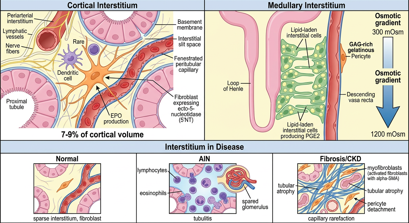
Renal Interstitium Diagram
Here's a breakdown of what the diagram shows:
Cortical Interstitium (Top Left)
  • The interstitium is very sparse (7-9% of cortical volume) - tubules are tightly packed
  • Stellate fibroblasts (orange) with long extensions span between tubule basement membranes and peritubular capillaries - they produce EPO and ECM (collagen, fibronectin)
  • Dendritic cells are the dominant immune cell in health
  • Periarterial interstitium surrounds arteries with lymphatics and nerves
Medullary Interstitium (Top Right)
  • Much larger volume fraction than cortex
  • Lipid-laden interstitial cells (green) arranged in a ladder-like pattern crossing between loops of Henle and vasa recta - produce PGE₂ and help maintain the osmotic gradient (300 → 1200 mOsm)
  • Rich GAG matrix creates gelatinous ground substance that stabilizes medullary osmolality
  • Pericytes wrap around capillaries and regulate tone
Disease States (Bottom)
  • Normal: sparse, quiet fibroblasts
  • AIN: interstitial edema + lymphocyte/eosinophil infiltrate + tubulitis; glomerulus spared
  • CKD/Fibrosis: pericytes detach → become myofibroblasts (αSMA+) → collagen deposition → tubular atrophy + capillary rarefaction

Tell me about acute tubular necrosis every small concept for everything why it's occurring

Finding Sources
Finding Sources
Reading File
Reading File
Reading File
Reading File
Reading File
Loading Image
Loading Image
Finding Sources
Finding Sources
Reading File
Loading Image
Reading File
Loading Image

Acute Tubular Necrosis (ATN) - Complete Deep Dive

What Is ATN? (Terminology First)

The term "acute tubular necrosis" is actually a misnomer - frank necrosis is rarely seen on biopsy. The modern preferred term is Acute Tubular Injury (ATI). The condition is characterized by damage to tubular epithelial cells causing AKI. However, "ATN" is still universally used clinically and on Step 1.
ATN is the most common cause of AKI in hospitalized patients (accounts for just under half of all inpatient AKI cases, and more than half of ICU AKI).
  • Robbins & Kumar Basic Pathology, p. 519
  • Frameworks for Internal Medicine, p. 454

Two Fundamental Types of ATN

TypePrimary CauseSegment Most Affected
Ischemic ATNDecreased blood flow → hypoxiaS3 (straight) segment of proximal tubule + medullary thick ascending limb (mTAL)
Nephrotoxic ATNDrugs, toxins, pigmentsPrimarily proximal tubule (S1, S2) - more uniform, more frank necrosis

WHY THE PROXIMAL TUBULE AND mTAL?

This is the most important conceptual question - why do these two segments die and not others?

The Proximal Tubule (especially S3 segment) is vulnerable because:

  1. High metabolic demand - it performs the most energy-demanding work (reabsorbs 60-70% of all filtered load). Requires massive amounts of ATP via oxidative phosphorylation. Cannot switch to anaerobic glycolysis effectively.
  2. Minimal mitochondrial reserve - when oxygen delivery drops, ATP falls rapidly → failure of Na⁺/K⁺-ATPase pumps → cell swelling and death
  3. High concentration of luminal toxins - as the filtrate is concentrated by water reabsorption, drugs and toxins accumulate in the tubular lumen at high concentrations, directly toxic to the epithelium
  4. High intracellular concentration of reabsorbed molecules - substances like organic acids, heavy metals, and aminoglycosides accumulate inside proximal tubular cells after uptake from the lumen

The Medullary Thick Ascending Limb (mTAL) is vulnerable because:

  1. Physiologically hypoxic environment - the outer medulla operates at the lowest PO₂ in the kidney due to the countercurrent exchange system (oxygen diffuses from descending to ascending vasa recta, "shunting" away from the medullary tubules)
  2. High metabolic demand - the mTAL actively reabsorbs NaCl (Na⁺/K⁺/2Cl⁻ cotransporter) and requires high ATP even under normal conditions
  3. No ability to extract more O₂ - already extracting nearly 100% of delivered oxygen; any reduction in delivery = immediate ischemia
  • Robbins & Kumar Basic Pathology, p. 519
  • Goldman-Cecil Medicine, p. 1265

STEP 1: THE COMPLETE PATHOGENESIS - HOW DOES ATN CAUSE AKI?

There are four interconnected mechanisms that simultaneously reduce GFR and urine output:

Mechanism 1: Tubuloglomerular Feedback (TGF) → Afferent Vasoconstriction

This is the dominant mechanism explaining the drop in GFR.
Step-by-step:
  1. Ischemia/toxin → proximal tubule injury → failure of Na⁺ reabsorption
  2. More Na⁺ and Cl⁻ reaches the macula densa at the distal tubule
  3. Macula densa detects ↑ luminal NaCl → signals the juxtaglomerular apparatus
  4. Tubuloglomerular feedback activates → afferent arteriolar vasoconstriction
  5. ↓ Glomerular blood flow → ↓ GFR → further ischemia (a vicious cycle)
This is a normal autoregulatory mechanism gone wrong - the kidney is "trading away" GFR to reduce tubular work and protect the medulla from worsening ischemia.

Mechanism 2: Vascular Factors - Endothelial Injury
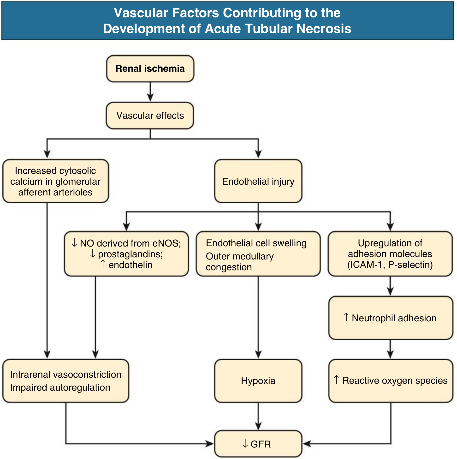
Vascular factors contributing to ATN - Comprehensive Clinical Nephrology
Ischemia directly injures the endothelium of peritubular capillaries and vasa recta:
  • ↓ eNOS → ↓ NO (normally vasodilatory) → vasoconstriction persists
  • ↓ Prostaglandins (PGE₂, PGI₂) → loss of vasodilation
  • ↑ Endothelin → potent vasoconstriction
  • Endothelial cell swelling → outer medullary congestion → compresses tubules → hypoxia worsens
  • Upregulation of ICAM-1 and P-selectin → neutrophil adhesion and infiltration → neutrophils release reactive oxygen species (ROS) and proteases → more tubular injury

Mechanism 3: Tubular Cell Injury - Loss of Polarity and Cast Formation
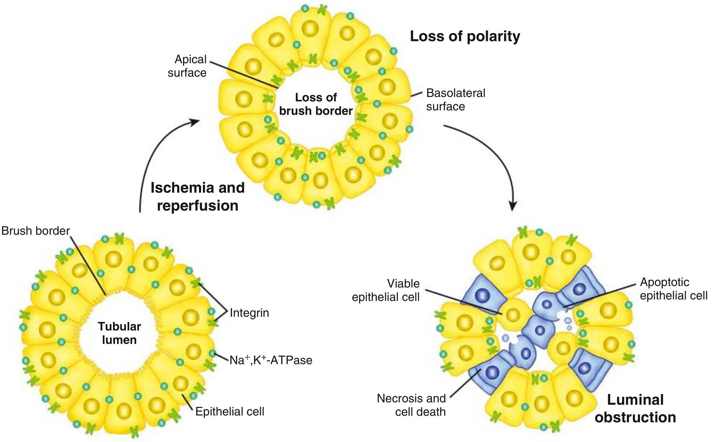
Tubular factors in ATN - loss of polarity leading to luminal obstruction
Normal tubular cell polarity:
  • Na⁺/K⁺-ATPase lives on the basolateral surface (pumps Na out into blood)
  • Integrins and adhesion molecules keep the cell attached to the basement membrane
After ischemia or toxin injury:
  1. ATP falls → Na⁺/K⁺-ATPase fails → intracellular Na⁺ and Ca²⁺ rise → cell swelling
  2. Loss of brush border (apical microvilli slough off) - first detectable change
  3. Loss of cell polarity - Na⁺/K⁺-ATPase redistributes from basolateral to apical surface → Na⁺ is now pumped backward into the lumen → more Na⁺ reaches the distal tubule (worsening TGF above)
  4. Integrins lose function → cells detach from the basement membrane
  5. Detached cells, cell debris, and Tamm-Horsfall protein aggregate in the tubular lumen → form "muddy brown" granular casts
  6. Casts obstruct tubular outflow → ↑ intratubular pressure → opposes glomerular filtration pressure → ↓ GFR

Mechanism 4: Backleak of Glomerular Filtrate

When cells detach, gaps appear in the tubular epithelium:
  • Glomerular filtrate leaks back through the denuded basement membrane into the interstitium
  • Filtrate is "lost" - never makes it to the urine
  • Creates interstitial edema → ↑ interstitial pressure → compresses capillaries → more ischemia
  • Results in oliguria despite ongoing filtration
  • Robbins & Kumar Basic Pathology, p. 520
  • Comprehensive Clinical Nephrology, 7th Ed., p. 990
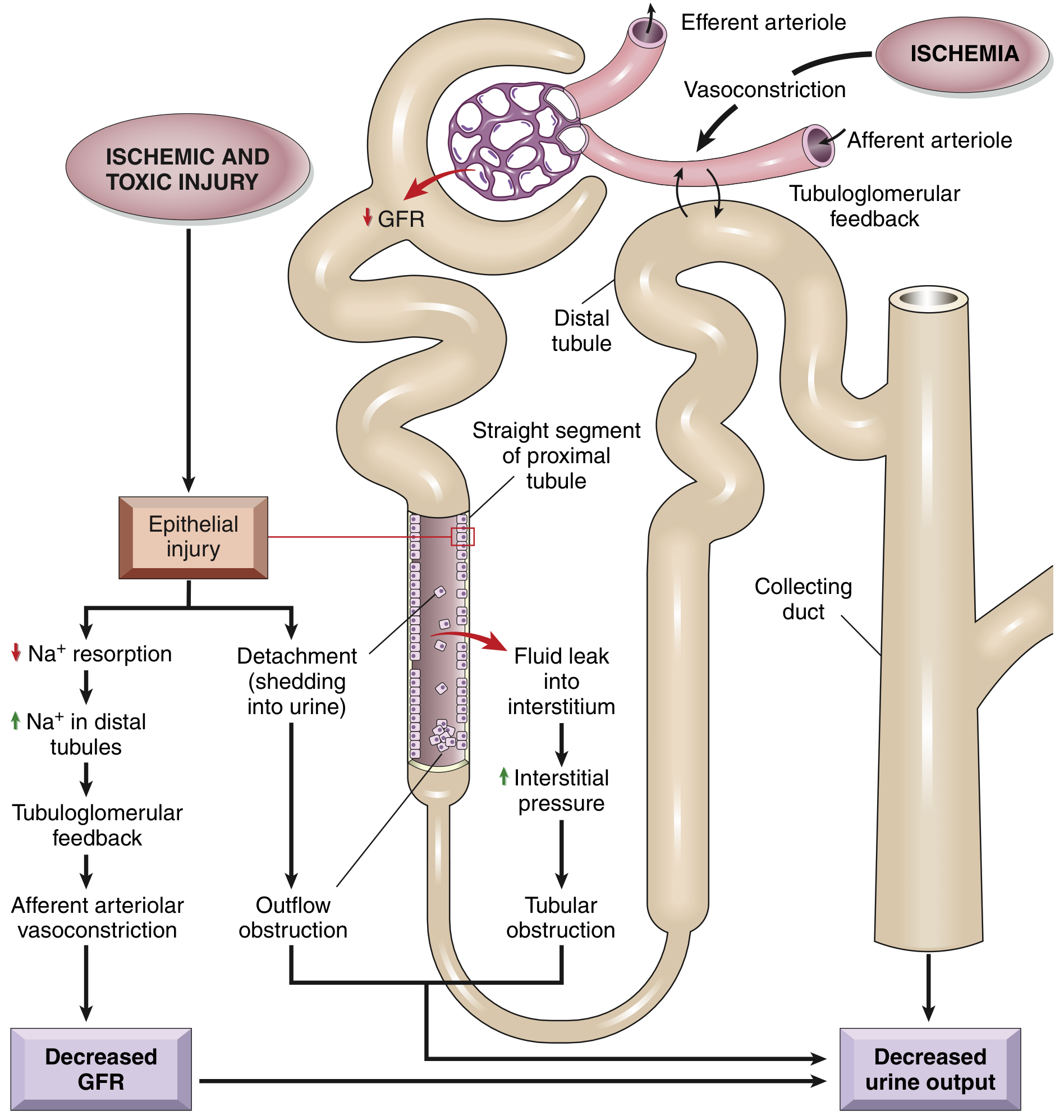
Summary diagram of all four mechanisms:
Postulated sequence in ischemic and toxic ATI - Robbins & Kumar

CAUSES OF ATN - EVERY CATEGORY

A. Ischemic ATN (= prerenal → intrarenal continuum)

When prerenal AKI is severe and prolonged enough, it transitions to ATN. The trigger is inadequate oxygen delivery to tubular epithelial cells.
CategoryExamples
Hypovolemia / Hemorrhagic shockTrauma, surgery, massive GI bleed, aortic surgery
Cardiogenic shockMI, severe heart failure, cardiac arrest
Septic shockGram-negative bacteremia (most common cause of ATN in the ICU)
Distributive shockAnaphylaxis, neurogenic shock
Obstetric emergenciesPlacental abruption, eclampsia (→ cortical necrosis risk)
Large vessel diseaseRenal artery thrombosis, embolism
MicrovascularMalignant hypertension, HUS/TTP, vasculitis

B. Nephrotoxic ATN

Exogenous toxins:
AgentMechanism / Notes
Aminoglycosides (gentamicin, tobramycin)Accumulate in proximal tubule lysosomes → direct toxicity; non-oliguric ATN is characteristic; delayed onset (5-7 days)
Contrast agents (iodinated)Direct tubular toxicity + vasoconstriction; risk ↑ with dehydration, DM, CKD, multiple myeloma, NSAIDs use
Cisplatin, carboplatinDNA damage in proximal tubule cells
Amphotericin BPore-forming in tubular cell membranes + vasoconstriction
VancomycinOxidative stress in tubular cells
NSAIDsBlock prostaglandin-mediated afferent dilation → ischemia (especially when dependent on PGs for GFR, e.g., CHF, CKD, elderly)
Heavy metals (mercury, lead, cisplatin, arsenic)Direct cytotoxicity to proximal tubule
Ethylene glycolMetabolized to oxalate → calcium oxalate crystals in tubules + direct toxicity
Carbon tetrachlorideFree radical-mediated injury
Endogenous toxins (Pigment nephropathy):
AgentSourceMechanism
MyoglobinRhabdomyolysis (crush injury, extreme exertion, statins, cocaine, hyperthermia)Direct tubular toxicity + cast obstruction + vasoconstriction
HemoglobinHemolytic crisis, mismatched blood transfusion, G6PD deficiencySame as myoglobin
Uric acidTumor lysis syndromeCrystal deposition and tubular obstruction
Light chainsMultiple myelomaCast nephropathy - obstructive + direct toxicity
Step 1 clue for pigment nephropathy: Urine dipstick positive for blood but no RBCs on microscopy = myoglobin or free hemoglobin in urine.

MORPHOLOGY - WHAT YOU SEE ON BIOPSY

Ischemic ATN:

  • Patchy - lesions skip areas; not all tubules involved
  • S3 segment of proximal tubule and mTAL are most affected
  • Proximal tubule brush border loss - earliest change
  • Cell blebbing, vacuolization, nuclear pyknosis
  • Cells detach and slough → bare tubular basement membrane (key finding)
  • Proteinaceous ("muddy brown") granular casts in distal tubules and collecting ducts
    • Composed of: Tamm-Horsfall protein + plasma proteins + cell debris
  • Basement membrane intact (allows regeneration!)
  • Interstitial edema with mild neutrophil/lymphocyte infiltrate
  • Glomeruli SPARED

Nephrotoxic ATN:

  • More uniform, more proximal - concentrated in proximal tubule (S1/S2 segments)
  • Frank necrosis more prominent than ischemic ATN (though still relatively sparse)
  • Similar cast formation, basement membrane largely intact
Key histologic distinction: In ischemic ATN, injury is patchy and affects straight segment + mTAL. In nephrotoxic ATN, injury is more uniform and predominantly proximal.
  • Robbins & Kumar Basic Pathology, p. 519-520

CLINICAL PHASES OF ATN

ATN classically evolves through 3 phases:

Phase 1: Initiation Phase (Hours to Days)

  • The inciting event occurs (hypotension, toxin exposure)
  • Oliguria begins
  • BUN and creatinine start rising but are not yet markedly elevated (lag time)
  • Histologic injury is occurring but clinical recognition may be delayed
  • Key: If the trigger is removed NOW, tubular injury can be prevented

Phase 2: Maintenance Phase (Days to Weeks - typically 1-3 weeks)

  • Established ATN - tubular cells are dead/dysfunctional
  • Oliguria (urine output <400 mL/day) in ~50% - may be non-oliguric (especially aminoglycosides, contrast)
  • Rising BUN and creatinine - azotemia
  • Electrolyte complications:
    • Hyperkalemia (↑K⁺ - tubules can't excrete K⁺; also released from injured cells)
    • Metabolic acidosis (↓ NH₄⁺ excretion, ↓ H⁺ secretion)
    • Hyperphosphatemia (↓ phosphate excretion)
    • Hyponatremia (dilutional, if giving free water)
    • Hypocalcemia (reciprocal to hyperphosphatemia)
  • Fluid overload → pulmonary edema, hypertension
  • Uremic symptoms: nausea, vomiting, lethargy, pericarditis (in severe/prolonged cases)
  • Even if the cause is removed, GFR does not improve immediately - this is what distinguishes ATN from prerenal AKI

Phase 3: Recovery Phase (Weeks)

  • Tubular epithelial cells regenerate from surviving cells that dedifferentiate, proliferate, and re-cover the basement membrane
  • Surviving tubular cells (not stem cells) drive regeneration
  • Polyuria (diuretic phase) - urine output increases dramatically (can be 3-5 L/day)
    • WHY: regenerating tubular cells cannot yet concentrate urine properly → pass large volumes of dilute urine
    • Risk: patients can become severely dehydrated and hypokalemic during this phase
    • Electrolyte losses (Na⁺, K⁺, Mg²⁺) can be life-threatening if not replaced
  • BUN and creatinine begin to fall
  • Function returns over weeks to months
  • If pre-existing CKD or severe prolonged injury: incomplete recovery → progression to CKD
  • M1 macrophages (pro-inflammatory) are replaced by M2 macrophages (reparative) during recovery
  • Robbins & Kumar Basic Pathology, p. 580
  • Comprehensive Clinical Nephrology, p. 959

LABORATORY FINDINGS & DIAGNOSIS

Urinalysis - The Most Important Test

FindingATNPrerenal AKIAIN
"Muddy brown" granular castsPRESENT - hallmarkAbsent (hyaline casts only)WBC casts
Tubular epithelial cell castsPresentAbsentAbsent
ProteinuriaMild-moderateAbsent/traceMild
HematuriaAbsentAbsentMild
WBC castsAbsentAbsentPresent
RBC castsABSENTAbsentAbsent
"Muddy brown" granular casts = ATN until proven otherwise. ATN is highly likely when ≥6 granular casts are seen.
Muddy brown granular cast on urine microscopy
Figure: "Muddy brown" granular cast from patient with ATN.

Fractional Excretion of Sodium (FENa)

$$\text{FENa} = \frac{U_{Na} / P_{Na}}{U_{Cr} / P_{Cr}} \times 100%$$
ResultInterpretation
FENa <1%Prerenal AKI - tubules intact, avidly reabsorbing Na⁺
FENa >2%ATN - damaged tubules can't reabsorb Na⁺, Na⁺ is wasted
Why FENa >2% in ATN?
  • Tubular cells are dead/dysfunctional → Na⁺/K⁺-ATPase fails
  • Na⁺ reabsorption is impaired → Na⁺ "spills" into the urine
  • Also: Na⁺/K⁺-ATPase mislocalizes to apical surface → actively pumps Na⁺ INTO the lumen
Important caveats - FENa can be falsely low (<1%) in ATN with:
  • Contrast nephropathy (early)
  • Myoglobinuria/hemoglobinuria
  • Recent diuretic use (use FEUrea instead - >35-50% suggests ATN)
  • ATN superimposed on prerenal disease

Other Labs

TestATN Finding
Serum creatinine↑↑ (rises 0.5-1 mg/dL/day in oliguria)
BUN↑↑ (BUN:Cr ratio ~10-15 in ATN; >20 suggests prerenal)
K⁺↑ (hyperkalemia)
HCO₃⁻↓ (metabolic acidosis)
Phosphate
Ca²⁺
Urine osmolality~300 mOsm/kg (isosthenuria) - tubules can't concentrate
Urine Na⁺>40 mEq/L (tubules can't reabsorb Na⁺)

INFLAMMATION IN ATN (High-Yield Mechanism)

The immune system actively participates in ATN injury:
Innate immune activation:
  • Injured tubular cells release DAMPs (damage-associated molecular patterns)
  • TLR2 and TLR4 are upregulated on tubular cells → recognize DAMPs → produce chemokines
  • Complement system activated via the alternative pathway → TNF-α, IL-6, IL-1β, MCP-1 release
Cellular response:
  • Neutrophils adhere to endothelium via ICAM-1 → enter peritubular spaces → release proteases and ROS → amplify injury
  • M1 macrophages infiltrate kidney → pro-inflammatory → worsen injury acutely
  • CD4+ and CD8+ T cells contribute to injury
  • Regulatory T cells help limit injury
Recovery:
  • M1 macrophages convert to M2 phenotype → support epithelial repair
  • Surviving tubular cells dedifferentiate, proliferate, migrate, and redifferentiate to cover denuded basement membrane
  • Comprehensive Clinical Nephrology, p. 959

SPECIAL TYPES OF ATN

Contrast-Induced Nephropathy (CIN)

  • Develops 24-48 hours after contrast exposure
  • Usually recovers within 7-10 days
  • Prevention: IV hydration (0.9% saline or isotonic NaHCO₃) before procedure; stop NSAIDs/metformin; minimize contrast volume
  • Risk factors: CKD, DM, dehydration, CHF, myeloma, concurrent nephrotoxins

Rhabdomyolysis-induced ATN (Pigment Nephropathy)

  • Myoglobin is filtered → precipitates in tubules → cast obstruction
  • Myoglobin is directly toxic (oxidative injury) + vasoconstricts
  • Classic findings: elevated CK (often >10,000 U/L), dark/tea-colored urine, dipstick positive for blood, no RBCs on microscopy, elevated LDH, hyperkalemia (K⁺ released from necrotic muscle), hyperphosphatemia, hypocalcemia (Ca²⁺ deposits in damaged muscle)
  • Treatment: aggressive IV fluid resuscitation (key!) to flush casts; urine alkalinization (prevents myoglobin precipitation); dialysis if needed

Aminoglycoside ATN

  • Non-oliguric ATN - urine output is preserved (important distinction)
  • Delayed onset: 5-7 days after starting drug
  • Aminoglycosides accumulate in proximal tubule lysosomes → lysosome rupture → cell death
  • Risk increased with: prolonged therapy, older age, CKD, volume depletion, concurrent loop diuretics

TREATMENT

  1. Remove/treat the cause - stop offending drug, restore perfusion
  2. Fluid management - correct hypovolemia; avoid overload; strict ins/outs
  3. Electrolyte management:
    • Hyperkalemia: calcium gluconate (cardiac protection), insulin+dextrose, kayexalate, dialysis
    • Metabolic acidosis: bicarbonate if severe
  4. Nutritional support - adequate calories to minimize catabolism
  5. Renal replacement therapy (dialysis) - indications: A-E-I-O-U:
    • Acidosis (refractory)
    • Electrolytes (hyperkalemia refractory to treatment)
    • Ingestion (toxins)
    • Overload (fluid refractory to diuretics)
    • Uremia (encephalopathy, pericarditis, bleeding)
  6. During recovery phase: Replace electrolyte losses (Na⁺, K⁺) during polyuric phase; avoid diuretics which can worsen hypovolemia

PROGNOSIS AND RECOVERY

  • Uncomplicated ATN with supportive care: complete recovery is common
  • Time to recovery: days to weeks for mild injury; months for severe
  • Non-oliguric ATN has a better prognosis than oliguric ATN
  • Poor prognostic factors: older age, pre-existing CKD, multiorgan failure, prolonged oliguria
  • Severe/prolonged injury → maladaptive repair → IFTA → CKD/ESRD

COMPLETE STEP 1 HIGH-YIELD SUMMARY

FeatureATN
Most common cause of AKI (inpatient)Yes - most common intrinsic AKI
Two typesIschemic (patchy, S3 + mTAL) and Nephrotoxic (uniform, proximal tubule)
Why S3 and mTAL?High O₂ demand + can't do anaerobic glycolysis + medullary hypoxia
Key pathogenesisEpithelial injury → ↓Na reabsorption → TGF → afferent vasoconstriction; + cast obstruction; + backleak; + endothelial injury
Hallmark urine finding"Muddy brown" granular casts
FENa>2% (tubules can't reabsorb Na⁺)
Urine osmolality~300 mOsm (isosthenuria)
Urine Na⁺>40 mEq/L
BUN:Cr ratio~10-15 (vs. >20 in prerenal)
Oliguric vs. non-oliguricBoth occur; aminoglycosides/contrast → non-oliguric
Recovery phase dangerPolyuria → dehydration + hypokalemia + hyponatremia
HistologyPatchy tubular cell loss, brush border loss, granular casts in distal tubules, intact basement membrane, spared glomeruli
Key difference from prerenalDoesn't improve with fluids; FENa >2%; muddy brown casts; azotemia persists

Tell

It looks like your message got cut off! Could you finish your question? What would you like to know about?

Answers previous question

The previous question was "Tell me about acute tubular necrosis - every small concept for everything why it's occurring" - and I already gave you the full, comprehensive answer above! Here's a quick recap of what was covered:

What Was Covered in the ATN Answer:

  1. Definition & Terminology - Why "necrosis" is a misnomer; modern term is Acute Tubular Injury (ATI)
  2. Two Types - Ischemic ATN vs. Nephrotoxic ATN
  3. WHY the Proximal Tubule and mTAL are vulnerable - High O₂ demand, can't do anaerobic glycolysis, medullary hypoxia
  4. Complete Pathogenesis (4 mechanisms):
    • Tubuloglomerular feedback → afferent vasoconstriction → ↓GFR
    • Vascular/endothelial injury → ↓NO, ↑endothelin, neutrophil ROS
    • Tubular cell polarity loss → cast formation → luminal obstruction
    • Backleak of filtrate → oliguria
  5. All Causes - Ischemic shock types, all nephrotoxic drugs, pigment nephropathy, contrast
  6. Morphology - Patchy tubular necrosis, brush border loss, muddy brown casts, intact basement membrane, spared glomeruli
  7. 3 Clinical Phases - Initiation → Maintenance (oliguria, hyperkalemia, acidosis) → Recovery (polyuric phase with risk of hypokalemia/dehydration)
  8. Lab Findings - Muddy brown casts, FENa >2%, isosthenuria, urine Na >40, BUN:Cr ~10-15
  9. Inflammation in ATN - TLRs, complement, M1→M2 macrophage shift, neutrophil ICAM-1
  10. Special Types - Contrast nephropathy, rhabdomyolysis, aminoglycoside ATN
  11. Treatment - Fluids, electrolytes, AEIOU dialysis indications, recovery phase management

Scroll up to see the full detailed answer with all diagrams! Is there a specific part you want me to go deeper on? For example:
  • The FENa calculation in detail
  • The polyuric recovery phase
  • Rhabdomyolysis specifically
  • Comparing ATN vs. prerenal vs. AIN
  • Treatment/dialysis indications

Didn't understand tell in complete detail

Asking for Preferences

Don't tell in short like you did in pathogensis form complete sentences I don't understand this way

Finding Sources
Finding Sources
Reading File
Reading File
Reading File
Now I have everything I need. Let me write the complete, paragraph-form explanation.

Acute Tubular Necrosis (ATN) - Complete Explanation in Full Sentences

Starting From the Very Beginning - What Is ATN?

To understand ATN, you first need to understand what the renal tubules do. Every minute, about 125 mL of fluid is filtered out of the blood at the glomerulus. This filtrate then travels through a long tube called the tubule, which has different segments - the proximal tubule, loop of Henle, distal tubule, and collecting duct. The job of the tubule cells is to work very hard to reabsorb most of that filtered fluid back into the blood, keeping only what the body needs to excrete as urine. This job requires enormous amounts of energy.
Acute Tubular Necrosis (also now called Acute Tubular Injury, or ATI) is a condition in which the epithelial cells lining these tubules are damaged or killed. When enough tubular cells die or stop functioning, the kidneys can no longer do their job properly, and the result is acute kidney injury (AKI) - a rapid decline in kidney function. ATN is the single most common cause of AKI in hospitalized patients, responsible for nearly half of all cases, and more than half of cases in the intensive care unit.
The word "necrosis" in the name is actually misleading because frank cell death (frank necrosis) is rarely seen on kidney biopsy. Most of the injury involves cell dysfunction, swelling, detachment, and partial death, not complete destruction. Despite this, the term "ATN" has stuck and is still used everywhere including on exams.
  • Robbins & Kumar Basic Pathology, p. 519

Why Do Tubular Cells Get Injured? The Two Root Causes

There are two fundamental reasons why tubular cells die, and these define the two types of ATN.
The first type is ischemic ATN, where the cells do not receive enough blood, and therefore not enough oxygen. Tubular cells - especially those in the proximal tubule and the medullary thick ascending limb of Henle - are extremely metabolically active. They cannot generate energy without oxygen because they rely almost entirely on aerobic metabolism (oxidative phosphorylation in their mitochondria). Unlike most cells in the body, they cannot meaningfully switch to anaerobic glycolysis to survive even brief periods without oxygen. So when blood supply drops - due to shock, hemorrhage, severe dehydration, sepsis, or any cause of low blood pressure - these cells run out of ATP very quickly and begin to fail.
The second type is nephrotoxic ATN, where a drug, toxin, or endogenous substance directly poisons the tubular cells. Some toxins are taken up from the filtered urine as the tubular cells reabsorb fluid, exposing the cells to concentrated toxic substances. Others enter cells through specific transporters and cause chemical damage inside the cell. The result is the same as with ischemia - the tubular cells die or become dysfunctional.
  • Guyton and Hall Medical Physiology, p. 429

Why Are the Proximal Tubule (S3 Segment) and the Medullary Thick Ascending Limb (mTAL) the Most Vulnerable?

Of all the tubule segments, two are particularly vulnerable to injury: the S3 segment of the proximal tubule (also called the straight portion of the proximal tubule, which dips into the outer medulla) and the medullary thick ascending limb of Henle (mTAL). Understanding why these two segments are vulnerable is one of the most important concepts in ATN.
The proximal tubule is where the majority of the tubular work happens - about 60-70% of all filtered sodium, glucose, amino acids, bicarbonate, and water are reabsorbed here. This requires massive amounts of ATP generated by mitochondria using oxygen. The S3 segment sits in the outer medulla where oxygen tension is already relatively low compared to the cortex, making it immediately sensitive to any further reduction in oxygen delivery. Additionally, as water is reabsorbed from the filtrate, any toxic substances that were filtered become progressively more concentrated in the lumen, meaning the proximal tubule cells are exposed to higher concentrations of nephrotoxins than any other segment. Furthermore, proximal tubule cells actively endocytose many substances from the luminal fluid through receptors called megalin and cubilin - this is how aminoglycosides, myoglobin, radiocontrast, and cadmium enter and damage these cells.
The medullary thick ascending limb sits deep in the outer medulla and is in an even more precarious position with respect to oxygen. The outer medulla is the most hypoxic zone of the kidney under normal conditions. This is because of the countercurrent exchange mechanism between the descending and ascending vasa recta - oxygen diffuses from the arterial blood in the descending vasa recta directly into the venous blood in the ascending vasa recta before it can reach the tubules, a process called "countercurrent shunting." Despite this chronically low oxygen environment, the mTAL actively and continuously reabsorbs sodium chloride via the Na⁺/K⁺/2Cl⁻ (NKCC2) cotransporter, which is energetically expensive. The mTAL is already extracting nearly 100% of available oxygen, so any further reduction in oxygen delivery immediately tips the balance toward ischemia and cell death.
  • Goldman-Cecil Medicine, p. 1265
  • Comprehensive Clinical Nephrology, 7th Ed., p. 957

The Pathogenesis - How Does Tubular Cell Injury Actually Reduce GFR and Cause AKI?

This is the most important part to understand, and there are four interconnected mechanisms working simultaneously. Think of them as four different ways in which injured tubular cells cause the glomerular filtration rate (GFR) to fall.

First Mechanism: Loss of Na⁺ Reabsorption → Tubuloglomerular Feedback → Afferent Arteriolar Vasoconstriction

Under normal conditions, the proximal tubule reabsorbs the vast majority of the filtered sodium before the fluid reaches the distal tubule. This keeps the sodium concentration at the macula densa (a group of specialized cells in the distal tubule that monitor luminal sodium and chloride) very low.
When proximal tubular cells are injured by ischemia or toxins, they lose their ability to reabsorb sodium. This happens because the Na⁺/K⁺-ATPase pump on the basolateral membrane of tubular cells requires ATP to function. When ATP falls, this pump fails. Sodium accumulates inside the cell and the reabsorption of sodium from the tubular lumen is severely impaired. The consequence is that an abnormally large amount of sodium now passes all the way to the distal tubule and reaches the macula densa.
When the macula densa detects this increased sodium delivery, it sends a signal through the juxtaglomerular apparatus to constrict the afferent arteriole of the same nephron. This reflex is called tubuloglomerular feedback (TGF), and it is actually a normal protective mechanism - the body is trying to reduce filtration in that nephron to lower the tubular workload and protect the medulla from worsening ischemia. However, in ATN, this protective reflex is maladaptive at the whole-kidney level, because now many nephrons simultaneously constrict their afferent arterioles, dramatically reducing glomerular blood flow across the kidney and slashing the GFR. This vicious cycle then causes more ischemia, more tubular injury, and more vasoconstriction.

Second Mechanism: Endothelial Injury and Vascular Dysfunction

Ischemia does not only injure tubular epithelial cells. It also damages the endothelial cells of the peritubular capillaries and the vasa recta. In a normal kidney, the endothelium produces nitric oxide (NO), which keeps the vessels dilated and maintains blood flow. It also produces vasodilatory prostaglandins. When endothelial cells are injured, eNOS (the enzyme that makes NO) is downregulated, so NO production falls and vasodilation is lost. At the same time, injured endothelial cells produce more endothelin-1, which is one of the most potent vasoconstrictors known. Together, these changes lead to sustained intrarenal vasoconstriction that persists even after the original cause of ischemia is corrected, which is part of why ATN does not immediately reverse when blood pressure is restored.
In addition, injured endothelial cells upregulate surface adhesion molecules called ICAM-1 and P-selectin. These molecules attract and trap neutrophils from the circulation. Neutrophils stick to the endothelium, migrate into the peritubular space, and release toxic reactive oxygen species (ROS) and proteases that directly injure the nearby tubular cells, worsening the damage. Endothelial cell swelling also causes physical congestion of the outer medullary capillaries, mechanically reducing blood flow to the already oxygen-deprived mTAL. This outer medullary congestion is one of the earliest and most consistent findings in ischemic ATN.

Third Mechanism: Loss of Tubular Cell Polarity → Cast Formation → Luminal Obstruction

A healthy tubular epithelial cell has a precise, polarized structure. The apical (luminal) surface faces the tubular fluid and has a brush border of microvilli that increase surface area for reabsorption. The basolateral surface faces the blood and contains the Na⁺/K⁺-ATPase pump that actively moves sodium from inside the cell into the interstitium, driving the reabsorption of water and solutes. Integrins on the basal surface anchor the cell firmly to the underlying basement membrane.
When ischemia or toxins strike, the first visible change is that the brush border membrane blebs and sheds from the apical surface into the tubular lumen. This is the earliest detectable injury. Then, as ATP continues to fall, the tight junctions between cells break down, and cell polarity is disrupted. The Na⁺/K⁺-ATPase pump, which is normally confined to the basolateral surface, redistributes to the apical surface. This is catastrophic - instead of pumping sodium out of the cell into the blood, the mislocated pumps now push sodium into the tubular lumen. This worsens the sodium delivery to the macula densa and amplifies the tubuloglomerular feedback vasoconstriction described above.
As injury progresses, the integrins lose their adhesive function, and tubular cells detach from the basement membrane. Both living and dead cells fall into the tubular lumen. These detached cells, along with shed brush border fragments and a protein called Tamm-Horsfall protein (which is normally secreted by tubular cells), aggregate in the tubular lumen to form casts. These casts physically plug the tubular lumen, blocking the outflow of urine from that nephron. The increased pressure inside the blocked tubule acts against the filtration pressure at the glomerulus, further reducing GFR in that nephron. This is why ATN can produce oliguria (very low urine output) even when the glomeruli themselves are completely normal and uninjured.
The casts that form in ATN have a characteristic appearance - they are coarse, granular, and brownish in color. They are called "muddy brown" granular casts and are the hallmark finding of ATN on urine microscopy. When you see ≥6 of these casts, ATN is highly likely.

Fourth Mechanism: Backleak of Glomerular Filtrate

When tubular cells detach from the basement membrane, bare gaps appear in the tubular epithelium. The glomerular filtrate - which is still being produced - can now leak backwards through these gaps out of the tubular lumen, through the denuded basement membrane, and into the renal interstitium. This filtrate never makes it to the collecting duct as urine, so even though filtration is occurring, urine output falls. The leaked fluid also accumulates in the interstitium, raising interstitial pressure, which compresses the peritubular capillaries and worsens local ischemia in a self-perpetuating cycle.
  • Robbins & Kumar Basic Pathology, p. 520
  • Goldman-Cecil Medicine, p. 1266

The Causes of ATN - Explained

Ischemic ATN occurs whenever blood supply to the kidney is severely and prolonged reduced. The most common causes in clinical practice are septic shock (especially from gram-negative bacteria, which is the dominant cause of ATN in the ICU), hemorrhagic shock from trauma or surgery, cardiogenic shock from a large myocardial infarction, severe dehydration, and any major surgery involving the aorta. All of these reduce the cardiac output or blood pressure enough to reduce renal perfusion to the point where tubular cells cannot survive.
Nephrotoxic ATN is caused by substances that directly damage tubular cells. Among drugs, the aminoglycoside antibiotics such as gentamicin and tobramycin are classic causes. These drugs are filtered at the glomerulus and then endocytosed by the proximal tubule cells via the megalin receptor system, accumulating in lysosomes. Over 5-7 days, they cause lysosomal damage and tubular cell death. Importantly, aminoglycoside ATN is typically non-oliguric, meaning urine output is maintained despite tubular injury - this makes it easy to miss without checking creatinine. Cisplatin, used in chemotherapy, damages tubular cell DNA and causes mitochondrial toxicity. Amphotericin B inserts into tubular cell membranes like pores, disrupting their ion transport. NSAIDs cause ATN indirectly by blocking prostaglandin synthesis - in certain states (heart failure, CKD, dehydration) the kidney depends on vasodilatory prostaglandins to maintain the afferent arteriolar tone, and blocking them causes sudden vasoconstriction and ischemia.
Pigment nephropathy is a special form of nephrotoxic ATN caused by myoglobin or hemoglobin. In rhabdomyolysis (crush injury, extreme exertion, statin toxicity, cocaine, heat stroke), massive amounts of myoglobin are released from destroyed muscle cells into the blood and filtered by the glomerulus. Myoglobin is directly toxic to tubular cells through oxidative mechanisms, and it also precipitates in the tubular lumen (especially in acidic, concentrated urine) to form casts that obstruct the tubule. A critical diagnostic clue is that the urine dipstick tests strongly positive for blood because myoglobin reacts with the same hemoglobin-detecting reagent, but on microscopy there are no red blood cells in the urine.
Contrast-induced nephropathy is another common form, occurring 24-48 hours after iodinated contrast agent administration. The contrast causes both direct tubular toxicity and vasoconstriction. Most patients recover within 7-10 days with adequate hydration.
  • Goldman-Cecil Medicine, p. 1265-1266

The Three Clinical Phases - The Full Story

The Initiation Phase begins at the moment the injurious event occurs. During the first hours to perhaps a day, the tubular cells are being damaged but have not yet died in large numbers. The GFR begins to decline and urine output starts to fall, but the serum creatinine and BUN have not yet risen dramatically because it takes time for waste products to accumulate. This phase is the most critical from a treatment standpoint - if the cause is identified and corrected immediately (restoring blood flow, stopping the offending drug), tubular injury can be limited and progression to full ATN may be prevented.
The Maintenance Phase is the period of established ATN, lasting from days to about 1-3 weeks in most cases. By this point, a sufficient number of tubular cells have died or become dysfunctional that kidney function is severely impaired. In approximately half of patients, urine output drops below 400 mL per day - this is called oliguria. In the other half (especially with aminoglycoside or contrast nephropathy), urine output is preserved despite poor tubular function - this is called non-oliguric ATN, and it generally has a better prognosis.
During this phase, waste products, electrolytes, and fluid accumulate in the body because the kidneys cannot excrete them. Serum creatinine and BUN rise progressively - creatinine typically rises by 0.5-1 mg/dL per day in oliguria. Because the tubules cannot secrete potassium properly, hyperkalemia develops, which is life-threatening because it can cause fatal cardiac arrhythmias. The inability to excrete acid leads to metabolic acidosis. Phosphate accumulates (hyperphosphatemia), and reciprocally calcium falls (hypocalcemia). Fluid accumulates because the kidneys cannot excrete the salt and water patients are receiving through IV fluids or eating, leading to hypertension, pulmonary edema, and peripheral edema. In severe or prolonged ATN, uremic toxins accumulate enough to cause nausea, vomiting, confusion, pericarditis, and bleeding from platelet dysfunction.
The Recovery Phase begins when the tubular cells start to regenerate. The source of new cells is not stem cells, as was once thought - rather, the surviving tubular epithelial cells undergo dedifferentiation (reverting to a less specialized state), begin proliferating, migrate along the denuded basement membrane to cover the gaps, and then re-differentiate into mature, functional epithelial cells. This is why an intact basement membrane is so important for recovery - it serves as a scaffold for new cells to migrate along. In ischemic ATN, the basement membrane is usually intact, which is why recovery is generally possible. In some severe toxic injuries (for example, certain heavy metals), the basement membrane itself is destroyed, preventing regeneration.
During the recovery phase, the newly formed tubular cells can proliferate and cover the basement membrane but initially cannot yet concentrate urine normally or reabsorb sodium efficiently. This results in a period of polyuria - sometimes called the diuretic phase - where urine output suddenly increases dramatically, sometimes to 3-5 liters per day. This sounds like good news, and the rising urine output does signal that the kidneys are recovering, but it creates a new danger. The patient can become severely volume-depleted and develop dangerous hypokalemia (low potassium) and hyponatremia as electrolytes pour out in the urine before the tubules can fully reabsorb them. This phase requires very careful monitoring and electrolyte replacement. Over weeks to months, tubular function normalizes completely in most patients. However, if the patient had pre-existing CKD, or if the ATN was very severe and prolonged, recovery may be incomplete, and the patient may progress to chronic kidney disease.
During recovery, the early inflammatory infiltrate of neutrophils and M1 macrophages (which were amplifying injury) is gradually replaced by M2 macrophages, which have a reparative phenotype that supports epithelial regeneration. When the injury is too severe or persists too long, maladaptive repair occurs - instead of recovering normal tubular architecture, fibroblasts and myofibroblasts are activated and begin depositing collagen, leading to interstitial fibrosis and permanent scarring.
  • Comprehensive Clinical Nephrology, p. 959
  • Guyton and Hall Medical Physiology, p. 429

What You Find in the Lab and Why

When you test a patient with ATN, the findings on urinalysis are diagnostic. Because tubular cells are dying and shedding into the urine along with their brush border fragments and Tamm-Horsfall protein, you see the muddy brown granular casts described earlier. You will also see free tubular epithelial cells in the urine. Because the tubular damage is non-inflammatory in nature (unlike AIN), you will not see white blood cell casts or red blood cell casts. The absence of RBC casts is important because it tells you the glomeruli are not being attacked.
Proteinuria is typically mild in ATN (less than 2 grams per day) because the glomerular filtration barrier is intact. The urine is dilute - it cannot be concentrated because the medullary concentration gradient is disrupted and the tubular cells cannot properly reabsorb water.
The fractional excretion of sodium (FENa) is one of the most useful tests for distinguishing ATN from prerenal AKI. In prerenal AKI, the tubular cells are still intact and functional, so they avidly reabsorb sodium to try to restore blood volume. The FENa in prerenal AKI is therefore less than 1% - meaning more than 99% of filtered sodium is reabsorbed. In ATN, the tubular cells are damaged and cannot reabsorb sodium properly, so sodium spills into the urine. The FENa rises to above 2% (often 3-5%). Also remember that in ATN, the mislocated Na⁺/K⁺-ATPase on the apical surface is actually pumping sodium into the lumen, making sodium wasting even worse. The FENa formula is: (Urine Na / Plasma Na) ÷ (Urine Creatinine / Plasma Creatinine) × 100%. It only works reliably in oliguric ATN and is unreliable in patients who have received diuretics - in those cases, the fractional excretion of urea (FEUrea >35-50% suggests ATN) is used instead.
The urine osmolality is approximately 300 mOsm/kg (equal to plasma osmolality - called isosthenuria) because the damaged tubules can neither concentrate nor dilute urine. The urine sodium is typically above 40 mEq/L (high, because of impaired reabsorption). The BUN-to-creatinine ratio is around 10-15 in ATN, compared to >20 in prerenal AKI (in prerenal, urea is reabsorbed more than creatinine because tubules remain functional).
  • Frameworks for Internal Medicine, p. 454

Treatment - Explained Logically

The most fundamental treatment of ATN is to identify and remove the cause. If it is ischemic ATN, blood pressure must be restored and blood volume replenished. If it is nephrotoxic ATN, the offending drug must be stopped. Once the cause is removed, the kidney is given time to recover on its own.
Because the kidneys cannot excrete fluids and electrolytes during the maintenance phase, all management is about preventing the complications of accumulation. Hyperkalemia is the most immediately life-threatening complication - if potassium rises above dangerous levels, calcium gluconate is given to stabilize the cardiac membrane, insulin and dextrose are given to shift potassium into cells, and if these measures fail, dialysis is needed. Fluid overload is managed by restricting fluid input and using diuretics, and if that fails, dialysis.
Dialysis (either intermittent hemodialysis or continuous renal replacement therapy) is used when any of the following occur: severe refractory acidosis, dangerous electrolyte abnormalities that cannot be medically controlled, fluid overload that cannot be managed with diuretics, signs of uremia such as encephalopathy or pericarditis, or ingestion of a dialyzable toxin. Dialysis is a supportive measure - it keeps the patient alive while the kidneys recover on their own.
During the recovery/polyuric phase, the management flips completely - instead of restricting fluids and electrolytes, the focus shifts to replacing the massive urinary losses of water, sodium, and potassium to prevent dangerous dehydration and hypokalemia.
  • Brenner and Rector's The Kidney
  • Goldman-Cecil Medicine

The Bottom Line - One Paragraph Summary

Acute tubular necrosis is an injury to the energy-dependent epithelial cells of the renal tubules, caused either by ischemia (insufficient blood and oxygen delivery) or nephrotoxins (drugs, heavy metals, myoglobin, contrast). The cells most vulnerable are those in the S3 segment of the proximal tubule and the medullary thick ascending limb, because both operate in a low-oxygen environment while performing high-energy work. When these cells are injured, four things happen simultaneously to reduce GFR: the tubuloglomerular feedback reflex vasoconstricts the afferent arteriole; the endothelium becomes dysfunctional causing sustained vasoconstriction; dead cells and debris form casts that obstruct the tubular lumen; and filtrate leaks backward through bare gaps in the tubular epithelium into the interstitium. The clinical result is acute kidney injury with oliguria, rising creatinine, hyperkalemia, metabolic acidosis, and fluid overload. The diagnostic hallmark on urine microscopy is muddy brown granular casts, and the FENa is characteristically above 2%. The disease progresses through three phases - initiation, maintenance (oliguria), and recovery (polyuria) - and treatment is supportive while the tubular cells regenerate from surviving cells that re-cover the basement membrane scaffold. Most cases are reversible if caught early.

Explain same like this overflow incontinence urgency incontinence stress incontinence

Finding Sources
Finding Sources
Reading File
Reading File
Reading File
Reading File
Reading File
Finding Sources
Reading File
Reading File
I now have all the material needed. Here is the full explanation in complete, flowing sentences - the same style as the ATN answer.

Urinary Incontinence - Complete Explanation in Full Sentences

Starting From the Very Beginning - How Does the Bladder Normally Work?

To understand why different types of incontinence happen, you first need to understand how the normal bladder keeps urine in and releases it at the right time. The bladder has two jobs that it must perform in perfect coordination. Its first job is to act as a reservoir - it must relax and expand as urine fills it, storing urine without leaking it out. Its second job is to contract strongly and completely at the right moment to empty all the urine when you choose to void.
The wall of the bladder is made of smooth muscle called the detrusor muscle. During filling, the detrusor stays relaxed, allowing the bladder to stretch and accommodate increasing volumes of urine without the pressure inside rising significantly. At the same time, the urethral sphincter - which consists of the internal sphincter (smooth muscle, involuntary) and the external sphincter (skeletal muscle, voluntary) - remains firmly closed, keeping urine from escaping. The internal sphincter is controlled automatically by the sympathetic nervous system, which keeps it contracted (closed) during bladder filling. The external sphincter is under your voluntary control and adds an extra layer of protection, especially during physical activities that raise abdominal pressure.
When the bladder fills to a certain volume (usually around 150-300 mL), stretch receptors in the bladder wall send signals to the brain. The brain acknowledges the sensation but, assuming the time and place are not appropriate, sends inhibitory signals back down through the spinal cord to actively suppress the detrusor from contracting. When you finally decide to void, the brain releases this inhibition, the detrusor contracts, the sphincters relax, and urine flows out.
The key takeaway is this: continence requires two things to be intact simultaneously - the bladder must be able to store urine under low pressure, and the outlet (urethra + sphincters) must be able to stay closed. When either of these two elements fails, incontinence results. Each type of incontinence represents a different failure mode of this system.
  • Smith and Tanagho's General Urology, 19th Ed., p. 513

STRESS URINARY INCONTINENCE (SUI)

What Is It?

Stress urinary incontinence is the involuntary leakage of urine that occurs during physical activities that increase the pressure inside the abdomen. The classic examples that every medical student should know are coughing, sneezing, laughing, jumping, or lifting heavy objects. In each of these activities, the diaphragm pushes down, the abdominal muscles contract, and the pressure inside the abdomen spikes suddenly and forcefully. This increased abdominal pressure is transmitted directly to the bladder (which sits inside the abdomen), suddenly squeezing it from outside. If the outlet cannot resist this sudden squeezing pressure, urine escapes.
The critical point here is that the bladder itself is not contracting during stress incontinence - there is no bladder spasm, no detrusor overactivity, and no urge sensation. The bladder is simply being squeezed from outside by the sudden rise in abdominal pressure, and the outlet is too weak to resist it. This is what makes stress incontinence mechanically and conceptually different from urgency incontinence.

Why Does the Outlet Fail?

Under normal circumstances, when you cough or sneeze, two protective mechanisms work together to prevent leakage. First, the increased abdominal pressure is transmitted not just to the bladder but also equally to the urethra, because the proximal urethra is also positioned inside the abdominal cavity. This means the squeezing force is applied equally to both sides of the system - the bladder gets pushed and so does the upper urethra - so the pressure gradient that would drive urine out does not develop. Second, a reflex contraction of the pelvic floor muscles and external sphincter occurs just before and during the cough, actively increasing urethral closing pressure to resist the incoming pressure wave.
In stress incontinence, one or both of these mechanisms are lost. There are two primary anatomic reasons why this happens.
The first and more common mechanism is urethral hypermobility. In women who have had vaginal deliveries, or in those with weakened pelvic floor muscles from aging, the connective tissue and ligaments that normally anchor the proximal urethra and bladder neck in their correct position become lax and loose. Normally, the proximal urethra lies inside the abdominal cavity, so when abdominal pressure rises, it is transmitted equally to the bladder and the upper urethra. When the pelvic floor support weakens, the proximal urethra and bladder neck descend out of their normal position and rotate downward and backward. They effectively fall out of the abdominal pressure zone. Now, when abdominal pressure rises during a cough, the pressure is transmitted to the bladder but not to the urethra anymore - the urethra has slipped below the effective pressure transmission zone. This creates a pressure imbalance: the bladder is suddenly squeezed hard, but the urethra is not receiving the same counterforce, so urine is pushed out.
The second mechanism is intrinsic sphincter deficiency (ISD). In this condition, the sphincter mechanism itself is inherently weak - the smooth muscle and collagen of the urethra are atrophied or damaged, so the urethra cannot generate enough closing pressure to resist any transmitted abdominal pressure at all. This is seen most commonly in women who have had multiple previous pelvic surgeries (which damage the sphincter), in postmenopausal women (where estrogen loss leads to urethral atrophy), and occasionally after radiation therapy. In ISD, the bladder neck may actually be open at rest, and even minimal activity causes leakage.

Who Gets Stress Incontinence?

Stress incontinence is predominantly a condition of women, for anatomical reasons. The female urethra is short (approximately 4 cm), and the female pelvic floor is under tremendous stress during vaginal childbirth, when the baby's head stretches and sometimes tears the pelvic floor muscles and their connective tissue attachments. With each subsequent vaginal delivery, this damage accumulates. Estrogen normally helps maintain the urethral mucosa and pelvic floor connective tissue - when estrogen levels fall at menopause, the tissues weaken and atrophy, making postmenopausal women particularly vulnerable. Obesity also plays a role because excess abdominal fat chronically elevates intra-abdominal pressure. In men, stress incontinence is rare because the male urethra is long and the prostate normally contributes to outlet resistance, but it can occur after radical prostatectomy, which removes the prostate and can damage the sphincter.

Treatment of Stress Incontinence

The treatment logic follows directly from the mechanism. Since the problem is a weak pelvic floor and poor urethral support, conservative treatment focuses on pelvic floor muscle training (Kegel exercises) - these exercises strengthen the levator ani and other pelvic floor muscles, improving urethral support and enhancing the reflex protective contraction during coughing. Weight loss, reducing caffeine and alcohol intake, and avoiding activities that repeatedly spike abdominal pressure are also helpful. When conservative measures fail, the surgical approach involves creating a support structure under the urethra - procedures like the tension-free vaginal tape (TVT) place a synthetic mesh sling under the midurethra to give it a backboard to compress against during increases in abdominal pressure, effectively restoring continence in the majority of patients.
  • Smith and Tanagho's General Urology, 19th Ed., p. 610-615

URGENCY URINARY INCONTINENCE (UUI)

What Is It?

Urgency urinary incontinence is a completely different type of incontinence from stress incontinence, and understanding this difference is fundamental. In urgency incontinence, the problem is not with the outlet at all - the sphincter and pelvic floor may be perfectly normal. The problem is with the bladder itself, which contracts suddenly, powerfully, and involuntarily when it is not supposed to. This abnormal involuntary detrusor contraction generates a sudden, overwhelming urge to void that the person cannot suppress or defer, and urine leaks out before they can get to the bathroom.
This involuntary detrusor contraction is called detrusor overactivity, and when it is severe enough to cause leakage, the result is urgency urinary incontinence. When it causes urinary frequency, urgency, and nocturia (waking at night to urinate) without necessarily causing leakage, the condition is called overactive bladder (OAB). OAB "wet" refers to OAB with actual leakage (urgency incontinence), and OAB "dry" refers to OAB with urgency and frequency but without leakage.
The key clinical feature that distinguishes urgency incontinence from stress incontinence is the sensation of sudden, compelling, irresistible urgency that comes before the leakage. Patients often describe feeling a sudden, overpowering need to void, being unable to hold it, and leaking on the way to the bathroom or before they can reach it. There is no specific trigger like coughing or laughing - it can happen at rest, upon hearing running water, putting keys in the door, or for no apparent reason at all.

Why Does the Detrusor Become Overactive?

Understanding why the detrusor contracts at the wrong time requires understanding how the brain normally keeps it quiet. During bladder filling, the brain (specifically the frontal lobe and the pontine micturition center) continuously sends inhibitory signals down through the spinal cord to the detrusor, preventing it from contracting until voiding is socially and physiologically appropriate. This central inhibitory control is what allows humans to ignore the urge to void for hours even when the bladder is full.
When this central inhibitory control is weakened or lost, the detrusor becomes "overactive" - it starts firing spontaneously during filling, generating inappropriate contractions that the person cannot override. This is why urgency incontinence is so common in neurological conditions that damage the frontal cortex or the descending spinal pathways. Stroke patients frequently develop urgency incontinence because the stroke damages the frontal lobe inhibitory areas. Multiple sclerosis, Parkinson's disease, and spinal cord injuries also frequently cause detrusor overactivity through the same mechanism - loss of descending inhibition allows the bladder to contract unchecked.
However, neurological damage is not the only cause. The detrusor muscle itself can become overactive through local changes in the bladder wall. In men with benign prostatic hyperplasia (BPH), the enlarged prostate chronically obstructs urinary outflow. The detrusor muscle has to work harder and harder against this obstruction over many years, and as a result it becomes hypertrophied and "irritable" - it develops overactivity even though there is no neurological problem. In women, local bladder irritation from recurrent urinary tract infections, interstitial cystitis, bladder stones, or even tumors can trigger detrusor overactivity. In many patients, no cause is ever found - this is called "idiopathic" detrusor overactivity, and it is very common in aging adults because the neurological inhibitory pathways naturally become less efficient as we age.

Treatment of Urgency Incontinence

The treatment of urgency incontinence targets the overactive detrusor directly. The first approach is behavioral therapy - bladder training involves gradually extending the interval between voids, training the patient to suppress the urge consciously and resist running to the bathroom immediately, which slowly re-establishes cortical control over the detrusor. Reducing caffeine and alcohol intake helps because both are diuretics and bladder irritants that can trigger detrusor contractions.
When behavioral therapy is insufficient, antimuscarinic (anticholinergic) drugs are the first-line pharmacological treatment. The detrusor contracts in response to acetylcholine binding to muscarinic (M3) receptors on the detrusor muscle. Drugs like oxybutynin, solifenacin, tolterodine, and trospium block these receptors, reducing the muscle's ability to contract spontaneously. The side effects of these drugs - dry mouth, constipation, blurred vision, and urinary retention - are all consequences of blocking muscarinic receptors elsewhere in the body, which is why they are sometimes poorly tolerated, especially in the elderly where anticholinergic burden can impair cognition. For this reason, mirabegron - a beta-3 adrenergic receptor agonist - has become an important alternative. It works by stimulating beta-3 receptors in the detrusor, which causes the muscle to relax during filling rather than blocking the contraction receptors. It has a much more favorable side effect profile and is now preferred in the elderly. For refractory cases, onabotulinumtoxin A (Botox) injected directly into the detrusor via cystoscope blocks acetylcholine release at the neuromuscular junction, effectively paralysing the overactive muscle for 6-9 months, after which re-injection is needed.
  • Smith and Tanagho's General Urology, 19th Ed., p. 717-754

OVERFLOW INCONTINENCE

What Is It?

Overflow incontinence is the third major type, and it arises from the exact opposite problem to urgency incontinence. In urgency incontinence, the bladder contracts too much. In overflow incontinence, the bladder does not empty at all, or empties very poorly, because either there is a blockage at the outlet that urine cannot get past, or because the bladder muscle itself has become so weak that it cannot generate enough contractile force to expel the urine.
When the bladder cannot empty, urine accumulates continuously. The bladder stretches to enormous volumes - sometimes 500, 800, or even over 1000 mL - far beyond its normal capacity of 300-500 mL. At some point, when the pressure of the accumulated urine inside the overfull bladder exceeds the pressure that the weakened or obstructed outlet can maintain, urine dribbles out passively - not in a strong stream, not with any urgency sensation, but in a constant low-pressure trickle or drip. This is overflow incontinence - urine simply overflows from a chronically overfull bladder.
The classic description that every medical student needs to know is constant dribbling of small amounts of urine, without urgency, without a recognizable trigger, and often without the patient feeling any particular urge at all. This lack of sensation can be because the chronically overstretched bladder wall has reduced sensory function, so the patient may genuinely not feel how full their bladder is.

Why Does the Bladder Not Empty?

There are two fundamentally different reasons the bladder fails to empty, and both lead to overflow incontinence.
The first reason is bladder outlet obstruction. Here, something is physically blocking the path that urine would need to take to exit the body. The bladder is trying to contract and push urine out, but it cannot overcome the obstruction. In men, this is almost always due to benign prostatic hyperplasia (BPH). The prostate surrounds the urethra just below the bladder neck, and as it enlarges with age, it compresses the urethra from all sides like a hand squeezing a garden hose. Initially, the bladder compensates by contracting more forcefully, which is why BPH first causes symptoms like hesitancy (having to wait a long time before urine starts flowing), a weak stream, and incomplete emptying. Over years, if the obstruction is not treated, the detrusor muscle becomes exhausted and eventually decompensates - it can no longer overcome the obstruction, and chronic urinary retention develops, which then leads to overflow incontinence. Other causes of outlet obstruction include urethral stricture (scarring of the urethra from prior infection, trauma, or catheterization), bladder neck contracture (scarring of the bladder neck after prostate surgery), and in women, cystocele or pelvic organ prolapse (where the bladder herniates forward into the vaginal canal, kinking the urethra).
The second reason is an underactive or acontractile detrusor (also called detrusor underactivity). Here, the outlet may be completely normal, but the bladder muscle itself is so weakened that it cannot generate enough contractile force to empty. This most commonly occurs because of nerve damage that disconnects the motor supply to the detrusor. The most classic and important cause is diabetic cystopathy - in long-standing diabetes mellitus, the autonomic nerves that supply the bladder (particularly the parasympathetic fibers that drive detrusor contraction) are progressively damaged by diabetic neuropathy. The sensory nerves that tell the patient the bladder is full are also damaged, so the patient does not feel the need to void, the bladder fills to massive volumes silently, and eventually overflows. Other neurological causes include spinal cord injury below the level of S2-S4 (which damages the sacral micturition center), cauda equina syndrome, pelvic nerve injury from radical hysterectomy or abdominoperineal resection, and multiple sclerosis. Certain medications can also cause detrusor underactivity - especially anticholinergic drugs, opioids, calcium channel blockers, and tricyclic antidepressants, all of which impair detrusor contractility.

How Do You Diagnose Overflow Incontinence?

The key diagnostic test is measuring the post-void residual (PVR) urine volume. This is done by ultrasound (preferred) or catheterization immediately after the patient has urinated. Normally, the bladder empties almost completely, leaving less than 50 mL. In overflow incontinence, the PVR is typically greater than 200 mL, and in severe cases it can be several hundred milliliters or more. This single measurement is what distinguishes overflow incontinence from the other types - both stress and urgency incontinence have a normal (low) PVR because the bladder empties completely between episodes.

Treatment of Overflow Incontinence

The treatment follows directly from the cause. If the problem is outlet obstruction in men with BPH, the obstruction must be relieved - either medically with alpha-1 blockers (tamsulosin, alfuzosin), which relax the smooth muscle in the prostate and bladder neck, or surgically with transurethral resection of the prostate (TURP). If the obstruction is from a urethral stricture, dilation or surgical urethrotomy is needed.
If the problem is an underactive detrusor that cannot contract, the approach is different. Decompressing the bladder with an indwelling catheter or clean intermittent catheterization (CIC) for one to two weeks is often the first step, allowing the overstretched bladder wall to recover some contractility. Reversible causes like anticholinergic medications are stopped. If the detrusor remains too weak to empty voluntarily, clean intermittent catheterization (CIC) - where the patient or caregiver regularly inserts a catheter to drain the bladder at set intervals - becomes the long-term management of choice, as it is far safer than a permanent indwelling catheter in terms of reducing infection and preserving bladder function.
  • Smith and Tanagho's General Urology, 19th Ed., p. 792-812

Side-by-Side Comparison of All Three Types

To make the distinctions absolutely clear, here is how the three types differ from each other in every important way.
Stress incontinence occurs because the outlet is mechanically weak. The detrusor is not contracting abnormally and the patient is not full. Leakage is triggered by a sudden rise in abdominal pressure from coughing, sneezing, laughing, or exercise. The patient feels no urgency beforehand - they simply experience a squirt of urine at the moment of the physical stress. The post-void residual is normal because the bladder empties completely. This condition is most common in women after childbirth or at menopause, and the treatment targets the weak pelvic floor and urethral support.
Urgency incontinence occurs because the bladder contracts involuntarily and powerfully at the wrong time. The outlet is often normal. Leakage is preceded by a sudden, irresistible, overwhelming urge to void that cannot be deferred. The patient knows they need to void but cannot get to the bathroom in time. The post-void residual is normal. Treatment targets the overactive detrusor with bladder training and antimuscarinic drugs or mirabegron.
Overflow incontinence occurs because the bladder is chronically overfull and cannot empty. There is typically no urgency sensation and no specific trigger - urine dribbles out continuously in small amounts because the overfull bladder overflows passively. The post-void residual is very high (greater than 200 mL). The cause is either outlet obstruction (BPH, stricture) or a weak, underactive detrusor (diabetic neuropathy, nerve damage). Treatment targets the obstruction or assists bladder emptying with catheterization.
The single most important diagnostic test that separates overflow incontinence from the other two types is the post-void residual. Always measure it when evaluating any patient with urinary incontinence.

Explain renal cysts disorder

Finding Sources
Finding Sources
Reading File
Reading File
Reading File
Reading File
Reading File
Reading File
Loading Image
Reading File

Explain again

I had just started searching the library for renal cysts - I hadn't written the full explanation yet when you sent this message. Let me now write the complete explanation in full sentences, the same way I explained ATN and the incontinence types.

Renal Cystic Disorders - Complete Explanation in Full Sentences

Starting From the Very Beginning - What Is a Renal Cyst?

A renal cyst is simply a fluid-filled sac that forms within the kidney. To understand why these form and why some are harmless while others destroy the kidneys, you need to first understand something about the structure of the renal tubule. The tubules of the kidney are long, hollow tubes whose walls are lined by epithelial cells. Under normal conditions, these epithelial cells are tightly regulated - they grow only as needed, they maintain the tubular structure, and they do not produce excessive fluid. A cyst forms when a small segment of a tubule starts to behave abnormally - the cells lining that segment begin to proliferate and multiply, the segment balloons outward, and fluid accumulates inside it. Over time, this fluid-filled pouch separates from the parent tubule and becomes an independent cyst that grows progressively larger.
The medical importance of renal cysts depends entirely on how many there are, how large they grow, whether they compress surrounding kidney tissue, whether they are inherited or acquired, and whether they affect other organs. A single cyst sitting quietly on the kidney surface in a 55-year-old is completely different from hundreds of progressive cysts replacing the entire kidney architecture in a young adult.
  • Goldman-Cecil Medicine, p. 1294

The Big Picture - Classification of Renal Cystic Disorders

Renal cysts can be divided into two broad categories. The first category is simple (non-hereditary) cysts, which are isolated fluid-filled sacs that develop spontaneously, are extremely common in older adults, cause no problems, and require no treatment. The second category is polycystic kidney disease (PKD), which refers to a group of genetically inherited disorders in which hundreds or thousands of cysts form throughout both kidneys, progressively replacing normal kidney tissue and leading to kidney failure. There are also several other rarer forms of cystic kidney disease caused by different genetic mutations, each with their own pattern of inheritance, affected kidney segment, and associated features.

SIMPLE RENAL CYSTS

Simple renal cysts are the most common type of kidney cyst seen in clinical practice and are present in approximately 50% of all individuals over the age of 40. By the time a person is in their 70s or 80s, the prevalence reaches nearly 90%. These cysts are almost always benign, do not cause symptoms, do not impair kidney function, and are almost always discovered incidentally when an ultrasound or CT scan is done for another reason.
A simple cyst appears as a round, smooth, fluid-filled sac that bulges outward from the surface of an otherwise normal-sized kidney. On ultrasound, it looks like a perfect dark circle with clean edges and no internal complexity. This simple appearance is reassuring - it tells the doctor the cyst is just fluid with no solid components, no internal walls (septations), and no calcification that might suggest something more dangerous like renal cell carcinoma.
The question radiologists ask themselves when they find a renal cyst is whether it looks simple and therefore benign, or whether it has features that raise concern for malignancy. This is classified using the Bosniak classification system - a Bosniak I cyst is a perfectly simple cyst (benign), while higher Bosniak categories (II, IIF, III, IV) have increasingly worrying features such as internal septations, calcification, or solid components, with Bosniak IV being essentially a cystic renal cancer until proven otherwise.

AUTOSOMAL DOMINANT POLYCYSTIC KIDNEY DISEASE (ADPKD)

What Is It and How Common Is It?

Autosomal dominant polycystic kidney disease, universally abbreviated as ADPKD, is the most important and most common hereditary kidney disease in humans. It affects 1 in every 400 to 1000 people - making it one of the most common single-gene disorders known. It accounts for approximately 8-10% of all cases of end-stage renal disease (kidney failure requiring dialysis) in the United States. Because it is autosomal dominant, every child of an affected parent has a 50% chance of inheriting the disease, regardless of sex.
The word "dominant" in autosomal dominant means that you only need one defective copy of the gene to develop the disease. You inherit one mutated copy from your affected parent, and under the classical view, a "second hit" - a somatic mutation occurring in a single tubular cell during life that inactivates the one remaining normal copy - triggers that particular cell to begin forming a cyst. This is why ADPKD cysts do not appear at birth but instead accumulate progressively throughout life, appearing in the 20s and 30s and growing larger with each passing decade.

The Genetic Cause - PKD1 and PKD2

ADPKD is caused by mutations in one of two genes. PKD1 (located on chromosome 16) is mutated in approximately 80% of cases, and PKD2 (located on chromosome 4) is mutated in approximately 15% of cases. PKD1 encodes a protein called polycystin-1, and PKD2 encodes a protein called polycystin-2. These two proteins form a complex that sits in the primary cilium of renal tubular cells - a tiny antenna-like projection on the surface of the cell that normally detects fluid flow through the tubular lumen and transmits mechanical signals into the cell.
Understanding why the primary cilium matters is central to understanding the entire field of cystic kidney disease. When urine flows through the tubule and bends the primary cilium, polycystin-1 and polycystin-2 together generate a calcium signal that enters the cell. This calcium signal activates downstream pathways that suppress cell proliferation and fluid secretion, keeping tubular cells in a quiescent, normal state. When polycystin-1 or polycystin-2 is lost due to a PKD1 or PKD2 mutation, this calcium signaling is absent. Without the calcium brake on cell proliferation, tubular cells begin to divide and multiply abnormally. Additionally, without proper calcium signaling, the cAMP (cyclic AMP) pathway in the cell becomes overactive - and elevated cAMP has two critical effects: it stimulates further cell proliferation, and it activates chloride channels (specifically CFTR channels) on the apical (luminal) surface of the cell, which drives chloride secretion into the cyst lumen. Water follows the chloride by osmosis, so fluid pours into the cyst and it expands. This is why ADPKD cysts grow even after they have separated from the parent tubule and have no connection to the filtrate flow - they are actively secreting their own fluid.
So in summary, the loss of polycystin function leads to: (1) uncontrolled cell proliferation of the cyst lining, and (2) active fluid secretion driven by the cAMP-CFTR pathway, both of which combine to make cysts grow progressively larger year after year.

Why Do PKD1 Mutations Cause More Severe Disease Than PKD2?

This is an important comparison. Although both PKD1 and PKD2 mutations cause ADPKD, PKD1 disease is significantly more severe than PKD2 disease. Patients with PKD1 mutations develop kidney failure at a median age of approximately 53-58 years, while patients with PKD2 mutations typically reach kidney failure much later, often in their 70s. The reason for this difference is not in how fast cysts grow once they form - the rate of cyst expansion is similar in both. The difference is in cyst initiation: PKD1 mutations lead to far more cysts initiating in the first place, meaning more nephrons become involved. Because more nephrons are progressively destroyed, the decline in kidney function is faster and more severe.

How Do Cysts Destroy Kidney Function?

A common misconception is that cysts directly kill nephrons by invading them. In reality, the mechanism of kidney damage is more indirect but equally devastating. As cysts grow - and they can eventually reach the size of grapes, then oranges - they physically compress the surrounding normal tubules that lie between them. This compression blocks the flow of filtrate through those normal tubules and causes them to dilate and eventually die from the back pressure. The cystic epithelium also secretes chemokines and growth factors that attract macrophages and fibroblasts into the surrounding tissue, driving progressive interstitial inflammation and fibrosis. This fibrosis further destroys the architecture of the kidney. So the kidney is attacked from two directions simultaneously - physical compression from expanding cysts, and inflammatory fibrosis triggered by those cysts.
As the kidneys become progressively larger and more filled with cysts, the normal functioning nephrons become fewer and fewer. The kidneys that in a normal adult weigh 150 grams each can reach 1-2 kilograms or more in advanced ADPKD - these massively enlarged kidneys can actually be palpated in the abdomen and sometimes cause such severe pain and pressure that patients need pain management long before their kidney function deteriorates significantly.
Gross pathology of simple cysts (A), ADPKD kidney cross-section (B), and ARPKD kidney (C)
Fig. 112-1: A - Simple cysts bulging from a normal-sized kidney. B - ADPKD: massively enlarged kidney completely replaced by macroscopic cysts, with evidence of prior hemorrhage (dark areas). C - ARPKD: small kidney with multiple tiny slit-like radially oriented cysts replacing the entire cortex and medulla.

Clinical Features of ADPKD

ADPKD is called the "adult" form of polycystic kidney disease because cysts develop slowly throughout adult life. Most patients first develop symptoms in their 30s and 40s, though cysts begin forming in childhood and can sometimes be detected in utero or in childhood with sensitive ultrasound. The disease affects men and women equally.
The most common symptom is flank or abdominal pain, which comes from the enlarging kidneys stretching their capsule, from cyst hemorrhage, or from kidney stones. Hematuria (blood in the urine) is very common and can be either macroscopic (visibly bloody urine) or microscopic. Hematuria typically results from cyst rupture, from cyst hemorrhage bleeding into the collecting system, or from the concurrent development of kidney stones, which are more common in ADPKD because tubular dysfunction impairs citrate excretion and acidification.
Hypertension is one of the earliest and most consistent features of ADPKD, appearing often before any significant loss of kidney function. It occurs because the expanding cysts compress intrarenal arteries and activate the renin-angiotensin-aldosterone system (RAAS), leading to renin-mediated hypertension. Controlling blood pressure aggressively with ACE inhibitors or angiotensin receptor blockers is one of the most important interventions in ADPKD because it slows the progression of both kidney damage and cardiovascular complications.
Urinary tract infections are more common in ADPKD patients because the cysts create pockets where bacteria can colonize and flourish. When a cyst itself becomes infected (cyst infection), this is a particularly serious problem because many antibiotics penetrate cysts poorly - only lipophilic antibiotics like fluoroquinolones and trimethoprim-sulfamethoxazole reach adequate concentrations inside cysts.
Kidney stones occur in approximately 20-30% of ADPKD patients, primarily uric acid and calcium oxalate stones, driven by low urinary citrate, urinary acidification defects, and urinary stasis from the distorted kidney architecture.

Extra-Renal Manifestations - ADPKD Is a Systemic Disease

One of the most important things to understand about ADPKD is that it is not just a kidney disease. Because polycystin-1 and polycystin-2 are expressed in the primary cilia of many types of epithelial cells throughout the body, their loss causes problems in multiple organs. This is critical for Step 1.
Liver cysts are the most common extrarenal manifestation, present in the majority of patients by middle age. Liver cysts in ADPKD arise from the bile duct epithelium. They do not impair liver function in most patients - the liver parenchyma is not replaced by cysts the way the kidneys are. However, in some women (especially those who have had multiple pregnancies or taken estrogen), the liver cysts can grow to massive size, causing abdominal distension, pain, and compression of surrounding structures, a condition called polycystic liver disease. Estrogen stimulates cyst growth in the liver, which is why women are advised against estrogen-containing contraceptives and to limit pregnancies if they have severe liver involvement.
Intracranial aneurysms (berry aneurysms) are present in approximately 5-10% of ADPKD patients, compared to only about 1-2% in the general population. These are located at arterial branch points in the Circle of Willis, exactly like sporadic berry aneurysms. The concern is rupture, which causes subarachnoid hemorrhage - a catastrophic neurological emergency. About 5% of all ADPKD patients die from ruptured intracranial aneurysms. For this reason, patients with a personal or family history of aneurysm or subarachnoid hemorrhage should be screened with magnetic resonance angiography (MRA). Patients in high-risk professions like airline pilots should also be screened.
Mitral valve prolapse occurs in approximately 25% of ADPKD patients - the connective tissue abnormality driven by polycystin dysfunction affects the valve leaflets. Most cases are mild and asymptomatic, but it can occasionally cause significant mitral regurgitation.
Colonic diverticula are more common in ADPKD patients than in the general population, again reflecting the underlying connective tissue and smooth muscle abnormality.

Diagnosis of ADPKD

The diagnosis is most commonly made by ultrasound, which is cheap, safe, and highly effective. The ultrasound diagnostic criteria for ADPKD in patients with a known family history depend on age: in patients aged 15-39 years, the presence of at least 3 cysts total (in one or both kidneys combined) is diagnostic; in patients aged 40-59 years, at least 2 cysts in each kidney are required; in patients over 60, at least 4 cysts per kidney are required. The reason the criteria increase with age is that simple benign cysts also accumulate with aging, so younger patients with cysts are more likely to have ADPKD, while older patients need more cysts to distinguish ADPKD from the very common simple cysts of aging. In atypical or ambiguous cases, CT or MRI is more sensitive and can detect smaller and more numerous cysts. Genetic testing (mutation analysis of PKD1 and PKD2) is available for cases where imaging is inconclusive or for preimplantation or prenatal diagnosis.

Treatment of ADPKD

For many years there was no disease-modifying treatment for ADPKD - management was entirely aimed at controlling complications. This changed significantly with the approval of tolvaptan, a vasopressin V2 receptor antagonist. The rationale for tolvaptan is elegant: vasopressin (ADH) elevates cAMP levels in the kidney, which as described above drives both cyst cell proliferation and cyst fluid secretion. Blocking the vasopressin V2 receptor reduces cAMP in cystic epithelial cells, thereby slowing both cyst growth and fluid accumulation. Multiple clinical trials have confirmed that tolvaptan significantly slows the increase in total kidney volume and slows the rate of decline in GFR, making it the first and so far only approved disease-modifying therapy for ADPKD. However, tolvaptan has significant side effects including severe thirst, polyuria (because blocking V2 receptors also reduces urine concentration), and potentially serious liver toxicity.
Other management includes aggressive blood pressure control with ACE inhibitors or ARBs (RAAS inhibitors are preferred because they also reduce intrarenal angiotensin effects beyond just lowering blood pressure), treatment of UTIs and cyst infections with appropriate antibiotics (fluoroquinolones for cyst infections), management of kidney stones, screening and management of intracranial aneurysms, and ultimately renal replacement therapy (dialysis or kidney transplantation) when kidney failure develops.
  • Goldman-Cecil Medicine, p. 1295-1300

AUTOSOMAL RECESSIVE POLYCYSTIC KIDNEY DISEASE (ARPKD)

What Is It and How Is It Different From ADPKD?

Autosomal recessive polycystic kidney disease, abbreviated ARPKD, is a completely different disease from ADPKD despite having a similar name. It is also called "infantile" polycystic kidney disease because unlike the adult-onset ADPKD, ARPKD presents at or before birth. It is much rarer than ADPKD, affecting approximately 1 in 10,000 to 40,000 live births. Because it is autosomal recessive, both parents must be carriers of the mutated gene, and each child of two carriers has a 25% chance of being affected.
ARPKD is caused by mutations in the PKHD1 gene on chromosome 6, which encodes a protein called fibrocystin (also called polyductin). Fibrocystin is also located in the primary cilium of tubular cells, but it is expressed predominantly in the collecting duct and the distal nephron, which is why ARPKD cysts arise specifically from those segments. This is in contrast to ADPKD where cysts can arise from any part of the nephron.

How Does ARPKD Present?

The presentation of ARPKD is dramatically different from ADPKD. Because the disease is already active in the developing fetus, the kidneys are already massively enlarged and filled with cysts at birth. On imaging or autopsy, the kidneys contain multiple tiny, slit-like cysts arranged radially (perpendicular to the kidney surface) throughout both the cortex and the medulla. The cysts are much smaller than those seen in ADPKD - instead of large grape-sized cysts, ARPKD cysts are narrow, elongated channels, giving the kidney a spongy appearance on cross section.
In the most severe cases, the massively enlarged kidneys compress the developing lungs in utero, preventing normal lung development. This results in pulmonary hypoplasia - underdeveloped lungs that cannot support life after birth. This is the dominant cause of death in the most severely affected infants. Approximately 25% of cases are associated with severe pulmonary hypoplasia, and these infants die shortly after birth despite all supportive measures. Historically, ARPKD was considered uniformly fatal in infancy, but with the advent of modern neonatal intensive care including postnatal dialysis and kidney transplantation, more than 80% of affected infants now survive beyond 1 year.
The Potter sequence is a constellation of findings seen in severe ARPKD (and in other conditions causing severe oligohydramnios) - the markedly reduced amniotic fluid (because fetal kidneys produce urine that is the main source of amniotic fluid) leads to compression of the developing fetus, resulting in characteristic features including a flattened face with a beaked nose (Potter facies), limb contractures (club foot, bowing), and pulmonary hypoplasia.

The Liver Connection - Congenital Hepatic Fibrosis

An absolutely critical feature of ARPKD that distinguishes it from ADPKD is that virtually all patients with ARPKD also develop congenital hepatic fibrosis. Fibrocystin is expressed not only in renal tubule cells but also in the bile duct epithelium. When fibrocystin is absent, the intrahepatic bile ducts do not develop normally - they remain dilated and dysplastic, and the surrounding portal tracts become progressively fibrotic. This leads to portal hypertension (increased pressure in the portal venous system), which then causes the complications of portal hypertension: splenomegaly (enlarged spleen), hypersplenism (low blood counts due to the enlarged spleen trapping blood cells), esophageal varices (dilated veins in the esophagus that can bleed catastrophically), and ascites (fluid accumulation in the abdomen). Importantly, the liver cell function itself is usually preserved - these patients do not develop liver failure or jaundice from the hepatic fibrosis, but the portal hypertension complications can be life-threatening.
This combination of kidney disease and liver disease in ARPKD is a key distinguishing feature. In ADPKD, the liver cysts are fluid-filled pockets that generally do not cause fibrosis or portal hypertension. In ARPKD, it is fibrous change in the portal tracts, not cysts, that causes the liver problem.
  • Robbins & Kumar Basic Pathology, p. 516
  • Goldman-Cecil Medicine, p. 1300

NEPHRONOPHTHISIS (NPHP)

Nephronophthisis is an autosomal recessive cystic kidney disease that is actually the leading genetic cause of end-stage renal disease in children and young adults, accounting for about 10-15% of renal failure in pediatric patients despite being individually quite rare (approximately 1 in 500,000). It is caused by mutations in any of numerous genes (at least 20 identified, collectively called NPHP1 through NPHP20) that encode proteins called nephrocystins, which again are primarily located in the primary cilium.
What makes nephronophthisis conceptually and morphologically distinct from ADPKD and ARPKD is the following. In ADPKD, the kidneys are massively enlarged. In ARPKD, the kidneys are also enlarged. In nephronophthisis, the kidneys are normal in size or even shrunken. The cysts in nephronophthisis are small and located preferentially at the corticomedullary junction - the border between the cortex and the medulla. In addition to cysts, there is prominent tubulointerstitial fibrosis and tubular atrophy, which is the dominant histological finding. The clinical presentation is one of slowly progressive renal failure in childhood or adolescence, often accompanied by polyuria and polydipsia (because the damaged medullary tubules cannot concentrate urine) and anemia. Because the kidneys are not enlarged and there is no dramatic symptom at onset, the diagnosis is often delayed.
Nephronophthisis is part of a group of conditions called ciliopathies - diseases caused by dysfunction of primary cilia. Because primary cilia are present in many organs and developmental systems, nephronophthisis can be associated with extrarenal features. The most classic association is Joubert syndrome, where NPHP gene mutations cause not only kidney disease but also a distinctive brain malformation called the "molar tooth sign" on MRI (caused by abnormal development of the cerebellar vermis and the midbrain), along with intellectual disability, abnormal eye movements, and breathing irregularities.

MEDULLARY SPONGE KIDNEY

Medullary sponge kidney is a relatively benign and common condition - more common than ADPKD by some estimates - in which the collecting ducts in the renal pyramids are dilated and ectatic, creating a spongy appearance to the medullary papillae on imaging. It is usually not inherited (most cases are sporadic) and does not cause progressive kidney failure in most patients. The clinical significance is that the dilated collecting ducts create pockets where calcium can precipitate, leading to nephrocalcinosis (calcium deposits within the kidney substance visible on X-ray) and recurrent kidney stones. Patients also have an increased risk of urinary tract infections. Most patients live normal lives with conservative management of their stone disease.

ACQUIRED CYSTIC KIDNEY DISEASE

Acquired cystic kidney disease is a completely different entity from the inherited forms. It occurs in patients who have been on dialysis (renal replacement therapy) for years because their kidneys have already failed from some other cause. Approximately 90% of patients who have been on dialysis for 8 years or more develop numerous bilateral renal cysts in their shrunken, failed kidneys. The exact mechanism is not fully understood but is thought to relate to the chronic ischemia and tubular obstruction in the failing kidney stimulating aberrant tubular cell proliferation.
The most important clinical consequence of acquired cystic kidney disease is a significantly increased risk of renal cell carcinoma arising from the cystic epithelium. This risk is approximately 50 times higher than in the general population. For this reason, patients on long-term dialysis are sometimes screened periodically with imaging.

Quick Comparison - All the Key Differences

To cement everything together, here is how the major types of renal cystic disease differ from each other in the most important ways.
Simple cysts are isolated, benign, extremely common in older adults, require no treatment, and do not impair kidney function.
ADPKD is autosomal dominant, caused by PKD1 (80%) or PKD2 (15%) mutations, presents in adults in their 30s-40s, causes massively enlarged kidneys with large bilateral cysts arising from any nephron segment, leads progressively to kidney failure (median age 53-58 years for PKD1, 70s for PKD2), and has important extrarenal features including liver cysts, intracranial aneurysms, and mitral valve prolapse. The primary cilium is the key structure - loss of polycystin function in the cilium leads to uncontrolled cAMP-driven cell proliferation and fluid secretion. Treatment is tolvaptan (the only approved disease-modifying agent) plus aggressive blood pressure control.
ARPKD is autosomal recessive, caused by PKHD1 mutations, presents at birth or in utero, causes bilateral enlarged kidneys with tiny radially-oriented cysts specifically from the collecting duct, and is universally associated with congenital hepatic fibrosis causing portal hypertension. Pulmonary hypoplasia from massive renal enlargement in utero is the main cause of perinatal death. It is the "infantile" form of PKD.
Nephronophthisis is autosomal recessive, caused by nephrocystin gene mutations, presents in childhood or adolescence, causes small or normal-sized kidneys with cysts at the corticomedullary junction and prominent interstitial fibrosis, is the most common genetic cause of childhood renal failure, and is part of the ciliopathy spectrum - associated with Joubert syndrome (brain malformation) among other syndromes.
The unifying concept across ADPKD, ARPKD, and nephronophthisis is primary ciliary dysfunction. The proteins encoded by PKD1, PKD2, PKHD1, and the NPHP genes all live in or around the primary cilium. When ciliary function is lost, the normal mechanical signaling that keeps tubular cells quiescent is disrupted, cells proliferate abnormally, cysts form, and kidney function is progressively destroyed. This is why all of these conditions are grouped together under the term ciliopathies.
  • Goldman-Cecil Medicine, p. 1294-1305
  • The Developing Human - Clinically Oriented Embryology, p. 699

Renal cysts disorder

I gave you a very detailed full-sentence explanation of renal cystic disorders in the previous message — scroll up to read it! Here's a quick guide to what was covered:

What was explained (in full sentences, same style as ATN):
  1. What a renal cyst is - how a tubular segment balloons out and fills with fluid
  2. Simple renal cysts - benign, present in 50% of people over 40, Bosniak classification
  3. ADPKD (Autosomal Dominant PKD)
    • Most common hereditary kidney disease (1:400-1000)
    • PKD1 (chr 16, 80%) vs PKD2 (chr 4, 15%)
    • Role of primary cilium - polycystin proteins detect flow
    • Why cysts form: loss of calcium signaling → cAMP rises → cell proliferation + chloride/fluid secretion
    • Why PKD1 is worse than PKD2
    • Clinical features: flank pain, hematuria, hypertension, UTIs, kidney stones
    • Extrarenal features: liver cysts, intracranial berry aneurysms (5%), mitral valve prolapse, colonic diverticula
    • Treatment: tolvaptan (V2 receptor blocker, only approved disease modifier) + ACE inhibitors
  4. ARPKD (Autosomal Recessive PKD)
    • PKHD1 gene → fibrocystin
    • Presents at birth / in utero
    • Tiny radially-oriented cysts from collecting duct
    • Pulmonary hypoplasia → perinatal death in 25%
    • Universally associated with congenital hepatic fibrosis → portal hypertension
    • Potter sequence
  5. Nephronophthisis
    • Most common genetic cause of renal failure in children
    • Normal/small kidneys, corticomedullary cysts + fibrosis
    • Ciliopathy - associated with Joubert syndrome (molar tooth sign)
  6. Medullary sponge kidney - dilated collecting ducts → nephrocalcinosis + stones, benign
  7. Acquired cystic kidney disease - develops after years on dialysis → risk of renal cell carcinoma

Is there a specific type you want me to go deeper on, or something you didn't understand? Let me know and I'll explain it further.

Tell me in full explanation like you was telling yesterday

Finding Sources
Finding Sources
Reading File
Reading File
Reading File
Reading File

Tell me about renal cysts disorder in full paragraph every small explained

Reading File
Reading File
Reading File
Reading File
Reading File
Loading Image
Reading File
Reading File
Here is the complete, detailed, full-paragraph explanation of every renal cystic disorder - every concept explained from scratch, the same way as ATN.

RENAL CYSTIC DISORDERS - Complete Explanation


PART 1 - What Is a Renal Cyst and Why Does One Form?

Before you can understand any of the cystic kidney diseases, you need to understand what a renal cyst actually is at the cellular level, because that understanding will make every disease in this chapter fall naturally into place.
The kidney is made up of roughly one million nephrons, and each nephron is a long, continuous hollow tube lined by epithelial cells. These epithelial cells have a very specific job - they absorb or secrete certain substances as the filtrate passes through the tubule. Under normal conditions, these cells sit quietly and do their job. They do not divide unnecessarily, they do not secrete fluid in the wrong direction, and they maintain the structural integrity of the tubule. The entire inner surface of the tubular cells faces the lumen (the hollow center of the tube), and projecting from the top of each cell into that lumen is a tiny microscopic antenna-like structure called the primary cilium. This primary cilium is not used for movement - it is a sensory organelle. When urine flows through the tubule and bends the primary cilium, a mechanical signal is transmitted into the cell. The key protein complex that mediates this signal is formed by polycystin-1 and polycystin-2, which sit in the membrane of the primary cilium. When the cilium is bent by flow, polycystin-2 opens and allows calcium to enter the cell. This calcium signal activates downstream pathways that keep the cell in a resting, non-dividing, non-secreting state.
Now here is the critical concept: if you disable the primary cilium - either by mutating the polycystin proteins or by mutating any of the other proteins that maintain the cilium's structure - this calcium signal is lost. Without the calcium brake on cell behavior, two things happen simultaneously. First, the cAMP (cyclic AMP) pathway inside the cell becomes overactive. Elevated cAMP has two effects - it drives cell proliferation (the cells start multiplying and the tubule segment begins to balloon out), and it activates CFTR chloride channels on the luminal surface of the cell, which secrete chloride into the tubular lumen. Water follows the chloride by osmosis, so fluid pours into the swelling tubule segment. Second, normal cell polarity is disrupted - instead of the Na-K-ATPase sitting on the basolateral surface of the cell where it belongs, it gets mislocated to the apical surface. This means the cell pumps sodium into the lumen rather than out of it, further driving fluid accumulation. The net result is that a small segment of the tubule begins to expand outward into a balloon, then keeps growing as cells proliferate on its walls and fluid accumulates inside it. Eventually this balloon completely separates from the parent tubule and becomes an independent fluid-filled sac - a cyst. The cyst then continues to grow for the rest of the person's life, driven by ongoing cell proliferation and ongoing fluid secretion from its lining cells. This is why cysts never stop growing unless treated.
All of the major hereditary renal cystic diseases - ADPKD, ARPKD, and nephronophthisis - are caused by mutations in genes encoding proteins that work in or around the primary cilium. This is why they are collectively called ciliopathies. Understanding this unifying concept makes the entire chapter coherent.
  • Robbins & Kumar Basic Pathology, p. 522
  • Goldman-Cecil Medicine, p. 1294-1295

PART 2 - SIMPLE RENAL CYSTS

Simple renal cysts are the most common type of kidney cyst and the most important thing to know about them is that they are almost always completely benign and require no treatment. They are present in approximately 50% of all individuals over the age of 40, and by the age of 70-80, nearly everyone has at least one. They are usually found incidentally on an ultrasound or CT scan done for an unrelated reason - the patient comes in for abdominal pain, the doctor orders a scan, and the report mentions a 2 cm simple cyst on the right kidney. The patient panics, thinking it might be cancer. Understanding what a simple cyst looks like on imaging and why it is not dangerous is therefore a basic clinical skill.
On gross examination, a simple cyst is a round, smooth, translucent sac ranging from 1 to 5 cm in diameter, filled with clear watery fluid. It bulges outward from the kidney surface rather than growing inward. On histology, the wall of the cyst is lined by a single thin layer of flattened or cuboidal epithelium, often so atrophic it is barely visible. There is no cellular proliferation, no solid component, and no vascularity inside the cyst. On ultrasound, this translates to the classic appearance of a perfectly round anechoic (dark) structure with posterior acoustic enhancement (the sound waves pass straight through fluid and hit the tissue behind it with extra force, making it look brighter beyond the cyst), with no internal echoes and clean smooth walls. This pattern is so characteristic that when a cyst looks like this - perfectly round, purely dark, sharp borders, no internal complexity - it can be called benign with near-100% certainty.
The clinical question that arises in practice is: how do we distinguish a benign simple cyst from a potentially malignant cystic tumor, specifically a cystic renal cell carcinoma? The answer is the Bosniak classification system, which is a standardized system for categorizing renal cysts on CT scan based on how complex they look. A Bosniak I cyst is a perfectly simple cyst with no complexity - it is benign, requires no follow-up, and the patient can be reassured. A Bosniak II cyst has a few thin septa (thin internal walls) or a tiny amount of calcification, but is still almost certainly benign - minimal or no follow-up needed. A Bosniak IIF cyst has slightly more complexity - more septa, more calcification, or slight thickening - and needs periodic imaging follow-up to make sure it does not progress. A Bosniak III cyst has thick irregular walls or septa, irregular calcification, or measurable enhancement with contrast, and has roughly a 50% chance of being malignant - surgical removal is generally recommended. A Bosniak IV cyst has solid enhancing components inside it and is essentially a cystic renal cell carcinoma until proven otherwise - surgery is mandatory.
Simple cysts do not cause hypertension, do not impair kidney function, do not predispose to kidney failure, and do not require any change in the patient's lifestyle. The only time a simple cyst causes symptoms is if it grows very large and compresses adjacent structures, or if it bleeds internally (cyst hemorrhage), causing sudden severe flank pain. Even these complications are usually managed conservatively.
  • Robbins & Kumar Basic Pathology, p. 522

PART 3 - AUTOSOMAL DOMINANT POLYCYSTIC KIDNEY DISEASE (ADPKD)

What It Is and How Common It Is

Autosomal dominant polycystic kidney disease - universally called ADPKD - is the most important hereditary kidney disease in medicine and one of the most common single-gene disorders in all of human genetics. It affects between 1 in 400 and 1 in 1000 people, making it far more common than cystic fibrosis, sickle cell disease, or Huntington disease. In the United States, it accounts for approximately 8-10% of all end-stage renal disease, meaning that roughly 1 in every 10 people sitting in a dialysis center has ADPKD. Because it is autosomal dominant, only one mutated copy of the gene is needed to cause disease, and every child of an affected parent has a 50% chance of inheriting it regardless of sex - males and females are equally affected.

The Genetic Cause - PKD1 and PKD2

ADPKD is caused by a mutation in one of two genes. PKD1, located on chromosome 16p13.3, is mutated in approximately 85-90% of cases and encodes a large receptor-like transmembrane protein called polycystin-1 (PC1). PKD2, located on chromosome 4q21, is mutated in approximately 10-15% of cases and encodes a calcium channel protein called polycystin-2 (PC2). These two proteins work together as a complex in the primary cilium of renal tubular cells. Polycystin-1 acts as the mechanosensor that detects fluid flow and bends, while polycystin-2 is the calcium channel that opens in response to that bending. Together, they generate the calcium signal that keeps tubular cells quiescent and prevents cyst formation.
The key concept in ADPKD genetics is called the "two-hit" model (also called somatic second hit). A patient with ADPKD inherits one mutated (non-functional) copy of PKD1 or PKD2 from their affected parent in every single cell of their body - this is the "first hit." However, this one mutated copy alone is not enough to cause a cyst in most tubular cells, because the one remaining normal copy still produces enough polycystin to maintain ciliary function. A cyst only forms in a tubular cell where a second somatic mutation spontaneously occurs during the patient's lifetime, inactivating the one remaining normal copy. Once that happens, that single tubular cell has absolutely no functioning polycystin, ciliary signaling fails completely, and that cell begins to proliferate and form a cyst. Because somatic mutations are random events that accumulate over decades, cysts do not appear at birth but instead accumulate progressively throughout adult life. This explains why ADPKD is called an "adult onset" disease - cysts start forming in the 20s and 30s, become numerous and large by the 40s and 50s, and eventually cause kidney failure.
An important related concept is why PKD1 disease is more severe than PKD2 disease. Both genes produce the same phenotype - bilateral progressive kidney cysts - but PKD1 patients reach kidney failure roughly 10-20 years earlier than PKD2 patients (median ESRD age approximately 53-58 years for PKD1 versus 70s for PKD2). The reason is not that cysts grow faster in PKD1 - the rate of cyst expansion is similar once a cyst forms. The difference is in cyst initiation: because the PKD1 gene is much longer than PKD2 (4302 vs 968 amino acid protein) and has six similar pseudogenes on the same chromosome that increase the chance of gene conversion events, somatic second hits occur much more frequently in PKD1 tubular cells. More cysts initiate early in life, more nephrons are destroyed, and kidney failure comes sooner.

How Cysts Grow and Destroy the Kidney

Once a tubular cell loses all polycystin function, cAMP rises dramatically in that cell. Elevated cAMP does two things: it drives the cell to proliferate, and it activates CFTR chloride channels that pump chloride (and therefore water) into the expanding cyst lumen. The cyst grows and eventually pinches off from the parent tubule, becoming a completely isolated sphere. It keeps growing because its lining cells continue to proliferate and continue to secrete fluid into it. Some cysts can reach the size of grapes, then plums, then oranges over the course of years.
As cysts expand, they compress the surrounding normal tubules that lie between them. This compression blocks the flow of filtrate through normal tubules and causes them to dilate and eventually die. At the same time, the cyst wall produces chemokines and growth factors - including EGF, TNF-alpha, and various interleukins - that attract macrophages and fibroblasts into the surrounding interstitium, driving progressive inflammatory fibrosis. This fibrosis further destroys normal nephrons. So the kidney is being attacked simultaneously by mechanical compression from expanding cysts AND by inflammatory fibrosis spreading outward from those cysts. The end result is that normal kidney tissue is progressively replaced by a chaotic mass of fluid-filled sacs, and the kidneys can grow to enormous sizes - reaching weights of up to 4 kg each (the normal kidney weighs about 150g). These massively enlarged kidneys create visible and palpable abdominal masses. Despite the dramatically abnormal appearance, only about 1% of total nephrons actually form cysts - but the secondary destruction of neighboring nephrons through compression and fibrosis is what ultimately destroys most of the kidney's function.
ADPKD gross pathology: A and B show the external surface and bisected ADPKD kidney enlarged and filled with variably-sized cysts. C shows childhood ARPKD with tiny radial cysts. D shows liver cysts in PKD.
Fig. 12.23 - A & B: ADPKD kidney externally and on cut section. The kidney is massively enlarged, completely replaced by cysts up to 3-4 cm, filled with clear, turbid, or hemorrhagic fluid, with virtually no normal parenchyma visible between the cysts. C: ARPKD in a child - much smaller kidney with tiny elongated cysts arranged radially at right angles to the cortical surface. D: Liver cysts in PKD.

Clinical Features

ADPKD does not produce symptoms in most patients until the fourth decade of life, by which time the kidneys are already substantially enlarged. The condition can sometimes be identified simply by abdominal palpation - a physician pressing on the abdomen can feel the enlarged irregular kidney mass.
Flank pain or abdominal pain is the most common presenting symptom, and it arises from several mechanisms. The most common cause is the kidneys stretching their capsule as they enlarge - this produces a dull, constant ache. Another cause is acute cyst hemorrhage, where a cyst suddenly bleeds internally. When a cyst hemorrhages, the patient experiences sudden severe flank pain that may be accompanied by hematuria (visible blood in the urine) as blood from the ruptured cyst drains into the collecting system. Cyst hemorrhage is usually managed conservatively with rest and analgesia. A third cause of pain is nephrolithiasis (kidney stones), which occurs in 20-30% of ADPKD patients. Stones form because tubular dysfunction in ADPKD impairs urinary acidification and reduces citrate excretion (citrate is a natural stone inhibitor), leading to uric acid and calcium oxalate stone formation.
Hematuria (blood in urine) is very common, occurring in the majority of patients at some point. It can be microscopic (only detectable on urinalysis) or macroscopic (visibly bloody urine). The main causes are cyst hemorrhage, urinary tract infection, and kidney stones.
Hypertension is one of the earliest and most consistent features, developing in about 70% of ADPKD patients before any significant decline in GFR. This is a critical point - the hypertension is not a consequence of reduced kidney function. Instead, it is caused by the expanding cysts physically compressing intrarenal arteries, which reduces blood flow to nephrons and triggers the renin-angiotensin-aldosterone system (RAAS). The compressed nephrons release renin, which converts angiotensinogen to angiotensin I, then angiotensin II, which causes vasoconstriction and raises blood pressure. This renin-mediated hypertension is why ACE inhibitors and angiotensin receptor blockers (ARBs) are the preferred antihypertensive agents in ADPKD - they block the RAAS at the core of the problem. Uncontrolled hypertension accelerates the progression to kidney failure dramatically.
Urinary tract infections are more common in ADPKD patients because the multiple cysts create stagnant pockets of fluid where bacteria can colonize. A particularly dangerous form is cyst infection, where bacteria colonize the cyst fluid itself. Cyst infection is difficult to treat because most antibiotics do not penetrate the cyst wall well - only lipophilic (fat-soluble) antibiotics like fluoroquinolones (ciprofloxacin) and trimethoprim-sulfamethoxazole achieve adequate concentrations inside cysts. Standard beta-lactam antibiotics will not work for cyst infections even if the organism is sensitive to them in vitro.

Extrarenal Manifestations - ADPKD Is a Whole-Body Disease

This is one of the most tested aspects of ADPKD on USMLE Step 1. Because polycystin-1 and polycystin-2 are expressed in primary cilia of epithelial cells throughout the body - not just in the kidney - their absence causes problems in multiple organ systems. ADPKD must be thought of as a systemic disorder, not just a kidney disease.
Liver cysts are the most common extrarenal manifestation, developing in 30-80% of patients in an age-dependent fashion. Unlike the kidney cysts, liver cysts arise from the bile duct epithelium (intrahepatic bile ducts) rather than from nephrons, but the mechanism is the same - loss of polycystin function in biliary epithelial cilia leads to biliary cell proliferation and fluid secretion into expanding cysts. Liver cysts almost never impair liver function - the hepatocytes remain normal, liver enzymes are normal, and liver synthetic function (albumin, clotting factors, bilirubin processing) is preserved. The problem is purely mechanical - the cysts can grow to massive size, especially in women, causing severe abdominal distension, pain, early satiety, and compression of nearby structures. Estrogen stimulates cyst growth in the liver through mechanisms related to cAMP signaling in biliary epithelium. This is why women with ADPKD who take estrogen-containing contraceptives or who have had multiple pregnancies (elevated estrogen during pregnancy) tend to develop much more severe liver cysts. Women with significant liver cysts are advised to avoid estrogen-containing contraceptives and to limit pregnancies.
Intracranial (berry) aneurysms are present in approximately 4-8% of asymptomatic ADPKD patients, compared to only 1-2% in the general population - a 3-5 times higher prevalence. In patients who have a first-degree family member with a known intracranial aneurysm or prior subarachnoid hemorrhage, the risk rises to 10-20%. These aneurysms form at the branch points of the Circle of Willis, exactly like sporadic berry aneurysms. The concern is rupture, which causes subarachnoid hemorrhage - a catastrophic event presenting as the "worst headache of my life," sudden severe headache, nausea, vomiting, neck stiffness, and potentially death. Approximately 5% of all ADPKD patients die from ruptured intracranial aneurysm - it is a major cause of premature death in this disease. Importantly, ADPKD-associated aneurysms tend to rupture at a smaller size and at a younger age (on average 10 years younger) than sporadic aneurysms in the general population. Screening with MR angiography (MRA) is recommended for patients with a personal or family history of aneurysm or subarachnoid hemorrhage, for those in high-risk occupations like airline pilots, and for those with new severe headaches.
Mitral valve prolapse occurs in approximately 20-25% of ADPKD patients - nearly double the general population rate. The connective tissue abnormality driven by polycystin dysfunction affects the valve leaflets, causing the mitral valve to bulge backward into the left atrium during systole. Most cases are mild and asymptomatic, but occasional patients develop significant mitral regurgitation requiring treatment.
Colonic diverticula are also more prevalent in ADPKD, particularly in patients who have reached ESRD. Other less common extrarenal features include aortic root dilation, intracranial and aortic aneurysms, inguinal hernias, and seminal vesicle cysts. Cysts can also form in the pancreas, spleen, ovaries, epididymis, and other ductal organs, but these are usually small and asymptomatic.

Diagnosis

The standard first-line diagnostic test for ADPKD is renal ultrasound, which is cheap, safe, and highly effective for detecting cysts. The diagnostic criteria depend on the patient's age, because simple benign cysts also accumulate with normal aging, making it necessary to require more cysts in older patients to distinguish ADPKD from normal aging. The unified ultrasound criteria for patients with a known family history are: in patients aged 15-39 years, at least 3 cysts total (in one or both kidneys combined) is diagnostic; in patients aged 40-59 years, at least 2 cysts in each kidney (4 total) is required; in patients over 60 years, at least 4 cysts in each kidney (8 total) is needed. The reason the threshold increases with age is that in a 30-year-old, even a few cysts are suspicious because simple cysts are rare that young, whereas in a 65-year-old, simple cysts are nearly universal, so more cysts are required to call it ADPKD.
For patients where imaging is inconclusive or for preimplantation/prenatal genetic diagnosis (e.g., a couple where one partner has ADPKD who wants to avoid passing it to their embryos), genetic testing with sequencing of PKD1 and PKD2 is available and increasingly used. MRI with contrast is more sensitive than ultrasound for detecting small cysts and for measuring total kidney volume (TKV), which is used to stage disease severity and predict prognosis.

Treatment

For decades, there was no treatment that could slow ADPKD - management was entirely supportive. This changed with the approval of tolvaptan. The rationale for tolvaptan is based on the mechanism of cyst growth: vasopressin (ADH) binds to V2 receptors on collecting duct cells and raises intracellular cAMP. As explained earlier, elevated cAMP drives both cyst cell proliferation and fluid secretion. Tolvaptan is a vasopressin V2 receptor antagonist that blocks vasopressin from binding, thereby reducing cAMP in cystic epithelial cells. The result is slower cell proliferation and slower fluid secretion into cysts. Large clinical trials (the TEMPO and REPRISE trials) confirmed that tolvaptan significantly slows the rate of total kidney volume increase and slows the decline in GFR, making it the first and currently the only approved disease-modifying therapy for ADPKD. The side effects of tolvaptan are significant: because V2 receptors are responsible for urine concentration, blocking them causes an inability to concentrate the urine, resulting in severe thirst and polyuria (aquaresis). Patients must have continuous access to water or they will become severely dehydrated. Tolvaptan also has a serious risk of liver toxicity (hepatotoxicity), which has limited its use to patients with rapidly progressive disease who meet specific criteria.
Other aspects of treatment include aggressive blood pressure control with ACE inhibitors or ARBs as first-line agents (because they target the RAAS that drives hypertension in ADPKD, not just blood pressure lowering generally), adequate hydration (dilute urine suppresses vasopressin and may slow cyst growth), low-sodium diet, fluoroquinolones for cyst infections, management of kidney stones, MRA screening for intracranial aneurysms in high-risk patients, and ultimately dialysis or kidney transplantation when ESRD develops. ADPKD patients do very well after kidney transplantation, with outcomes comparable to other causes of ESRD.
  • Robbins & Kumar Basic Pathology, p. 522-524
  • Goldman-Cecil Medicine, p. 1295-1301
  • Comprehensive Clinical Nephrology, p. 656-670

PART 4 - AUTOSOMAL RECESSIVE POLYCYSTIC KIDNEY DISEASE (ARPKD)

What It Is

Autosomal recessive polycystic kidney disease - ARPKD - is a completely different disease from ADPKD despite the similar name, and understanding the differences between the two is a constant exam question. ARPKD is rarer, affecting approximately 1 in 20,000 live births, and because it is autosomal recessive, both parents must carry one mutated copy of the gene (they themselves are unaffected carriers), and each of their children has a 25% chance of receiving both mutated copies and developing full disease. It is called the "infantile" or "childhood" form of polycystic kidney disease because, unlike ADPKD which presents in adults, ARPKD is already active and severe at birth.

The Genetic Cause - PKHD1 and Fibrocystin

ARPKD is caused by mutations in the PKHD1 gene on chromosome 6p21.1. PKHD1 encodes a protein called fibrocystin (also called polyductin). Fibrocystin is, just like polycystin-1 and polycystin-2 in ADPKD, a protein that localizes to the primary cilium of epithelial cells. The crucial difference is that while polycystin is expressed in all segments of the nephron, fibrocystin is expressed predominantly in the collecting duct and the biliary tract (the intrahepatic bile ducts and bile duct epithelium). This is why ARPKD cysts arise specifically from collecting ducts in the kidney, rather than from any nephron segment as in ADPKD.
In ARPKD, both copies of PKHD1 are non-functional from conception. The child never had functional fibrocystin from the very beginning of kidney development. Because of this, cysts begin forming during fetal development in utero rather than accumulating gradually over adult life. By the time the baby is born, the kidneys are already massively enlarged and full of cysts.

How ARPKD Looks and Why It Is Different Morphologically

The gross morphology of ARPKD is dramatically different from ADPKD. In ADPKD, the cysts are large, variably sized spheres (grape to orange-sized) scattered throughout the kidney, arising from any nephron segment, giving the kidney a lumpy chaotic appearance. In ARPKD, the cysts are tiny, narrow, elongated channels arranged radially - they run perpendicular to the cortical surface, oriented at right angles to the outer edge of the kidney. On a cross-section of an ARPKD kidney, this gives the cut surface a sponge-like or sieve-like appearance, with tiny regular slits visible throughout. The cysts are all derived from collecting ducts, so they all have the same uniform cuboidal lining. Both kidneys are always involved (bilateral disease), and instead of being irregularly lumpy and huge like ADPKD kidneys, ARPKD kidneys are massively enlarged but retain the overall kidney contour - they look like enormous versions of a normal kidney shape rather than the irregular warty mass of ADPKD.

Clinical Presentation - Severity Depends on Age at Diagnosis

One of the most important things to understand about ARPKD is that its clinical presentation varies enormously depending on how early in life it becomes apparent, because the earlier the presentation, the more nephrons are involved.
In the most severe form - perinatal ARPKD - the kidneys are so massively enlarged in utero that they compress the developing fetal lungs, preventing them from growing normally. This leads to pulmonary hypoplasia - underdeveloped lungs that cannot sustain life after birth. This is the dominant cause of death in the most severely affected patients. Approximately 25-30% of affected infants die in the first days of life from respiratory failure due to pulmonary hypoplasia. The enlarged fetal kidneys also compress the fetus, reduce amniotic fluid production (because the cystic kidneys cannot produce adequate urine, which is the main source of amniotic fluid after 16 weeks of gestation), and lead to oligohydramnios - reduced amniotic fluid in the womb.
Oligohydramnios is extremely important because amniotic fluid does three critical things: it allows fetal movement that enables normal limb and joint development, it allows the fetus to breathe it in and out for lung development, and it provides a cushion preventing compression deformities. When amniotic fluid is severely reduced, the fetus is compressed against the uterine wall. This compression causes a constellation of physical findings called Potter sequence: a characteristic flattened face with a beaked or parrot-like nose and wide-set eyes (called Potter facies, from compression of the face), limb deformities such as clubfoot and joint contractures (from inability to move freely), and most critically, pulmonary hypoplasia (from inability to inhale amniotic fluid). Potter sequence is not exclusive to ARPKD - it occurs in any condition causing severe oligohydramnios, including bilateral renal agenesis (total absence of both kidneys, the original condition Potter described), severe obstructive uropathy, and any condition that severely reduces fetal urine output.
For infants who survive the neonatal period, the clinical picture shifts. Kidney function, though impaired, can partially compensate in the first months of life, and with modern neonatal intensive care including postnatal dialysis and kidney transplantation, more than 80% of ARPKD infants now survive beyond 1 year. The major clinical problems in survivors include hypertension (developing in 70-80% of patients within the first months of life), impaired urinary concentrating and diluting ability (because the damaged collecting ducts cannot respond normally to ADH), and progressive decline in GFR over years to decades. About 40% reach end-stage renal disease by age 20.
In children who present later - in mid-childhood or adolescence - the kidney disease is less severe but liver disease becomes the dominant clinical problem. This brings us to one of the most important distinguishing features of ARPKD.

The Liver in ARPKD - Congenital Hepatic Fibrosis

Every single patient with ARPKD also has congenital hepatic fibrosis - this is an invariable feature of the disease, not an occasional complication. The reason is that fibrocystin is expressed not only in renal collecting duct cells but also in the epithelial cells lining the intrahepatic bile ducts. In the developing fetus, these biliary cells need fibrocystin to remodel a primitive ductal plate structure into mature bile ducts. When fibrocystin is absent, this remodeling fails. The bile duct epithelium remains in a primitive configuration, and the portal tracts (the connective tissue structures in the liver that contain bile ducts and portal veins) become progressively fibrotic. Importantly, the hepatocytes themselves are normal - the liver parenchyma is not affected. This means liver cell function is preserved: the liver can still make albumin, clotting factors, and conjugate bilirubin normally. There is no liver failure in the traditional sense.
However, the fibrosis in the portal tracts compresses the portal veins that run through them. This raises resistance in the portal venous system, leading to portal hypertension - elevated pressure in the portal vein and its tributaries. Portal hypertension then causes all the classic downstream complications: splenomegaly (the spleen enlarges as blood backs up into the splenic vein), hypersplenism (the enlarged spleen traps and destroys blood cells, causing thrombocytopenia, anemia, and leukopenia), esophageal and gastric varices (dilated tortuous veins in the esophageal and gastric submucosa that form as portal blood finds alternative pathways to drain into the systemic circulation - these varices can rupture and cause massive, life-threatening upper gastrointestinal hemorrhage), and ascites (fluid accumulation in the abdominal cavity from a combination of portal hypertension and reduced oncotic pressure). In older children with ARPKD who present in adolescence, the portal hypertension complications can actually be more life-threatening than the kidney disease.
Another serious liver complication is ascending cholangitis - bacterial infection ascending up the bile ducts. Because the ductal plate malformation leaves behind abnormally configured, dilated intrahepatic bile ducts (a condition called Caroli disease when severe), bile stagnates in these ducts and bacteria can colonize them, causing recurrent episodes of fever, jaundice, and sepsis. Fulminant hepatic failure can occur from overwhelming cholangitis.
This combination of kidney cysts plus congenital hepatic fibrosis plus portal hypertension in a neonate or child is essentially diagnostic of ARPKD. When you see these three things together on an exam, think ARPKD.
  • Robbins & Kumar Basic Pathology, p. 524-525
  • Comprehensive Clinical Nephrology, p. 668-669

PART 5 - NEPHRONOPHTHISIS (NPHP)

Nephronophthisis is an autosomal recessive cystic kidney disease that is individually rare (approximately 1 in 50,000 to 1 in 500,000) but collectively represents the most common genetic cause of kidney failure in children and young adults - accounting for roughly 10-15% of renal failure in pediatric patients worldwide. It differs from ADPKD and ARPKD in several fundamental ways that make it a distinct disease.
The most important structural difference is the kidney size. In ADPKD, the kidneys are massively enlarged. In ARPKD, the kidneys are enlarged at birth then may become normal or small. In nephronophthisis, the kidneys are normal-sized or frankly small and contracted. This is because the dominant pathological process in nephronophthisis is not cyst formation per se, but rather progressive tubulointerstitial fibrosis and tubular atrophy. Cysts do form, but they are small and located specifically at the corticomedullary junction (the border between cortex and medulla). They are lined by flattened or cuboidal epithelium and are surrounded by a characteristic thickened, abnormal tubular basement membrane. The tubules throughout the kidney show progressive atrophy, and the interstitium is filled with chronic inflammatory cells and dense fibrosis.
Nephronophthisis is caused by mutations in any of at least 20 genes (named NPHP1 through NPHP20) encoding proteins called nephrocystins. Just as with polycystins and fibrocystin, nephrocystins are components of the primary ciliary apparatus. Their loss disrupts ciliary structure and function, causing the tubular cell dysfunction that leads to cyst formation and the progressive tubulointerstitial damage.
Because the medullary and corticomedullary tubules are the primary site of damage, the first symptoms are those of tubular dysfunction rather than glomerular failure. The tubules can no longer concentrate urine normally, so patients present with polyuria (making large volumes of dilute urine all day and night) and polydipsia (constantly thirsty and drinking). Nocturia (waking at night to urinate) is prominent. This tubular dysfunction begins years before GFR declines significantly. Over a period of 2-10 years, GFR gradually falls and the patient progresses to end-stage renal disease.
Nephronophthisis is one of the most difficult kidney diseases to diagnose. Unlike ADPKD where the cysts are huge and obvious on any imaging, the cysts in nephronophthisis are small - often too small to be seen on ultrasound or CT in early disease. There are no specific blood tests or antibodies. Urinalysis is often relatively bland. Proteinuria is typically minimal. The diagnosis requires a very high index of suspicion, a positive family history in a young person, and kidney biopsy showing the characteristic combination of cysts at the corticomedullary junction, tubular atrophy, thickened tubular basement membranes, and interstitial fibrosis.
Because nephronophthisis is a ciliopathy, it is associated with multiple extrarenal manifestations when the mutated gene affects cilia in organs other than the kidney. The most important association for Step 1 is Joubert syndrome, caused by NPHP mutations that affect ciliary function in the brain as well as the kidney. Joubert syndrome is characterized by a distinctive brain malformation called the "molar tooth sign" on axial MRI - the cerebellar vermis (the midline part of the cerebellum) fails to develop normally, and the superior cerebellar peduncles become abnormally elongated and horizontal, creating an appearance on axial MRI that looks strikingly like the roots of a molar tooth. Clinically, Joubert syndrome presents with intellectual disability, abnormal eye movements (oculomotor apraxia - inability to make smooth eye tracking movements), abnormal breathing patterns (episodic hyperpnea alternating with apnea in the neonatal period), and eventually kidney failure from nephronophthisis. Other associations include Senior-Loken syndrome (nephronophthisis plus retinitis pigmentosa causing progressive blindness) and Bardet-Biedl syndrome (ciliopathy with obesity, polydactyly, intellectual disability, hypogonadism, and nephronophthisis).
  • Robbins & Kumar Basic Pathology, p. 525

PART 6 - MEDULLARY SPONGE KIDNEY (MSK)

Medullary sponge kidney is a relatively common congenital condition (present in roughly 1 in 5000-20,000 people) that is almost always benign and incidentally discovered. The name accurately describes the appearance - the terminal collecting ducts in the renal pyramids (medullary papillae) are dilated and ectatic (abnormally wide), creating a spongy appearance to the medulla on imaging. On intravenous pyelogram (IVP) or CT with contrast, the collecting ducts fill with contrast and give the papillae a "paintbrush" or "bouquet of flowers" appearance that is virtually diagnostic.
Unlike the hereditary cystic diseases, medullary sponge kidney is usually sporadic (not clearly inherited) and does not progress to kidney failure. However, it is not completely harmless. The dilated collecting ducts create areas of urinary stasis where calcium can precipitate, leading to nephrocalcinosis (calcium deposits within the kidney substance) visible on plain abdominal X-ray or CT, and recurrent kidney stones (nephrolithiasis). Patients with medullary sponge kidney are much more prone to forming calcium oxalate and calcium phosphate stones than the general population, and recurrent stone episodes with associated pain, hematuria, and obstructive complications are the main clinical problem. They also have slightly higher rates of urinary tract infections. Kidney function is preserved in the vast majority of patients throughout life. Management is the same as for any patient with recurrent kidney stones - increased fluid intake, dietary calcium and oxalate modification, and specific medications depending on stone composition.

PART 7 - ACQUIRED CYSTIC KIDNEY DISEASE (ACKD)

Acquired cystic kidney disease is the one form of renal cystic disease that has nothing to do with inherited mutations. It is called "acquired" because it develops after the fact in patients whose kidneys have already failed from some other cause entirely. Approximately 90% of patients who have been on dialysis for 8 or more years develop multiple bilateral renal cysts in their shrunken, end-stage kidneys. The exact mechanism is not completely understood, but it is thought that the chronic ischemia, uremia, and tubular obstruction in the chronically failing kidney provide the stimulus for aberrant tubular cell proliferation and cyst formation.
The kidneys in ACKD are small and shrunken (unlike the massively enlarged kidneys of ADPKD), and the cysts are typically small, located in both the cortex and medulla. On their own, the cysts of ACKD do not cause symptoms. However, the most important clinical consequence of ACKD is a dramatically increased risk of renal cell carcinoma (RCC). Patients with ACKD have approximately 50-100 times the risk of developing renal cell carcinoma compared to the general population. This makes ACKD a significant pre-malignant condition in dialysis patients. The RCC arising in ACKD tends to be multifocal and bilateral, and it has a somewhat better prognosis than sporadic RCC. Because of this risk, some centers screen long-term dialysis patients with periodic renal imaging to detect RCC early.
  • Robbins & Kumar Basic Pathology, p. 522

PART 8 - VHL SYNDROME (Von Hippel-Lindau) - The Genetic Cyst/Tumor Syndrome

Von Hippel-Lindau (VHL) syndrome deserves mention in the context of renal cystic disease because it causes both renal cysts and renal cell carcinoma and is a classic autosomal dominant tumor suppressor syndrome. VHL is caused by mutations in the VHL tumor suppressor gene on chromosome 3p25. VHL protein normally tags the HIF-1alpha transcription factor for proteasomal degradation. HIF-1alpha is a master regulator that activates genes for angiogenesis (VEGF, PDGF), erythropoiesis (EPO), and cell proliferation - essentially the genes that help cells survive under low-oxygen conditions. When VHL is mutated, HIF-1alpha is not degraded and remains permanently active, driving uncontrolled angiogenesis and cell proliferation.
The clinical features of VHL syndrome are: bilateral renal cell carcinoma and renal cysts (the kidneys develop both clear cell RCC and benign cysts simultaneously - this combination on imaging is highly suggestive of VHL), cerebellar and spinal hemangioblastomas (highly vascularized tumors of the cerebellum/brainstem/spinal cord that cause headache, ataxia, and vomiting), retinal hemangioblastomas (vascular tumors on the retina that can cause blindness if untreated, also called retinal angiomas), pheochromocytoma (catecholamine-secreting adrenal tumors causing paroxysmal hypertension, palpitations, and headache), and pancreatic cysts and neuroendocrine tumors. When you see a young patient with bilateral kidney lesions (both cysts and tumors) plus a cerebellar tumor plus a family history, think VHL.

Summary Table - Key Differences at a Glance

To anchor everything you just read, here is how the major renal cystic diseases differ from each other in the facts that matter most:
Simple cysts are non-hereditary, present in 50% of adults over 40, completely benign, cortical, normal-sized kidney, no renal failure, no treatment needed. The Bosniak system categorizes them by malignancy risk.
ADPKD is autosomal dominant, PKD1 (chr 16, 85-90%) or PKD2 (chr 4, 10-15%), mutations in polycystin-1 or -2 (primary cilium proteins), adult onset (30s-40s), bilateral massively enlarged kidneys with large cysts from any nephron segment, leads to ESRD (PKD1 by 50s, PKD2 by 70s). Extrarenal features: liver cysts (most common), intracranial berry aneurysms (5%, can rupture), mitral valve prolapse. Hypertension develops early from RAAS activation. Treatment: tolvaptan (V2 receptor blocker), ACE inhibitors/ARBs, fluoroquinolones for cyst infections.
ARPKD is autosomal recessive, PKHD1 (chr 6) mutation, fibrocystin protein (primary cilium of collecting duct + biliary epithelium), presents at birth/in utero, bilateral enlarged kidneys with tiny radially-oriented collecting duct cysts, Potter sequence from oligohydramnios, pulmonary hypoplasia (25% perinatal mortality), AND always congenital hepatic fibrosis causing portal hypertension (esophageal varices, splenomegaly, hypersplenism). No liver failure - hepatocytes normal.
Nephronophthisis is autosomal recessive, NPHP genes (nephrocystins, ciliary proteins), childhood/adolescent onset, small/normal kidneys with corticomedullary cysts + prominent interstitial fibrosis, presents with polyuria/polydipsia from concentrating defect, most common genetic cause of ESRD in children, associated with Joubert syndrome (molar tooth sign on MRI), Senior-Loken syndrome (retinitis pigmentosa), Bardet-Biedl syndrome. Hard to diagnose - cysts too small to see on imaging.
Medullary sponge kidney is sporadic (not clearly inherited), dilated terminal collecting ducts in medullary papillae, usually benign, does NOT cause ESRD, causes recurrent nephrolithiasis and nephrocalcinosis. Paintbrush appearance on IVP.
Acquired cystic kidney disease is non-hereditary, develops in long-term dialysis patients (90% after 8 years), small/shrunken kidneys, most important complication is massively increased risk of renal cell carcinoma (50-100x).
VHL syndrome is autosomal dominant, VHL tumor suppressor gene (chr 3p), bilateral RCC + renal cysts + cerebellar/retinal hemangioblastomas + pheochromocytoma + pancreatic lesions.

The unifying concept to always remember: ADPKD, ARPKD, and nephronophthisis are all ciliopathies - diseases caused by dysfunction of the primary cilium. The primary cilium is the key sensory organelle that keeps tubular cells quiescent. When ciliary proteins are mutated, cAMP rises, cells proliferate, fluid is secreted into expanding cysts, and kidney function is progressively destroyed.
  • Robbins & Kumar Basic Pathology, pp. 522-526
  • Goldman-Cecil Medicine, pp. 1294-1305
  • Comprehensive Clinical Nephrology 7th Ed, pp. 656-671

Explain renal cell carcinoma

Your credits are exhausted. You can purchase a top-up or subscribe to a plan to continue using Orris. If you're already subscribed, your daily credits will reset tomorrow.

Make a presentation on renal cell carcinoma types and pathology

Your credits are exhausted. You can purchase a top-up or subscribe to a plan to continue using Orris. If you're already subscribed, your daily credits will reset tomorrow.

Purchase a credit top-up to continue using Orris

Your credits are exhausted. You can purchase a top-up or subscribe to a plan to continue using Orris. If you're already subscribed, your daily credits will reset tomorrow.
This is a shared conversation. Sign in to Orris to start your own chat.