
Volkman ichemic contracture
Reading File
Volkmann's ischemic contracture is a serious complication of compartment syndrome, most commonly following a supracondylar fracture of the humerus in children. It results from prolonged ischemia to the forearm muscles, leading to fibrosis and a characteristic flexed-wrist, extended-finger deformity.
Searching sources...
Finding Sources
Searching the Web
Volkmann ischemic contracture pathophysiology classification treatment
Reading File
Finding Sources
Reading File
Reading File
Reading File
Reading File
Loading Image
Loading Image
Reading File
Here is a comprehensive overview of Volkmann's Ischemic Contracture drawn from Campbell's Operative Orthopaedics (15th Ed 2026), Miller's Review of Orthopaedics, Sabiston Textbook of Surgery, and current web sources:
Volkmann's Ischemic Contracture
Definition and History
Richard von Volkmann, a 19th-century German surgeon, was the first to describe the sequelae of postischemic contracture, attributing it to trauma, swelling, and tight bandaging. Volkmann's ischemic contracture (VIC) is the classic late sequela of untreated or inadequately treated acute compartment syndrome, arising from prolonged ischemia resulting in myonecrosis and progressive fibrosis of the forearm musculature.
- Sabiston Textbook of Surgery, p. 781
Pathophysiology
- Trigger: Intracompartmental pressure rises above 30 mmHg (normal <10 mmHg), impairing arterial circulation.
- Mechanism: Sustained ischemia causes myonecrosis - fibroblastic proliferation - fibrosis and shortening of the cicatrix - myotendinous adhesions.
- Result: A fibrotic pull on the wrist and fingers producing the characteristic deformity.
- The flexor digitorum profundus (FDP) and flexor pollicis longus (FPL) are the most vulnerable muscles, being located deepest in the forearm compartment.
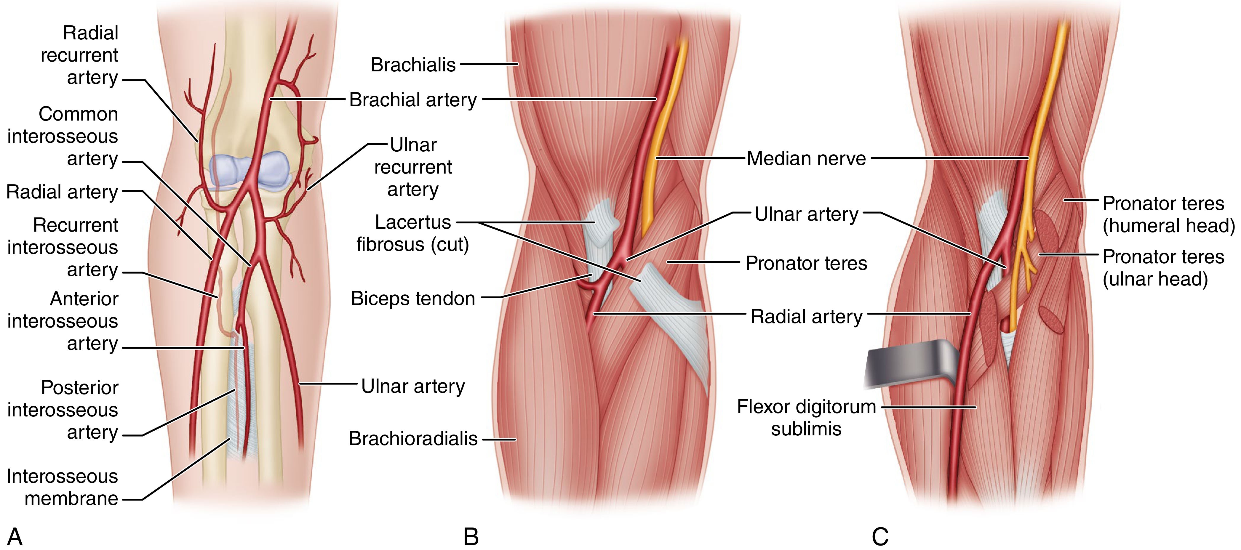
- The brachial artery and median nerve enter the forearm through a tight opening bounded by the biceps tendon laterally and pronator teres medially, covered by the lacertus fibrosus - this cruciate portal is particularly susceptible to compression.
The elbow collateral circulation does NOT communicate with vessels within the flexor compartment - joining the radial and ulnar arteries only proximal to the pronator teres.
Anatomy of Volkmann ischemia (Campbell's Operative Orthopaedics):
Causes / Etiology
- Most common: Supracondylar fracture of the humerus (children) - post-ischemic forearm swelling increases pressure within the enclosed osteofascial compartment
- Tight plaster casts or bandages
- Vascular injuries, crush injuries, burns
- Forearm fractures, elbow dislocations
- Any cause of acute compartment syndrome
Clinical Features
The typical clinical picture of established Volkmann contracture includes:
| Feature | Description |
|---|---|
| Elbow | Flexion |
| Forearm | Pronation |
| Wrist | Flexion |
| Thumb | Adduction |
| MCP joints | Extension |
| Fingers | Flexion (claw) |
- Earliest changes involve the flexor digitorum profundus in the middle third of the forearm
- Nerve involvement: median and ulnar nerve sensory and motor deficits
- Severe cases: intrinsic-minus (claw hand) deformity
Tsuge Classification (Grades of Severity)
Grade I - Mild (Localized)
- Partial ischemia of the profundus mass
- Flexion contractures of 2-3 fingers (usually middle and ring)
- Absent or minimal sensory changes
- No intrinsic or joint contractures
Grade II - Moderate
- Involves long finger flexors, FPL, and possibly wrist flexors
- Flexion contractures of all digits and thumb
- Median and ulnar nerve sensory changes
- Intrinsic minus deformities present
- May require complete release of wrist and finger flexors
Grade III - Severe
- Flexors and extensors both involved
- Forearm bone fractures and skin scars may coexist
- Severe sensory impairment (nerves strangulated by contracted, scarred muscles)
- Intrinsic-minus / claw hand deformity
Treatment
Treatment depends on the grade of contracture. The goal is always to prevent VIC through early recognition and fasciotomy; once established, treatment is reconstructive.
Prevention
- Urgent fasciotomy when compartment pressure >30 mmHg or within 30 mmHg of diastolic BP
- Reduce and stabilize fractures, correct circulatory problems
Established Contracture - by Grade
| Severity | Findings | Treatment |
|---|---|---|
| Mild | Flexion of some digits; absent/limited sensory changes | Physical therapy; dynamic splinting; tendon lengthening/release after 3 months |
| Moderate | All digits and thumb flexed; possible wrist flexors; median/ulnar sensory loss | Excision of necrotic muscle + neurolysis; muscle sliding operation (Page-Scaglietti); +/- carpal resection/bone shortening |
| Severe | All flexors and extensors involved; severe sensory loss; MCP extension, wrist flexion, forearm pronation | Excision of necrotic muscle + neurolysis; tendon transfers (brachioradialis to FPL, ECRL to FDP); free functioning gracilis muscle transfer |
- Campbell's Operative Orthopaedics, 15th Ed 2026, p. 4460
Key Surgical Procedures
1. Muscle Sliding Operation (Page-Scaglietti)
- First described by Page (1923), endorsed by Scaglietti (1957)
- Used for moderate contractures
- The origin of the flexor-pronator muscle mass is detached from the medial epicondyle and allowed to slide distally, lengthening the musculotendinous units without individual tendon cuts
2. Excision of Necrotic Muscle + Neurolysis (Severe)
- Extensive volar forearm incision
- Excise all avascular masses of FDP and FDS, preserve viable tissue
- Neurolysis of median and ulnar nerves (the median nerve classically shows an "hourglass deformity" at the midforearm)
- Neuroma excision and nerve grafting may be necessary
- Tendon transfers performed at a second-stage procedure
3. Two-Stage Free Gracilis Transfer (Oishi and Ezaki)
- For severe cases where no viable motors remain
- Stage 1: Wide forearm exposure, neurolysis, debridement of all fibrotic muscle
- Stage 2: Free innervated gracilis muscle transfer to restore M3-M4 finger flexion strength
- Good results reported with this approach
Key Points to Remember
- Artery and nerve compromised: Brachial artery (anterior interosseous artery) and median nerve - most commonly compressed
- Fracture most likely to cause VIC: Supracondylar fracture of the humerus
- Most vulnerable muscles: FDP and FPL
- 5 P's warning signs (precursors): Pain (especially on passive stretch), Pallor, Paresthesia, Paralysis, Pulselessness
- Most important prognostic factor: time between onset and surgical intervention
- Untreated VIC results in partial or complete loss of arm and hand function
Sources: Campbell's Operative Orthopaedics 15th Ed 2026; Miller's Review of Orthopaedics 9th Ed; Sabiston Textbook of Surgery; StatPearls - Volkmann Contracture; Acta Orthopaedica Belgica 2026 review
Anatomy of bone
Bone is a dynamic connective tissue composed of an organic matrix (osteoid, mainly type I collagen) and inorganic mineral (hydroxyapatite). Macroscopically, bones have compact (cortical) and cancellous (trabecular) regions. Key cell types include osteoblasts (bone formation), osteocytes (maintenance), and osteoclasts (resorption).
Searching sources...
Reading File
Finding Sources
Searching the Web
anatomy of bone structure macroscopic microscopic components
Finding Sources
Finding Sources
Finding Sources
Finding Sources
Excellent sources found. Let me read from the richest ones - Miller's Review (orthopaedics), Junqueira's Basic Histology, Histology A Text and Atlas, and THIEME Atlas.
Reading File
Reading File
Reading File
Reading File
Reading File
Reading File
Reading File
Now let me fetch the key diagrams to include visually.
Loading Image
Loading Image
Reading File
Reading File
I now have comprehensive material. Here is a detailed, well-structured answer.
Anatomy of Bone
Bone is a calcified, living connective tissue that forms the majority of the skeleton. It consists of an intercellular calcified matrix containing collagen fibers, plus several specialized cell types. - Gray's Anatomy for Students, p. 29
Functions of Bone
- Structural support for the body
- Protection of vital organs
- Reservoir for calcium and phosphorus
- Levers for muscle action and movement
- Container for blood-producing cells (bone marrow)
Classification of Bones by Shape
| Type | Description | Examples |
|---|---|---|
| Long bones | Tubular | Humerus, femur |
| Short bones | Cuboidal | Wrist (carpal), ankle (tarsal) |
| Flat bones | Two compact plates with spongy bone between | Skull, ribs |
| Irregular bones | Variable shapes | Vertebrae, facial bones |
| Sesamoid bones | Round/oval, develop within tendons | Patella, sesamoids of thumb |
Macroscopic Anatomy of a Long Bone
A long bone has three main regions:
| Region | Description |
|---|---|
| Diaphysis | The shaft; composed mainly of compact bone surrounding a medullary (marrow) cavity |
| Epiphysis | The wider ends; filled with spongy (cancellous) bone; covered by articular cartilage |
| Metaphysis | The flared zone between shaft and epiphysis; site of the growth plate (physis) in growing bone |
Supporting membranes:
- Periosteum: Dense fibrous connective tissue covering the outer bone surface (absent over articular cartilage). Has an outer fibrous layer and an inner cambium layer rich in osteoprogenitor cells and osteoblasts. Attached to bone by Sharpey's (perforating) fibers - bundles of type I collagen. Highly sensitive to injury (pain fibers) and essential for bone viability.
- Endosteum: A thin single layer of osteoprogenitor cells, osteoblasts, and osteoclasts lining all internal bone surfaces (Haversian canals, trabecular surfaces, medullary cavity).
Two Types of Bone Tissue
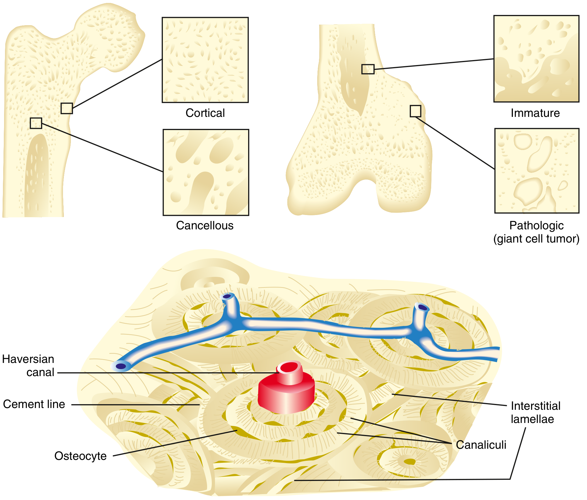
Fig. 1.1 - Types of bone. Cortical bone consists of tightly packed osteons. Cancellous bone consists of a meshwork of trabeculae. Miller's Review of Orthopaedics 9th Ed.
1. Cortical (Compact) Bone
- Makes up 80% of the skeleton
- Forms the outer shell (cortex) of all bones
- Slow turnover rate; higher Young's modulus (more stiff and rigid)
- Composed of tightly packed osteons (Haversian systems)
2. Cancellous (Spongy/Trabecular) Bone
- Makes up 20% of the skeleton
- Located deep to the cortex; porous lattice of trabeculae (struts and plates)
- 30-90% of its volume is pores filled with bone marrow
- Higher turnover rate; more elastic, less stiff than cortical bone
- Found prominently in epiphyses and metaphyses
Microscopic Anatomy
Microscopic Classification
| Type | Features | Examples |
|---|---|---|
| Lamellar bone | Normal, mature bone; organized parallel lamellae stress-oriented; strong | Femoral shaft cortex |
| Woven bone | Immature or pathologic; random collagen orientation; high turnover; weak | Embryonic skeleton, fracture callus, Paget's disease |
The Osteon (Haversian System)
The basic structural unit of compact bone:
- Haversian canal (central canal): runs longitudinally; contains arterioles, venules, capillaries, and nerves
- Concentric lamellae: rings of mineralized matrix arranged around the canal
- Lacunae: small spaces between lamellae, each housing one osteocyte
- Canaliculi: tiny channels radiating from each lacuna, interconnecting osteocytes with each other and with the Haversian canal - the route for nutrient/waste exchange
- Cement lines: define the outer boundary of each osteon; fibrils connect lamellae but do NOT cross cement lines
- Interstitial lamellae: remnants of old, remodeled osteons lying between current osteons
- Volkmann's canals: run perpendicularly (or obliquely) to the longitudinal axis, connecting adjacent Haversian canals to each other and to the periosteum
Cellular Biology of Bone
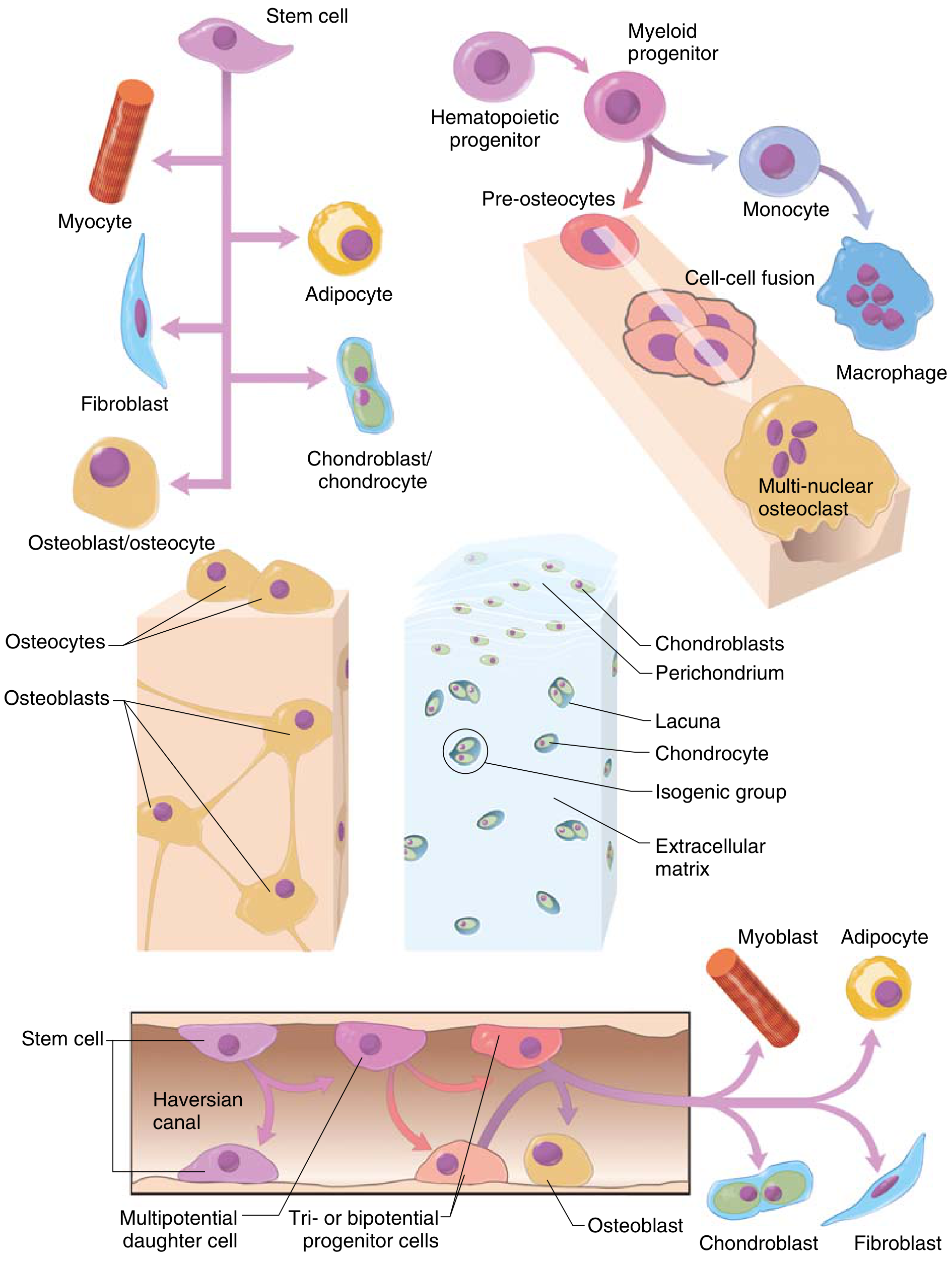
Fig. 1.2 - Cellular origins of bone and cartilage cells. Miller's Review of Orthopaedics 9th Ed.
Osteoblasts
- Derived from mesenchymal stem cells (MSCs) in bone marrow, periosteum, and endosteum
- Differentiation directed by transcription factors RUNX2 and BMP (bone morphogenetic protein)
- Cuboid cells aligned in layers along immature osteoid
- Functions: Synthesize and secrete osteoid (unmineralized bone matrix)
- Products: Type I collagen, alkaline phosphatase, osteocalcin, bone sialoprotein, RANKL, and osteoprotegerin (OPG)
- Stimulated by: intermittent/pulsatile PTH, Wnt proteins, BMPs
- Inhibited by: TNF-α, sclerostin (Scl), Dkk-1
Osteocytes
- Former osteoblasts entrapped within the lacunae of newly formed matrix - constitute 90% of cells in the mature skeleton
- Maintain bone matrix and regulate extracellular calcium and phosphorus
- Communicate via canalicular processes (dendritic extensions through canaliculi)
- Respond to mechanical loading - downregulate sclerostin under strain, promoting new bone formation
- Stimulated by calcitonin; inhibited by PTH
Osteoclasts
- Multinucleated giant cells derived from hematopoietic progenitors (monocyte/macrophage lineage) by cell-cell fusion
- Primary function: bone resorption
- Mechanism:
- Attach to bone via integrin (αvβ3 / vitronectin receptor)
- Form a ruffled border (brush border) - plasma membrane enfoldings that increase surface area
- Seal off a resorption space called Howship's lacuna
- Secrete H⁺ ions (via carbonic anhydrase) - lowers pH, dissolves hydroxyapatite
- Degrade organic matrix using cathepsin K (lysosomal enzyme)
- Produce tartrate-resistant acid phosphatase (TRAP)
- Regulation via RANK-RANKL axis:
- RANKL (on osteoblasts) binds RANK (on osteoclast precursors) → stimulates osteoclast differentiation
- OPG (secreted by osteoblasts) acts as decoy receptor, binds RANKL → inhibits osteoclastogenesis
- Inhibited by: calcitonin (direct receptor), IL-10
- Stimulated by: IL-1, IL-6
Bone Matrix Composition
| Component | % Dry Weight | Function | Key Constituents |
|---|---|---|---|
| Organic matrix | 40% | Tensile strength, scaffolding | Type I collagen (90%), proteoglycans, noncollagenous proteins |
| Inorganic matrix | 60% | Compressive strength | Calcium hydroxyapatite Ca₁₀(PO₄)₆(OH)₂ (primary); brushite |
Organic Matrix:
- Type I collagen (90% of organic matrix): triple helix of 2 α₁ + 1 α₂ chains; provides tensile strength. Mineralization occurs in the hole zones and pores within the collagen fibril.
- Proteoglycans: glycosaminoglycan-protein complexes; provide compressive strength; also inhibit mineralization
- Noncollagenous proteins:
- Osteocalcin: most abundant noncollagenous protein (10-20% of total); binds Ca²⁺; stimulated by 1,25(OH)₂D₃; measurable in serum/urine as a bone turnover marker
- Osteonectin (SPARC): regulates calcium and organizes mineral in matrix
- Osteopontin: cell-binding protein; attracts osteoclasts
- Growth factors / cytokines: TGF-β, IGF, BMPs, IL-1, IL-6
Inorganic Matrix:
- Calcium hydroxyapatite [Ca₁₀(PO₄)₆(OH)₂]: primary mineral; provides compressive strength. Deposited first in collagen hole zones, then on the periphery.
- Osteocalcium phosphate (brushite): remaining inorganic component
Bone Marrow
The medullary cavity and cancellous bone spaces contain marrow:
| Type | Composition | Location |
|---|---|---|
| Red marrow | Hematopoietic (40% water, 40% fat, 20% protein) | Epiphyses in adults; all bones in children |
| Yellow marrow | Primarily fat; contains mesenchymal stem cells | Diaphyses of long bones in adults |
Red marrow gradually converts to yellow marrow with aging. Yellow marrow can revert to red in states of increased hematopoietic demand.
Blood Supply and Innervation
- Each bone typically receives a nutrient artery (one per bone) that enters through the nutrient foramen and supplies the marrow, spongy bone, and inner cortex
- Periosteal vessels supply the outer cortex
- Epiphyseal and metaphyseal arteries supply those respective regions
- Nerves follow the vessels; most are vasomotor fibers. The bone itself has few sensory fibers - but the periosteum is richly innervated and very sensitive to trauma (explains pain in periostitis and fractures)
Bone Formation (Ossification)
| Type | Mechanism | Bones Formed |
|---|---|---|
| Intramembranous ossification | Osteoblasts differentiate directly from mesenchyme (no cartilage model) | Flat bones of skull, mandible, clavicle |
| Endochondral ossification | Hyaline cartilage model is replaced by bone via primary and secondary ossification centers | All other bones (long bones, vertebrae, etc.) |
Primary ossification center: forms in the diaphysis. Secondary ossification centers: form in the epiphyses. The epiphyseal growth plate (physis) between them is responsible for longitudinal bone growth in children.
Sources: Miller's Review of Orthopaedics 9th Ed; Gray's Anatomy for Students; Junqueira's Basic Histology 17th Ed; Histology: A Text and Atlas (Pawlina); TeachMeAnatomy - Ultrastructure of Bone; OpenStax Anatomy & Physiology - Bone Structure
This is a shared conversation. Sign in to Orris to start your own chat.