
What is the most hydrophilic opioid
Morphine is the most hydrophilic (water-soluble) opioid in common clinical use. Its low lipid solubility means it crosses the blood-brain barrier slowly, has a slower onset than lipophilic opioids like fentanyl, and is less likely to cause delayed respiratory depression when given neuraxially due to limited spinal cord uptake.
Searching sources...
Reading File
Finding Sources
Finding Sources
Reading File
Reading File
Morphine is the most hydrophilic opioid in common clinical use.
Here is a full breakdown, sourced directly from the textbooks:
Hydrophilicity Among Opioids
Opioids span a wide range of lipid solubility. At one end sit highly lipophilic agents like fentanyl and sufentanil; at the other end sit hydrophilic agents, with morphine being the prototype and most hydrophilic of the clinically used opioids. Hydromorphone is also considered hydrophilic but is less so than morphine.
Key Properties of Morphine (Hydrophilic Prototype)
| Property | Morphine (Hydrophilic) | Fentanyl/Sufentanil (Lipophilic) |
|---|---|---|
| Onset of analgesia | Delayed (30-60 min) | Rapid (5-10 min) |
| Duration of analgesia | Long (6-24 hr) | Shorter (2-4 hr) |
| CSF spread (neuraxial) | Extensive | Minimal |
| Primary site of action | Spinal > supraspinal | Spinal < systemic |
| Delayed respiratory depression | Yes (>6 hr possible) | Minimal |
| Metabolism | Hepatic conjugation (not CYP450) | CYP3A4 |
Why Hydrophilicity Matters Clinically
-
Slow BBB penetration - Morphine's poor lipid solubility slows its crossing of the blood-brain barrier, leading to a slower onset compared to fentanyl.
-
Neuraxial use - When given intrathecally or epidurally, morphine stays in the CSF much longer and spreads rostrally (upward toward the brain). This explains its risk of delayed respiratory depression hours after administration - a rare but serious complication.
-
Longer duration - Because it is not rapidly taken up by neural tissue or fatty tissue, morphine persists in the CSF and provides analgesia for up to 24 hours with a single intrathecal dose.
-
High intrathecal:IV potency ratio - The relative intrathecal-to-intravenous potency of morphine is 200-300:1, whereas for fentanyl it is only 10-20:1, reflecting how effectively morphine works at the spinal level.
-
Drug interactions - Because morphine is metabolized by hepatic glucuronidation rather than the CYP450 system, it has fewer pharmacokinetic drug-drug interactions than many other opioids.
Summary
Morphine = most hydrophilic opioid. Hydromorphone is the other clinically important hydrophilic opioid, though less so than morphine. All other commonly used opioids (fentanyl, sufentanil, alfentanil, remifentanil, oxycodone) are significantly more lipophilic.
- Miller's Anesthesia, 10e - Table 77.3 (Neuraxial Opioids) and Hydrophilic Opioids section
- Lippincott Illustrated Reviews: Pharmacology - Morphine hydrophilicity and metabolism
At what stage of labour intrathecal fentanyl can be given
Intrathecal fentanyl can be given at any stage of labour, but it is most commonly used in the active first stage. It is particularly useful as part of a combined spinal-epidural (CSE) technique. However, it is generally avoided in the second stage due to concerns about fetal bradycardia and uterine hyperstimulation.
Searching sources...
Finding Sources
Finding Sources
Reading File
Finding Sources
Finding Sources
Finding Sources
Reading File
Finding Sources
Finding Sources
Finding Sources
Finding Sources
Based on authoritative textbook sources and established clinical knowledge, here is a complete answer:
Intrathecal Fentanyl in Labour - When Can It Be Given?
Short Answer
Intrathecal fentanyl can be given at any stage of labour, but it is most commonly and appropriately used in the first stage of labour, particularly as part of a Combined Spinal-Epidural (CSE) technique. It can also be given just before delivery (end of first stage / approaching second stage) as a saddle block adjunct.
Stage-by-Stage Breakdown
First Stage of Labour (Most Common Indication)
This is the primary indication for intrathecal fentanyl in obstetrics. It is especially useful:
- Early first stage - when the patient has severe pain early in labour and desires fast-onset relief
- Active first stage - as part of a CSE technique, providing almost immediate analgesia while the epidural catheter is placed for subsequent top-ups
The CSE technique injects intrathecal opioid + small dose local anaesthetic, then threads an epidural catheter for continued labour analgesia. Typical dose: fentanyl 10-12.5 mcg intrathecally (or sufentanil 5 mcg), often combined with bupivacaine 2.5 mg or ropivacaine 3-4 mg.
Key advantage: the intrathecal component provides essentially immediate pain control with minimal effect on early labour progress, while the epidural catheter handles the rest of labour and potential caesarean section.
Approaching / Just Prior to Delivery (Second Stage / Operative Vaginal Delivery)
A saddle block given just before delivery can include fentanyl 12.5-25 mcg or sufentanil 5-7.5 mcg as an adjunct to intrathecal local anaesthetic (hyperbaric bupivacaine, tetracaine, or lidocaine). This significantly potentiates the block quality for operative vaginal delivery (forceps/vacuum).
Key Clinical Points
| Feature | Detail |
|---|---|
| Typical IT fentanyl dose (CSE) | 10-12.5 mcg |
| Typical IT fentanyl dose (saddle block) | 12.5-25 mcg |
| Onset | Near-immediate (~5 min) |
| Duration | ~1-2 hours (short, hence need for epidural catheter) |
| Effect on labour progress | Does NOT prolong labour or increase operative delivery when used in dilute combinations |
| Motor block | Minimal - patient can often ambulate ("walking epidural") |
| Side effects | Pruritus (most common), nausea, rare respiratory depression, fetal bradycardia (rare) |
Important Caveat: Fetal Bradycardia
There is a well-recognised association between intrathecal opioids (especially fentanyl/sufentanil) and fetal bradycardia, thought to be due to rapid pain relief causing a sudden drop in maternal catecholamines, leading to unopposed uterine activity (hyperstimulation) and reduced uteroplacental blood flow. This is a consideration at any stage but is monitored for with continuous CTG after administration.
Bottom line: Intrathecal fentanyl in labour is primarily a first-stage technique used via CSE for rapid pain relief, and also used as an adjunct in saddle blocks near delivery. It is not typically used as a standalone agent for the entire labour due to its short duration.
- Morgan and Mikhail's Clinical Anesthesiology, 7e - Combined Spinal & Epidural Analgesia section
- Miller's Anesthesia, 10e - Spinal Additives and Neuraxial Opioids
What happens if adenyl cyclase is inhibited
When adenylyl cyclase is inhibited, cAMP production falls. Since cAMP activates protein kinase A (PKA), reduced cAMP means less PKA activity, which downstream reduces phosphorylation of target proteins - blunting cellular responses like glycogenolysis, lipolysis, and cardiac contractility depending on the tissue.
Searching sources...
Reading File
Finding Sources
Reading File
Reading File
Finding Sources
Finding Sources
Finding Sources
Reading File
Finding Sources
Reading File
Loading Image
Here is a complete, textbook-backed answer:
What Happens When Adenylyl Cyclase is Inhibited
The Normal Pathway (for context)
Adenylyl cyclase is a membrane-bound enzyme that converts ATP → cyclic AMP (cAMP). cAMP then activates Protein Kinase A (PKA), which phosphorylates target proteins to produce a biological effect. This is the classical Gs-protein pathway.
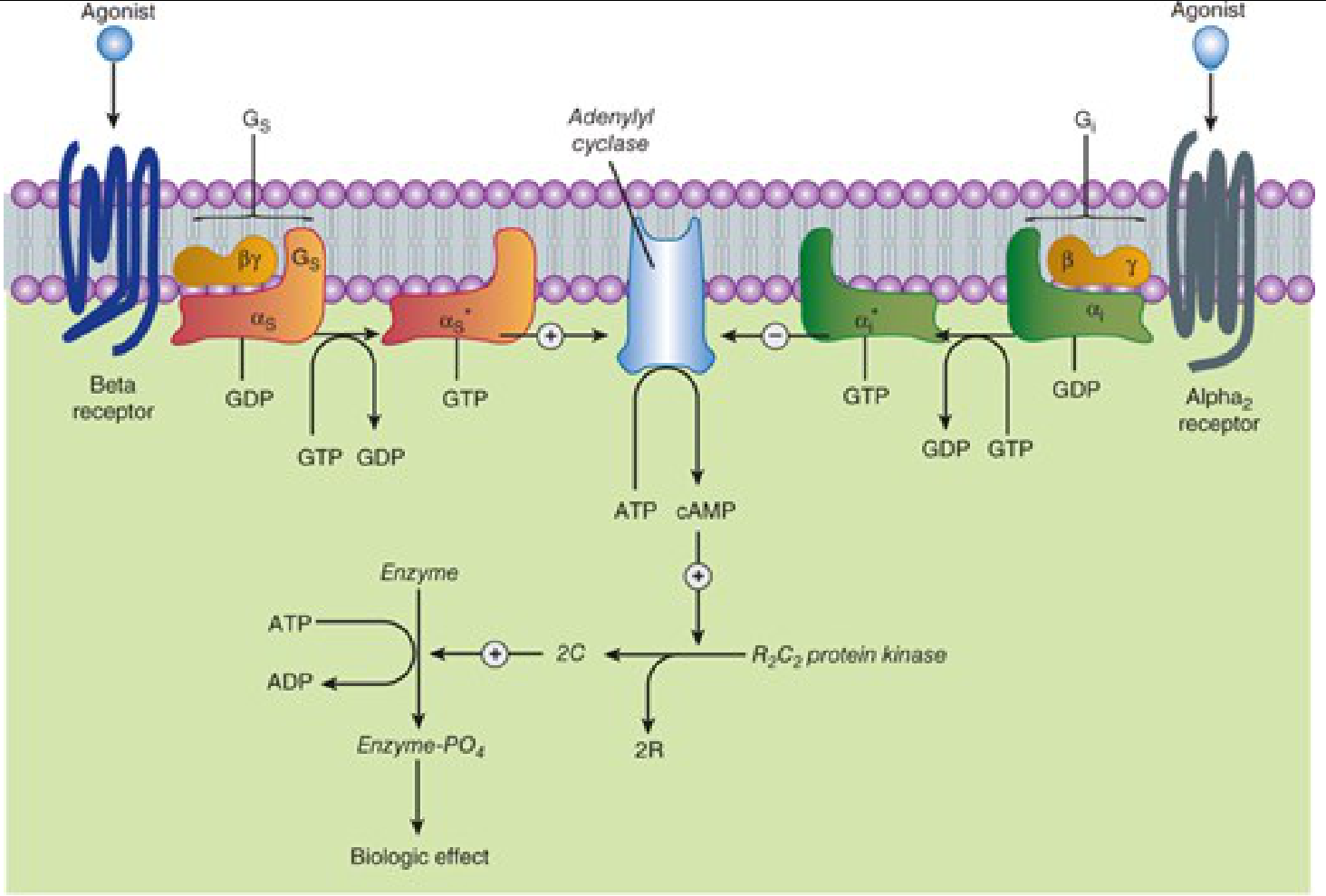
Left: Beta receptor activates adenylyl cyclase via Gs (stimulatory). Right: Alpha-2 receptor inhibits adenylyl cyclase via Gi (inhibitory). - Katzung's Basic and Clinical Pharmacology, 16e
Step-by-Step: What Inhibition of Adenylyl Cyclase Does
Receptor (e.g. α2, opioid, muscarinic M2) binds agonist
        ↓
Gi protein activated → αi subunit dissociates with GTP
        ↓
αi inhibits adenylyl cyclase
        ↓
↓ ATP → cAMP conversion  (less cAMP produced)
        ↓
↓ cAMP binds PKA regulatory subunits
        ↓
↓ Free catalytic subunits of PKA released
        ↓
↓ Phosphorylation of target proteins
        ↓
Tissue-specific effects (see below)
Tissue-Specific Consequences of Reduced cAMP
| Tissue / System | Normal cAMP Effect (via stimulation) | When Adenylyl Cyclase Inhibited |
|---|---|---|
| Liver | Activates glycogen phosphorylase → glycogenolysis | Reduced glycogenolysis → less glucose output |
| Adipose tissue | Activates hormone-sensitive lipase → lipolysis | Reduced lipolysis → less free fatty acid release |
| Heart (β1) | Increases Ca²⁺ influx → positive inotropy/chronotropy | Reduced contractility and heart rate |
| Smooth muscle | Phosphorylates myosin light-chain kinase (inactivates it) → relaxation | Smooth muscle contracts (or less relaxed) |
| Neurons | Modulates neurotransmitter release, excitability | Reduced neuronal excitability; decreased neurotransmitter release |
| Kidney (ADH/V2) | Inserts aquaporins → water reabsorption | Less water reabsorption (if adenylyl cyclase inhibited in collecting duct) |
| Thyroid (TSH) | Stimulates thyroid hormone synthesis | Reduced thyroid hormone synthesis |
Which Receptors Work by Inhibiting Adenylyl Cyclase?
All of these act through Gi/Go proteins to inhibit adenylyl cyclase:
| Receptor | Endogenous Ligand | Clinical Relevance |
|---|---|---|
| α2-adrenoceptor | Noradrenaline | Clonidine/dexmedetomidine act here (sedation, analgesia, ↓ sympathetic tone) |
| Opioid receptors (μ, δ, κ) | Endorphins, enkephalins | Morphine/fentanyl analgesia, reduced neuronal excitability |
| Muscarinic M2 | Acetylcholine | Slows heart rate (vagal tone on SA/AV nodes) |
| D2 dopamine receptor | Dopamine | Antipsychotics block this; also involved in emesis |
| Adenosine A1 | Adenosine | Slows AV conduction |
| GABA-B | GABA | CNS inhibition (baclofen acts here) |
Clinical Examples
-
Opioids (morphine, fentanyl): All opioid receptors couple to Gi/Go and inhibit adenylyl cyclase → reduced cAMP → decreased Ca²⁺ channel conductance, increased K⁺ channel opening → neuronal hyperpolarization → analgesia, sedation, and reduced pain transmission at all levels of the neuraxis.
-
Clonidine / Dexmedetomidine (α2 agonists): Activate Gi via α2 receptors → inhibit adenylyl cyclase → lower cAMP → presynaptically reduce noradrenaline release → sedation, analgesia, reduced sympathetic tone.
-
Pertussis toxin (Bordetella pertussis): Permanently ADP-ribosylates and inactivates Gi, so adenylyl cyclase can no longer be inhibited. This is a key pharmacological tool (and virulence mechanism).
-
Pseudohypoparathyroidism: Defective Gs (αs subunit mutation) prevents PTH from activating adenylyl cyclase, resulting in hypocalcaemia and hyperphosphataemia despite elevated PTH levels.
Summary
Inhibition of adenylyl cyclase → ↓ cAMP → ↓ PKA activity → reduced phosphorylation of downstream targets → the cell becomes less responsive to stimulatory signals. The downstream effects are highly tissue-specific but generally represent a braking or inhibitory signal on the cell's activity.
- Katzung's Basic and Clinical Pharmacology, 16e - Adrenoceptors, Figure 9-2
- Neuroscience: Exploring the Brain, 5e - Second Messenger Cascades, Figure 6.26
- Medical Physiology (Boron & Boulpaep) - G Proteins Coupled to Adenylyl Cyclase
- Miller's Anesthesia, 10e - Opioid receptor Gi/Go coupling
Inward rectifier potassium channels
Inward rectifier K⁺ channels (Kir channels) preferentially allow K⁺ to flow inward at membrane potentials negative to the K⁺ equilibrium potential, and strongly limit outward K⁺ flow during depolarization. This "rectification" helps stabilize the resting membrane potential and is critical in cardiac, neuronal, and epithelial physiology.
Searching sources...
Finding Sources
Finding Sources
Finding Sources
Finding Sources
Reading File
Finding Sources
Finding Sources
Finding Sources
Reading File
Finding Sources
Reading File
Finding Sources
Loading Image
Here is a comprehensive coverage of inward rectifier potassium channels:
Inward Rectifier Potassium Channels (Kir / IK1)
What is "Inward Rectification"?
Rectification means the channel conducts current more easily in one direction than the other. Inward rectifiers pass K⁺ inward (into the cell) more readily than outward. Paradoxically, this means at the resting membrane potential (near E_K), they actually allow a small outward K⁺ current that helps set the resting potential - but as soon as the cell depolarizes, outward K⁺ flow is blocked.
This is the opposite of delayed rectifiers (which open on depolarization to allow outward K⁺ flow during repolarization).
Mechanism of Rectification
The blockade of outward current is caused by intracellular Mg²⁺ and polyamines (spermine, spermidine, putrescine) plugging the channel pore from the cytoplasmic side during depolarization:
- At resting/hyperpolarized potentials: Mg²⁺/polyamines are electrostatically pulled away from the pore → channel open → K⁺ can flow inward (and small outward current is allowed)
- At depolarized potentials: Mg²⁺/polyamines are driven into the pore → channel blocked → outward K⁺ flow is prevented
This voltage-dependent block is the molecular basis of inward rectification, as confirmed in Braunwald's Heart Disease: "inward rectification appears to result from depolarization-induced internal blockage by Mg²⁺ and neutral or inwardly charged amino acid residues in the cytoplasmic channel pore."
Structure
- Kir channels are tetramers - four subunits, each with only 2 transmembrane domains (M1 and M2), unlike voltage-gated K⁺ channels which have 6
- They lack the classic S4 voltage-sensing domain
- A P-loop (H5) between M1 and M2 forms the ion selectivity filter
- Gene family: KCNJ genes (e.g., KCNJ2 encodes Kir2.1)
Major Subtypes and Their Roles
| Kir Subtype | Current Name | Key Location | Function | Clinical Relevance |
|---|---|---|---|---|
| Kir2.x (Kir2.1) | I_K1 | Atria, ventricles, His-Purkinje | Sets resting membrane potential; contributes to phase 3 repolarization | Andersen-Tawil syndrome (Kir2.1 loss-of-function) |
| Kir3.x (GIRK) | I_KACh | SA node, atria | Opened by Gβγ subunit (from Gi, via M2 muscarinic or adenosine A1 receptors) → hyperpolarization → slows heart rate | Vagal slowing of heart; bradycardia with high parasympathetic tone |
| Kir6.x + SUR | I_KATP | Pancreatic β-cells, cardiac muscle, smooth muscle, neurons | Closed by ATP (high energy state) → cell depolarizes → insulin secretion; opens in ischaemia (low ATP) | Sulfonylureas (glibenclamide) block KATP in β-cells → insulin release; neonatal diabetes from KATP gain-of-function |
| Kir4.x | - | Kidney tubules, astrocytes | Spatial K⁺ buffering in brain; K⁺ reabsorption in kidney | Mutations cause EAST/SeSAME syndrome |
| Kir7.1 | - | Thyroid, retinal pigment epithelium | TSH-regulated; retinal function | Loss-of-function → snowflake vitreoretinal degeneration |
Role in the Cardiac Action Potential
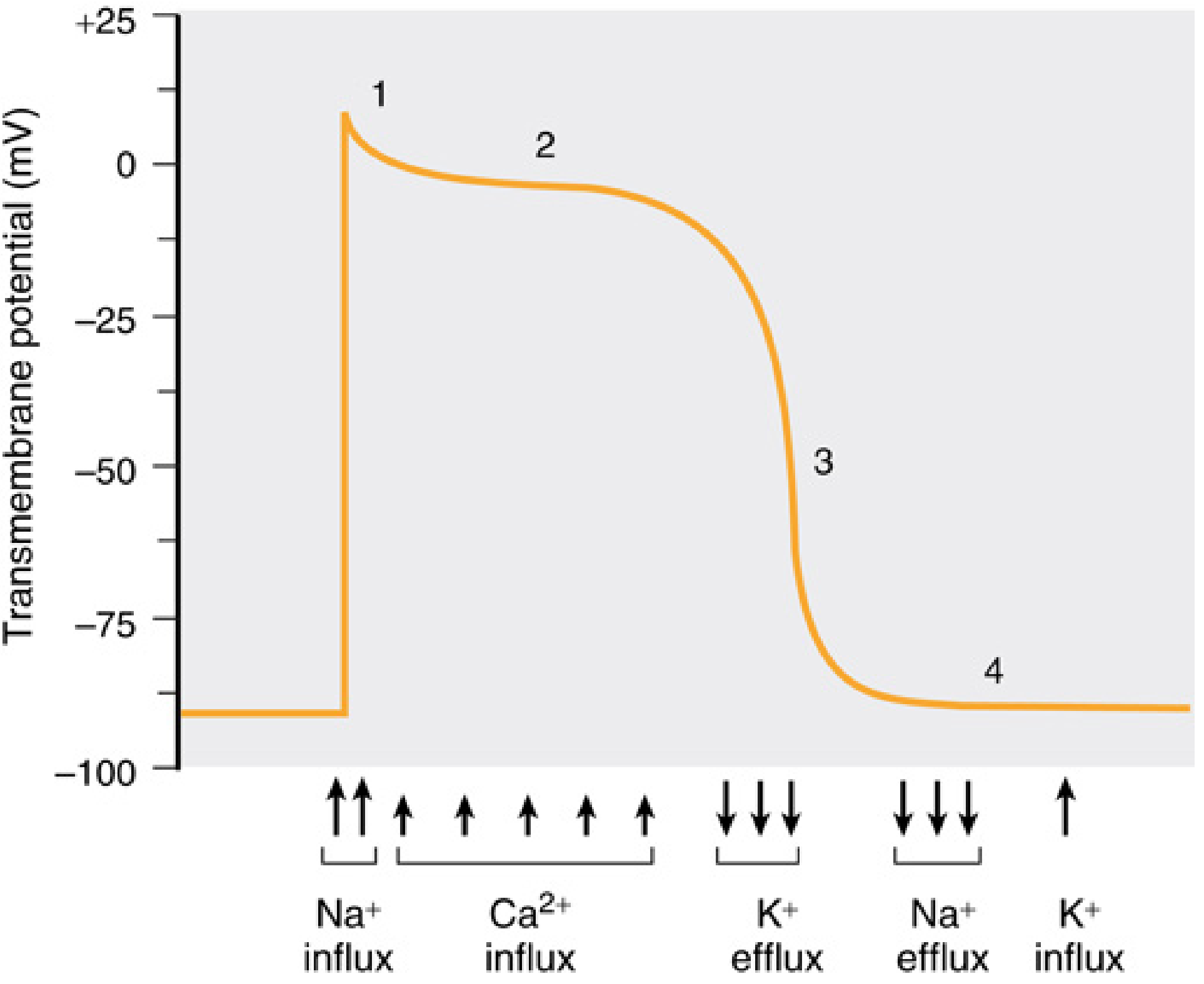
Phases of ventricular action potential - Miller's Anesthesia, 10e
- Phase 4 (resting): IK1 (Kir2.1) is active - provides the dominant K⁺ conductance that holds the cell near -85 mV
- Phase 0-2 (depolarization/plateau): IK1 is blocked by intracellular Mg²⁺/polyamines at depolarized voltages - this is critical for maintaining the long plateau (phase 2) of the cardiac action potential; if IK1 were not blocked, the plateau would collapse immediately
- Phase 3 (repolarization): As the membrane repolarizes, the Mg²⁺/polyamine block is progressively relieved → IK1 activates again → accelerates terminal repolarization back to resting potential
- Pacemaker cells (SA node): IK1 density is very low in pacemaker cells - this is why they spontaneously depolarize (phase 4 slope); without strong IK1 to anchor the resting potential, spontaneous depolarization driven by If (funny current) is possible
GIRK Channels - G Protein-Gated Inward Rectifiers
GIRK (G protein-coupled Inwardly Rectifying K⁺) channels, i.e., Kir3 family, are gated by Gβγ subunits released when Gi-coupled receptors are activated:
ACh → M2 muscarinic receptor → Gi activation
       ↓
Gβγ subunit released
       ↓
Gβγ binds and opens GIRK (IKACh) channel
       ↓
K⁺ flows out → hyperpolarization of SA node
       ↓
Slower phase 4 depolarization → reduced heart rate
This is the primary mechanism of vagal slowing of the heart.
KATP Channels - Metabolic Sensors
KATP channels (Kir6.x + SUR regulatory subunit) are unique in being gated by the ATP:ADP ratio:
- High ATP (fed state): Channel closed → cell depolarizes → β-cell secretes insulin
- Low ATP (ischaemia, fasting): Channel opens → K⁺ efflux → hyperpolarization → cell protected from Ca²⁺ overload (cardioprotective in ischaemia)
Drugs acting on KATP:
- Sulfonylureas (glibenclamide, glipizide): Block SUR1 on β-cells → close KATP → stimulate insulin secretion (type 2 diabetes treatment)
- Diazoxide: Opens KATP → inhibits insulin secretion (used in insulinoma/persistent hyperinsulinism)
- Nicorandil: Opens KATP in smooth muscle → vasodilation (anti-anginal)
Disease Associations
| Disease | Channel | Mechanism |
|---|---|---|
| Andersen-Tawil syndrome | Kir2.1 (IK1) | Loss-of-function → periodic paralysis, QT prolongation, dysmorphic features |
| Neonatal diabetes mellitus | Kir6.2 (KATP) | Gain-of-function → channel stays open → β-cell cannot depolarize → no insulin release; treatable with sulfonylureas |
| Familial hyperinsulinism | SUR1/Kir6.2 | Loss-of-function → channel stays closed → constant insulin secretion → hypoglycaemia |
| Bartter syndrome type 2 | Kir1.1 (ROMK) | Loss-of-function → impaired K⁺ recycling in thick ascending limb → salt wasting, hypokalaemia |
| EAST/SeSAME syndrome | Kir4.1 | Loss-of-function → epilepsy, ataxia, sensorineural deafness, tubulopathy |
Summary
Inward rectifier K⁺ channels are a diverse family of 2-TM domain channels that:
- Set and stabilize the resting membrane potential (Kir2/IK1)
- Mediate neurotransmitter/hormone-gated hyperpolarization (Kir3/GIRK - vagal heart slowing)
- Act as metabolic sensors linking ATP levels to membrane excitability (Kir6/KATP - insulin secretion, ischaemic protection)
- Allow a long cardiac action potential plateau by being blocked at depolarized voltages
- Braunwald's Heart Disease, 2e - Table 62.1, Transsarcolemmal Ionic Currents
- Miller's Anesthesia, 10e - Action Potential phases, IK1 and inwardly rectifying K⁺ current
- Yamada's Textbook of Gastroenterology, 7e - KIR channels in smooth muscle cells
This is a shared conversation. Sign in to Orris to start your own chat.