
Endopthalmitis
Reading File
Now let me search the medical library for endophthalmitis content in parallel.
Finding Sources
Searching PubMed
endophthalmitis treatment outcomes
Good. Kanski's Ophthalmology has the most comprehensive coverage. Let me read the key sections in parallel.
Reading File
Reading File
Reading File
I need the main postoperative bacterial endophthalmitis section from Kanski's Chapter 10. Let me search for it.
Finding Sources
Finding Sources
Reading File
Reading File
Reading File
Reading File
Reading File
Reading File
Reading File
Finding Sources
I now have comprehensive content. Let me also fetch the key clinical image and get the EVS (Endophthalmitis Vitrectomy Study) treatment section from Kanski.
Reading File
Loading Image
I now have comprehensive information to deliver a detailed, well-structured answer on endophthalmitis. Let me compile it.
Endophthalmitis
Endophthalmitis is a severe, sight-threatening inflammation of the intraocular contents (vitreous and aqueous humors), almost always caused by infection. It is one of the most feared complications in ophthalmology and requires immediate diagnosis and treatment.
Classification
| Type | Source | Common setting |
|---|---|---|
| Exogenous (>80%) | External inoculation | Post-surgical, post-traumatic, keratitis-associated |
| Endogenous (<20%) | Hematogenous seeding | Sepsis, IV drug use, immunocompromise |
Exogenous subtypes include:
- Acute postoperative (after cataract surgery, most common)
- Delayed/subacute postoperative
- Bleb-related (post-trabeculectomy)
- Post-traumatic (including retained IOFB)
Postoperative Endophthalmitis (Most Common Form)
Incidence & Pathogenesis
The contemporary incidence after cataract surgery is approximately 0.1%. Bacterial toxins and the host's own inflammatory response cause rapid, irreversible photoreceptor damage -- effects can continue long after the eye is sterilized.
Risk factors:
- Posterior capsule rupture
- Prolonged procedure time / combined surgery (e.g. with vitrectomy)
- Clear corneal sutureless incision, temporal incision
- Wound leak on the first postoperative day
- Adnexal disease (blepharitis, dacryocystitis), diabetes
- Topical anaesthesia
Clinical Features
Symptoms: Sudden onset pain, redness, and visual loss (typically days 2-7 after surgery).
Signs (vary with severity):
- Eyelid swelling, chemosis, conjunctival injection, discharge
- Relative afferent pupillary defect (common)
- Corneal haze
- Fibrinous exudate and hypopyon (hallmark sign)
- Vitritis with impaired fundal view
- Loss of red reflex
- Severe vitreous debris (see below)
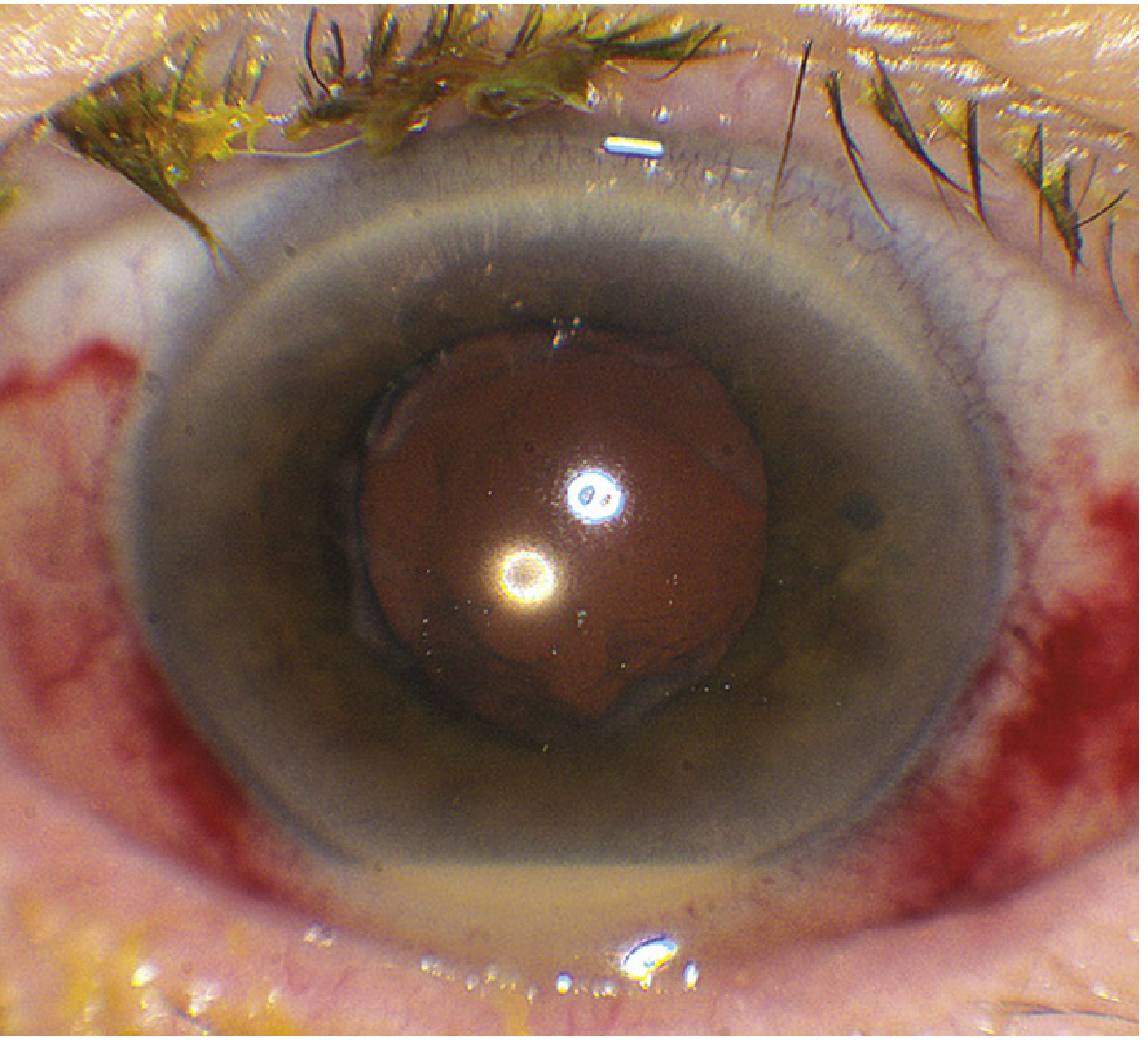
Clinical photo: Postoperative endophthalmitis showing hypopyon (Wills Eye Manual)
Causative Organisms
Acute Postcataract
| Organism | Notes |
|---|---|
| S. epidermidis (most common) | Gram-positive; reasonable prognosis with early treatment |
| S. aureus | More virulent |
| Streptococcal species | Often severe; poor prognosis |
| Gram-negatives (Pseudomonas, Klebsiella, Proteus, E. coli, etc.) | ~10% of isolates |
| Bacillus cereus | Very virulent; post-traumatic |
Overall: ~90% Gram-positive, ~10% Gram-negative isolates.
Other settings:
- Bleb-associated: Streptococcus or Gram-negatives (more virulent than post-cataract)
- Post-intravitreal injection: S. epidermidis and oral flora (notably Streptococcus)
- Post-traumatic: Staphylococcus spp. and Bacillus spp. (~90% of culture-positive cases)
- Subacute/delayed: Cutibacterium acnes (formerly Propionibacterium acnes), fungi
Differential Diagnosis
- TASS (Toxic Anterior Segment Syndrome): Occurs 6-24 hours post-cataract; diffuse corneal edema with KP; sterile, due to endotoxin on instruments. Responds to steroids.
- Acute noninfectious uveitis flare (HLA-B27 associated)
- Sterile endophthalmitis (e.g. after intravitreal triamcinolone or anti-VEGF)
- Retained lens particle uveitis
- Vitreous hemorrhage (especially depigmented blood)
Rule of thumb: If any doubt exists, treat as infectious endophthalmitis -- early recognition leads to better outcomes.
Management
Step 1: Sample First
Immediately obtain:
- Aqueous tap: 0.1-0.2 mL via limbal paracentesis (25G needle on tuberculin syringe)
- Vitreous tap: More likely to yield positive culture; 0.2-0.4 mL from mid-vitreous at 3.5 mm from limbus in a pseudophakic eye; use a disposable vitrector if available
- B-scan ultrasound before sampling if no clinical view (to exclude retinal detachment)
Send for: Gram stain, Giemsa, methenamine silver, and cultures (blood agar, chocolate agar, Sabouraud, thioglycolate, anaerobic). Use PCR if cultures are negative.
Note: C. acnes requires anaerobic culture held for 14 days - it will be missed otherwise.
Step 2: Intravitreal Antibiotics (Cornerstone of Treatment)
Injected immediately after sampling, into the mid-vitreous cavity via 25G needle:
| Drug | Dose | Coverage |
|---|---|---|
| Vancomycin | 2 mg in 0.1 mL (saline only) | Gram-positives including MRSA |
| Ceftazidime | 2 mg in 0.1 mL | Gram-negatives including Pseudomonas |
| Amikacin | 0.4 mg in 0.1 mL | Alternative to ceftazidime if definite penicillin/cephalosporin allergy - HIGHER retinal toxicity risk |
Intravitreal vancomycin used routinely as prophylaxis has been associated with haemorrhagic occlusive retinal vasculitis and should be avoided in prophylaxis.
Step 3: Adjuncts
- Oral fluoroquinolone: Moxifloxacin 400 mg/day x 10 days (excellent ocular penetration)
- Topical antibiotics: Vancomycin 5% or ceftazidime 5%, 4-6x daily (limited benefit, mainly to protect wounds)
- Subconjunctival antibiotics: Vancomycin 50 mg + ceftazidime 125 mg (doubtful additional benefit when intravitreal given)
- Topical steroids: Dexamethasone 0.1% 2-hourly; start after 48 hours (once fungal infection excluded)
- Topical mydriatic: Atropine 1% twice daily
- Oral steroids: Prednisolone 1 mg/kg/day in severe cases after 12-24 hours (fungal infection excluded); The EVS protocol used prednisone 60 mg/day x 5 days
Step 4: Pars Plana Vitrectomy (PPV)
The landmark Endophthalmitis Vitrectomy Study (EVS) established:
- Immediate PPV is indicated when VA = light perception only -- 50% reduction in severe visual loss
- Eyes with VA of hand movements or better do NOT benefit from immediate PPV over tap-and-inject alone
- EVS conclusions apply to post-cataract surgery; cannot be extrapolated to all forms
Step 5: Follow-up at 48 hours
- Relief of pain is a useful early sign of response
- Look for contraction of fibrinous exudate and reduction of hypopyon
- If worsening: review sensitivities, modify antibiotics, consider PPV if not yet done, repeat intravitreal antibiotics
- Avoid repeated amikacin (retinal toxicity risk)
Delayed-Onset (Subacute) Postoperative Endophthalmitis
- Onset: Weeks to months after surgery
- Classic organism: Cutibacterium acnes -- presents as recurrent anterior uveitis, granulomatous KP, white lens capsular plaque, minimal injection/pain, only transient response to steroids
- Other organisms: Fungi (Aspergillus, Candida, Cephalosporium), S. epidermidis
- Treatment: Similar initial approach; if C. acnes, may require removal of IOL and capsular remnants; if fungal, intravitreal amphotericin B (5-10 μg/0.1 mL) or voriconazole (100 μg/0.1 mL)
Bleb-Related Endophthalmitis (Post-Trabeculectomy)
- Organisms are significantly more virulent than post-cataract (streptococcal, Gram-negatives)
- Symptoms far more severe than blebitis
- Signs: White milky bleb, severe injection, large hypopyon, vitreous involvement, loss of red reflex
- Treatment: Vitreous + aqueous samples immediately; ceftazidime 2 mg + vancomycin 2 mg intravitreal; repeat at 48h; consider posterior vitrectomy; oral fluoroquinolones x 10-14 days; topical steroids after 48h
Post-Traumatic Endophthalmitis
- Risk in ~1/10 penetrating trauma cases with retained IOFB
- Prophylactic antibiotics (ciprofloxacin 750 mg twice daily or moxifloxacin 400 mg once daily) for open globe injuries
- Prompt IOFB removal is paramount
- Prophylactic intravitreal antibiotics for high-risk cases (agricultural injuries)
Endogenous Endophthalmitis
- Hematogenous seeding in patients with sepsis, IV drug use, immunosuppression, or indwelling catheters
- Klebsiella pneumoniae associated with liver abscess is the leading cause of endogenous bacterial endophthalmitis (especially in Asia)
- Fungal endogenous: almost exclusively Candida spp. (yeasts); Aspergillus also occurs
- Treatment: systemic antibiotics targeting the primary source + intravitreal antibiotics
Prognosis
Outcome depends on duration of infection before treatment and organism virulence:
| Scenario | Visual outcome |
|---|---|
| VA = light perception at presentation | ~30% achieve 6/12 after treatment |
| VA = better than LP at presentation | ~60% achieve 6/12 |
| Bacillus cereus infection | 70% achieve ≤6/60 (exotoxin-mediated retinopathy) |
| Streptococcal infection | 55% achieve ≤6/60 |
Late complications: Epiretinal membrane, cystoid macular edema, retinal ischemia, hypotony, retinal detachment, secondary glaucoma, phthisis bulbi.
Prophylaxis (Cataract Surgery)
- 5% povidone-iodine in conjunctival fornices for ≥3 minutes before surgery (strongest evidence)
- Intracameral cefuroxime 1 mg/0.1 mL at end of surgery (first choice)
- Intracameral moxifloxacin 0.5 mg/0.1 mL if cefuroxime unavailable
- Treatment of pre-existing blepharitis, dacryocystitis, conjunctivitis
- Early resuturing of leaking wounds
- Avoid intracameral vancomycin as routine prophylaxis (risk of HORV)
Sources:
- Kanski's Clinical Ophthalmology: A Systematic Approach, 10th ed., Ch. 10-11 (Acute & Delayed Postoperative Endophthalmitis; Bleb-Related & Post-Traumatic Endophthalmitis)
- The Wills Eye Manual, 8th ed., §12.13 (Postoperative Endophthalmitis)
- Tietz Textbook of Laboratory Medicine, 7th ed. (Eye Infections; Endophthalmitis)
- Robbins, Cotran & Kumar Pathologic Basis of Disease (Endophthalmitis)
- Recent meta-analysis (PMID: 39167582): Visual outcomes by organism in post-intravitreal injection and post-cataract endophthalmitis (2024, Retina)
This is a shared conversation. Sign in to Orris to start your own chat.