
Hepatic encephalopathy
I'll read the medical library skill first before answering.
Reading File
Now I'll search the library in parallel for hepatic encephalopathy content alongside a recent PubMed search.
Finding Sources
Searching PubMed
"hepatic encephalopathy"[MeSH Terms] AND management
Excellent. Now I'll read the most comprehensive sources in parallel - Sleisenger & Fordtran (the definitive GI/liver text) and Bradley & Daroff's Neurology.
Reading File
Reading File
Reading File
Excellent content. Now let me read the main Sleisenger & Fordtran block24 content on pathophysiology, clinical features, diagnosis and treatment - the core HE chapter.
Reading File
Reading File
I have very rich content from the textbooks. Let me fetch the pathophysiology diagram image and read more of the Sleisenger & Fordtran block for treatment details.
Reading File
Loading Image
I got displaced from the right offset. Let me read the treatment section directly, searching for it in the right block.
Finding Sources
Finding Sources
Now let me get the treatment and diagnosis paragraphs from the main HE chapter.
Finding Sources
Finding Sources
Reading File
Reading File
I need to read the treatment section specifically. Let me get it directly.
Reading File
I now have comprehensive content. Let me compile the full, detailed answer.
Hepatic Encephalopathy (HE)
Definition
Hepatic encephalopathy encompasses a wide array of transient, reversible neurologic and psychiatric manifestations in patients with chronic liver disease and portal hypertension, or with acute liver failure (ALF). HE develops in 50% to 70% of patients with cirrhosis and is a poor prognostic indicator - projected 1- and 3-year survival rates without liver transplant (LT) are 42% and 23%, respectively.
- Sleisenger and Fordtran's Gastrointestinal and Liver Disease, p. 1815
Pathophysiology
Multiple overlapping mechanisms operate simultaneously:
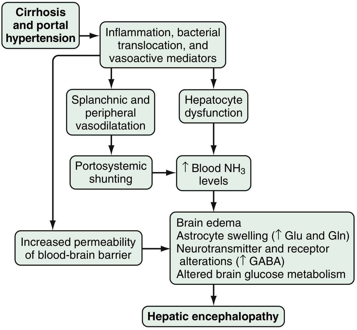
Fig. 94.1 - Proposed pathophysiology of HE (Sleisenger & Fordtran)
1. Ammonia Toxicity
- The best-described neurotoxin is ammonia, produced primarily in the colon by bacterial metabolism of proteins/nitrogen-based products, and by enterocyte synthesis from glutamine
- Under normal conditions, hepatocytes metabolize and clear portal ammonia; in cirrhosis, reduced hepatocyte function plus portosystemic shunting causes hyperammonemia
- Arterial hyperammonemia occurs in up to 90% of HE patients
- Increased blood-brain barrier permeability leads to enhanced uptake by the cerebellum and basal ganglia
- Ammonia directly causes brain edema, astrocyte swelling, and disrupts transport of neuroactive compounds (e.g., myoinositol)
- In ALF, ammonia increases astrocyte glutamine production (via glutamine synthetase), raising glutamine/glutamate levels - contributing to cytotoxic edema
2. GABA-Benzodiazepine System
- Increased sensitivity of the astrocyte peripheral-type benzodiazepine receptor enhances GABA-benzodiazepine activation
- A feed-forward loop: astrocytes produce neurosteroids (allopregnanolone, tetrahydrodeoxycorticosterone) that further activate GABA-A receptors
3. Other Neurotransmitter Changes
- Serotonin (5-HT), nitric oxide (NO), circulating opioid peptides, manganese, and excess oxygen free radicals also contribute
- Manganese causes dopaminergic dysfunction
- Other implicated agents: mercaptans, beta-phenylethylamine, tyramine, octopamine
4. Gut Microbiota
-
Differences in colonic mucosal microbiota in cirrhotic patients with vs. without HE influence production of ammoniagenic substances
-
Distinct allelic mutations in the glutaminase gene increase overt HE risk, potentially by increasing ammonia and glutamate levels
-
Sleisenger and Fordtran's Gastrointestinal and Liver Disease, p. 1815-1816
Classification
By Underlying Disease (Type)
| Type | Underlying Cause |
|---|---|
| Type A | Associated with acute liver failure (ALF) |
| Type B | Portosystemic shunts WITHOUT hepatocellular disease |
| Type C | Chronic/end-stage liver disease with portal hypertension (most common) |
By Severity - West Haven Criteria & SONIC Classification
| West Haven Grade | Intellectual Function | Neuromuscular Function | SONIC Category |
|---|---|---|---|
| 0 | Normal | Normal | Unimpaired |
| Minimal | Normal exam; subtle changes in work/driving | Minor abnormalities on psychometric/visual perception tests | Covert HE |
| 1 | Personality changes, attention deficits, irritability, depression | Tremor, incoordination | Covert HE |
| 2 | Sleep-wake cycle changes, lethargy, behavioral/mood changes, cognitive dysfunction | Asterixis, ataxic gait, slow/slurred speech | Overt HE |
| 3 | Somnolence, confusion, disorientation, amnesia | Rigidity, nystagmus, clonus, Babinski sign, hyporeflexia | Overt HE |
| 4 | Stupor and coma | Oculocephalic reflex, unresponsive to noxious stimuli | Overt HE |
- Covert/Minimal HE affects ~50% of all cirrhotic patients and is commonly overlooked
- Disorientation to time is the key feature distinguishing Grade 1 from Grade 2 (covert from overt HE)
By Time Course
- Episodic - recurrent HE (multiple episodes within 6 months)
- Persistent - altered behavior always present, with relapses
By Precipitating Factors
-
Spontaneous (no identifiable trigger)
-
Precipitated - by GI bleeding, electrolyte abnormalities (especially hypokalemia), infections, sedating medications (benzodiazepines, narcotics), dehydration, constipation
-
Sleisenger and Fordtran's, p. 1815-1816; Bradley and Daroff's Neurology in Clinical Practice, p. 3520-3525
Clinical Features
- Early/covert: Forgetfulness, altered handwriting, difficulty driving, reversal of sleep-wake cycle
- Progressive (overt): Asterixis (flapping tremor - characteristic sign), agitation, disinhibited behavior, disorientation, ataxia, slurred speech
- Severe: Stupor, seizures (10-30% of patients), coma, decerebrate/decorticate posturing
- ALF-specific: Cerebral edema and intracranial hypertension are invariably present in Stage 4, with brainstem herniation being the most common cause of death
Asterixis is pathognomonic but not exclusive to HE - it also occurs in other metabolic encephalopathies (uremia, CO2 narcosis, etc.)
Diagnosis
No Gold Standard Laboratory Test
- Blood ammonia levels are not sensitive or specific for HE
- False elevations: GI bleeding, tourniquet use during blood draw, delayed lab processing, diuretics, alcohol, narcotics, valproic acid
- Arterial ammonia offers no advantage over venous ammonia in chronic liver disease
Neuropsychometric Testing
- Portosystemic encephalopathy (PSE) syndrome test and Stroop test - evaluate attention, concentration, fine motor skills, orientation
- EncephalApp (smartphone-based Stroop test) - validated for covert/minimal HE detection
- Critical flicker frequency (CFF) test - reliable marker for minimal HE
Neurophysiological
- EEG: Early slowing of alpha rhythm progressing to delta frequencies; triphasic waves in later stages (poor prognosis)
- MR spectroscopy and T1 TAPIR mapping - research tools to quantify HE parameters
Differential Diagnosis of Hyperammonemia (Box 94.1)
Includes: ALF, chronic kidney disease, GI bleeding, urea cycle defects, medications (valproic acid, diuretics, alcohol, narcotics), muscle ischemia, portosystemic shunts
- Sleisenger and Fordtran's, p. 1816-1817
Treatment
1. Correct/Eliminate Precipitating Factors
This is the primary therapeutic approach: treat GI bleeding, infections (SBP, UTI), electrolyte disturbances (especially hypokalemia), dehydration, and remove offending medications.
2. Dietary Management
- Protein restriction is no longer recommended - it is harmful and does not benefit HE
- Vegetable and dairy proteins are preferable to animal proteins (more favorable calorie-to-nitrogen ratio)
- Branched-chain amino acid (BCAA) supplementation may benefit HE, but does not clearly reduce mortality
3. Reduce Intestinal Ammonia - First-Line
Lactulose (or lactitol)
- Non-absorbable disaccharide metabolized by colonic bacteria
- Mechanism: causes catharsis, acidifies the colon (inhibits ammonia absorption), and decreases transit time
- Effective for acute and chronic HE vs. placebo
- Dose: titrate to 2-3 soft stools/day
- Side effects: abdominal cramping, flatulence, diarrhea, electrolyte imbalance
- Can be given per rectum as an enema in patients at aspiration risk
4. Oral Antibiotics - Second-Line or Add-On
Rifaximin 550 mg twice daily (FDA-approved 2010)
- Used for chronic HE and prevention of recurrence of overt HE
- Superior tolerability vs. lactulose; main limitation is cost
- Other antibiotics studied (neomycin, metronidazole, vancomycin) - not clearly established for chronic HE
5. Other Agents
| Agent | Mechanism | Evidence |
|---|---|---|
| Probiotics | Modify gut flora, reduce ammonia generation | Probably improve recovery, overt HE, QoL, and plasma ammonia (Cochrane review) - not mortality |
| Acarbose | Alpha-glucosidase inhibitor; shifts colonic flora from proteolytic to saccharolytic | RCT shows benefit in mild HE with diabetes |
| L-ornithine L-aspartate (LOLA) | Activates urea cycle, enhances ammonia clearance | Possible benefit (Cochrane); NOT available in the USA |
| Zinc supplementation | Zinc deficiency common in cirrhosis; zinc activates ornithine transcarbamylase | Possible benefit; clear efficacy not established |
| Sodium benzoate/phenylacetate | Alternate ammonia excretion pathways | FDA-approved for urea cycle defects; efficacy in cirrhosis not well established; high sodium load |
| MARS (Molecular Adsorbent Recirculating System) | Extracorporeal albumin dialysis | Reduces ammonia in acute-on-chronic liver failure |
6. Liver Transplant (LT)
-
Definitive treatment - generally reverses HE
-
Should be considered before permanent neurologic damage
-
Sleisenger and Fordtran's, p. 1817-1818
Acute vs. Chronic HE - Key Distinguishing Features
| Feature | Acute Liver Failure | Portal Systemic Encephalopathy (Chronic) |
|---|---|---|
| Onset | Usually acute | Varies; may be insidious |
| Mental state | Mania evolving to deep coma | Blunted consciousness |
| Precipitant | Viral infection or hepatotoxin | GI hemorrhage, protein load, drugs, uremia, infection |
| Prior liver disease | No | Usually yes |
| Cerebral edema | Common, severe | Less prominent |
Special Considerations: Pediatric HE
-
In children, irritability and lethargy are the most common initial signs
-
Neurological findings: pyramidal signs → hypotonia → focal signs → early parkinsonism, ataxia, tremor, dysarthria
-
Asterixis is present but less prominent as a discriminating feature
-
Clinically evident HE is less common in children with chronic liver disease than in adults, but 50% of children with chronic liver disease have minimal HE on neuropsychological testing
-
Seizures occur in 10-30% of pediatric patients
-
Common pediatric causes: fulminant viral/autoimmune hepatitis, drug ingestion (acetaminophen, salicylates, valproate), Reye syndrome, Wilson disease, mitochondrial diseases
-
Bradley and Daroff's Neurology, p. 1252-1253
Prognosis
- Overt HE: 1-year survival 20-40%, 3-year survival ~15% without transplant
- Cerebral edema (Stage 4) - brainstem herniation is the leading cause of death
- Minimal HE predicts progression to overt HE and negatively impacts driving ability and quality of life
Recent Evidence Update (PubMed 2025-2026)
A 2025 meta-analysis (PMID 40812534) found that proton pump inhibitor (PPI) use is associated with increased risk of hepatic encephalopathy in cirrhotic patients - clinicians should avoid unnecessary PPI prescription in this population. A 2026 systematic review (PMID 41942881) evaluated risk prediction models for HE in liver cirrhosis, noting that MELD score, serum ammonia, and Child-Pugh score are the most commonly incorporated predictors.
This is a shared conversation. Sign in to Orris to start your own chat.