
Case study
Pneumonia.
pneumonia chest X-ray consolidation
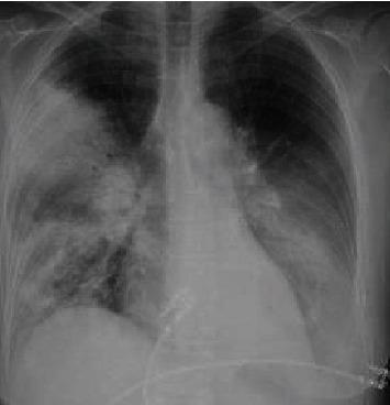
This diagnostic image is a posterior-anterior (PA) view chest X-ray demonstrating multifocal pneumonia with prominent consolidations. In the right lung, there is dense consolidation involving the upper lobe, characterized by increased radiopacity that obscures the regional vascular markings. Patchy, ill-defined airspace opacities are also visible extending into the right lower lobe. The left lung field reveals significant consolidation primarily localized within the lower lobe, partially obscuring the left hemidiaphragm silhouette. The cardiac silhouette appears normal in size, and the trachea is midline. The visual findings are consistent with severe pulmonary infection, such as atypical pneumonia (e.g., Legionella species). This clinical imaging serves as an educational example of multifocal bacterial consolidation and its radiographic distribution in the context of acute respiratory distress and sepsis.
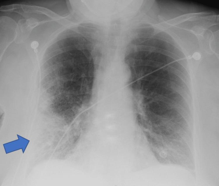
A frontal (anteroposterior) chest X-ray demonstrating clinical signs of pneumonia. A prominent blue arrow indicates a large area of increased opacity in the right lower lung field, suggestive of lobar consolidation or a dense pulmonary infiltrate. The opacity is heterogeneous with ill-defined borders, blending into the surrounding parenchyma. Beyond the focal area, there are diffuse bilateral reticulonodular interstitial markings, more pronounced in the mid and lower lung zones. The right costophrenic angle is relatively sharp, while the left shows mild blunting. Several medical devices are present: multiple radiopaque ECG leads/electrodes are positioned on the upper chest and shoulders, and a radiopaque enteric tube (nasogastric or feeding tube) is visible traversing the mediastinum toward the stomach. The image illustrates typical radiological findings of an infectious process in an acute clinical setting, highlighting focal consolidation against a background of generalized pulmonary congestion or inflammation.
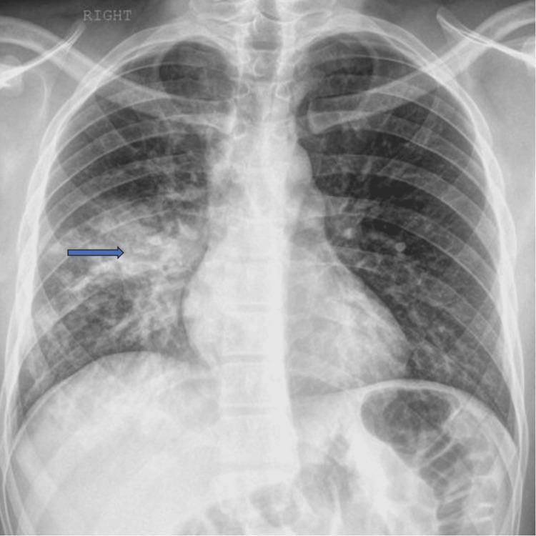
This diagnostic image is a posterior-anterior (PA) chest X-ray demonstrating common radiographic features of focal pneumonia. The primary finding is located in the right lung field, where patchy airspace opacities and an area of pulmonary consolidation are visible, highlighted by a blue arrow. The consolidation is characterized by increased density that obscures the underlying pulmonary vasculature, consistent with fluid or inflammatory exudate replacing air in the alveoli. The left lung field appears clear without similar focal opacities. The mediastinal contours, including the cardiac silhouette and trachea, are within normal limits. The costophrenic angles are sharp, suggesting the absence of significant pleural effusion. Bony structures, including the ribs, clavicles, and vertebrae, as well as the surrounding soft tissues, show no acute abnormalities. This image serves as a clinical example of localized infectious process, such as community-acquired pneumonia, in an adult patient.
community acquired pneumonia management guidelines
Pneumonia - Clinical Case Study
The Case
History of Presenting Illness
- Sudden-onset high-grade fever (39.8°C) with chills
- Productive cough with yellow-green sputum
- Worsening dyspnea on exertion
- Right-sided pleuritic chest pain
- Malaise and loss of appetite
Physical Examination
| Parameter | Finding |
|---|---|
| Temperature | 39.8°C |
| Heart Rate | 108 bpm |
| Respiratory Rate | 28/min |
| BP | 128/82 mmHg |
| SpO2 | 91% on room air |
| Mental status | Alert, slightly confused |
Investigations
- WBC: 18,200/mm³ (neutrophilia, 85%)
- CRP: 142 mg/L
- Blood urea nitrogen (BUN): 24 mg/dL
- Serum Na: 133 mEq/L
- Serum glucose: 218 mg/dL (elevated due to infection stress)
- Blood cultures: x2 sent before antibiotics
Questions for Discussion
Q1. What is the Diagnosis?
- Textbook of Family Medicine 9e, p. 321
Q2. Apply the CURB-65 Severity Score
| Criterion | This Patient | Score |
|---|---|---|
| C - Confusion (new) | Yes (slightly) | 1 |
| U - Urea > 7 mmol/L (BUN > 19 mg/dL) | BUN 24 mg/dL = Yes | 1 |
| R - Respiratory Rate ≥ 30/min | 28/min = No | 0 |
| B - BP <90 systolic OR ≤60 diastolic | No | 0 |
| 65 - Age ≥ 65 years | Age 58 = No | 0 |
| Total | 2 |
- Goldman-Cecil Medicine, PSI Table 85-3
Q3. What are the Common Causative Organisms?
- Streptococcus pneumoniae - most common overall
- Haemophilus influenzae - especially in COPD/smokers
- Staphylococcus aureus - aggressive, can follow viral illness (influenza)
- Mycoplasma pneumoniae - smoldering onset, dry cough, positive cold agglutinins
- Chlamydophila pneumoniae
- Legionella pneumophila - Legionnaire's disease; check urinary antigen; associated with multifocal consolidation
-
Immunocompromised/HIV: Pneumocystis jirovecii (PCP), Cryptococcus, CMV
-
Alcoholics: H. influenzae, anaerobes (aspiration), Klebsiella
-
Nursing home/healthcare: Gram-negative rods, MRSA, MDR organisms
-
Textbook of Family Medicine 9e, p. 321
Q4. How do Typical vs. Atypical Pneumonia Differ Clinically?
| Feature | Typical (e.g., S. pneumoniae) | Atypical (e.g., Mycoplasma) |
|---|---|---|
| Onset | Sudden | Gradual |
| Fever | High-grade, chills | Low-grade |
| Cough | Productive, purulent | Dry, non-productive |
| WBC | High (>15,000), neutrophilia | Mild elevation |
| CXR | Lobar consolidation | Bilateral interstitial pattern |
| Auscultation | Dullness, bronchial breathing, egophony | Diffuse crackles |
| Response to β-lactams | Yes | Incomplete (needs macrolide/quinolone) |
Q5. Management Plan
- Beta-lactam (e.g., IV Amoxicillin-Clavulanate or Ceftriaxone) + Macrolide (e.g., Azithromycin) - to cover both typical and atypical organisms
- OR Respiratory fluoroquinolone monotherapy (e.g., Moxifloxacin, Levofloxacin)
- Duration: minimum 5 days, and at least 48-72 hours after last fever/clinical instability
Although atypical pathogens commonly cause CAP, controlled trials show β-lactam antibiotics are as effective as macrolides and quinolones in most cases. In bacteremic pneumococcal pneumonia, dual therapy (β-lactam + atypical coverage) may reduce case-fatality rates vs. β-lactam alone. - Textbook of Family Medicine 9e
- Supplemental oxygen to maintain SpO2 ≥ 94%
- IV fluids for hydration
- Antipyretics (paracetamol)
- Blood glucose monitoring (DM patient)
- Chest physiotherapy
- Afebrile for >24 hours
- Stable vitals, HR <100, RR <24
- SpO2 ≥ 90% on room air
- Able to tolerate oral medications
Q6. Complications to Watch For
| Complication | Signs |
|---|---|
| Parapneumonic effusion / Empyema | Persistent fever, dullness, pleural fluid on CXR |
| Lung abscess | Fever >2 weeks, foul-smelling sputum, cavity on CXR |
| Septicaemia / Septic shock | Hypotension, altered consciousness, multiorgan failure |
| ARDS | Worsening hypoxia despite O2, bilateral infiltrates |
| Respiratory failure | SpO2 not maintaining, rising pCO2 - may need NIV/intubation |
Q7. Prevention
-
Annual influenza vaccine - for all patients >50 years and those with diabetes, chronic lung disease, or immunodeficiency
-
Pneumococcal vaccine (PPSV23 or PCV15/20) - for all adults ≥65 years; re-vaccination after 10 years if first dose given before age 65
-
Smoking cessation counseling
-
Good hand hygiene
-
Textbook of Family Medicine 9e, Key Points
Recent Evidence Note
Summary
| Domain | Key Teaching Points |
|---|---|
| Diagnosis | Clinical + CXR; lobar consolidation = typical bacterial |
| Severity | CURB-65 and PSI guide admission decisions |
| Organisms | S. pneumoniae most common; consider atypicals |
| Treatment | β-lactam + macrolide OR respiratory fluoroquinolone |
| Duration | Minimum 5 days, 48-72h post-defervescence |
| Prevention | Influenza + pneumococcal vaccines |
| Complications | Effusion, abscess, sepsis, ARDS |